

[Linarin inhibits microglia activation-mediated neuroinflammation and neuronal apoptosis in mouse spinal cord injury by inhibiting the TLR4/NF-κB pathway]
- PMID: 39276055
- PMCID: PMC11378057
- DOI: 10.12122/j.issn.1673-4254.2024.08.18
[Linarin inhibits microglia activation-mediated neuroinflammation and neuronal apoptosis in mouse spinal cord injury by inhibiting the TLR4/NF-κB pathway]
Abstract
Objective: To investigate the mechanism underlying the neuroprotective effect of linarin (LIN) against microglia activation-mediated inflammation and neuronal apoptosis following spinal cord injury (SCI).
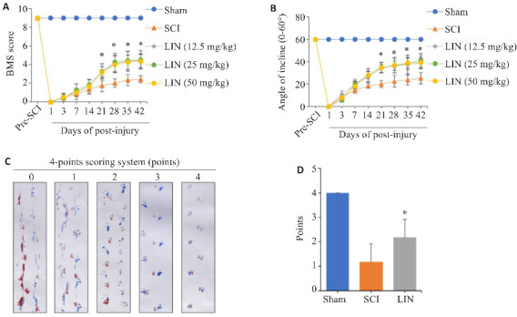
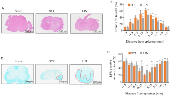
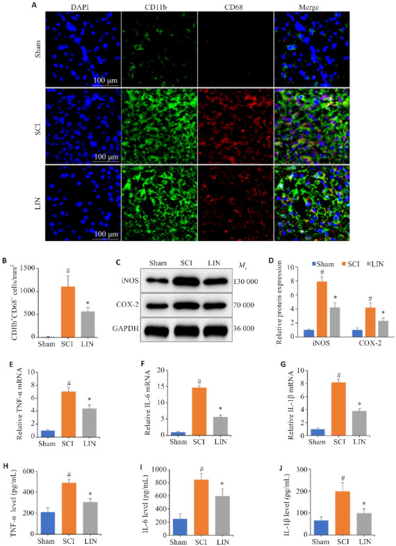
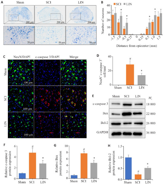
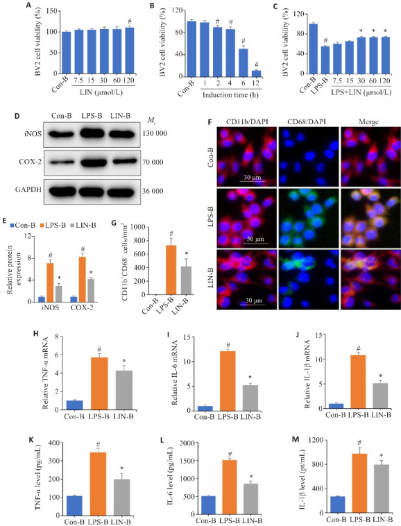
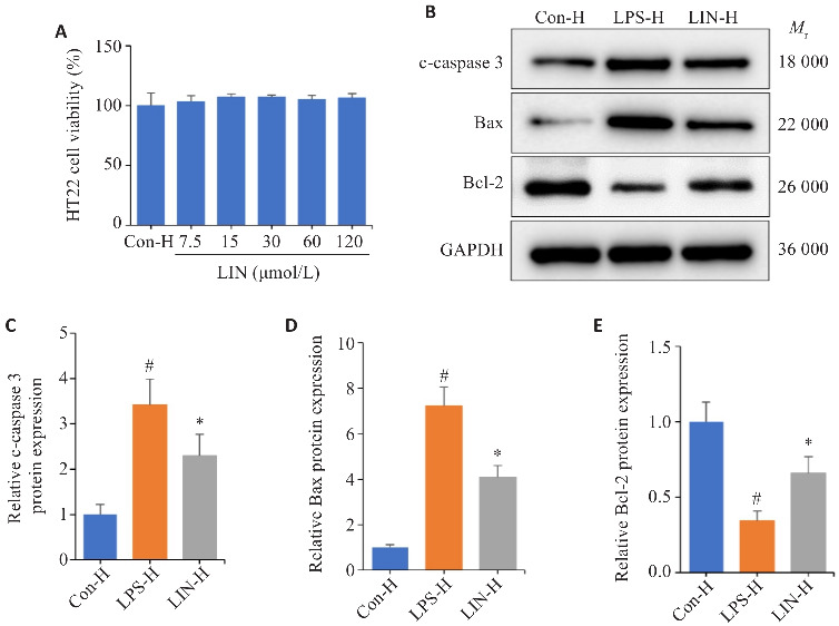
Methods: Fifty C57BL/6J mice (8- 10 weeks old) were randomized to receive sham operation, SCI and linarin treatment at 12.5, 25, and 50 mg/kg following SCI (n=10). Locomotor function recovery of the SCI mice was assessed using the Basso Mouse Scale, inclined plane test, and footprint analysis, and spinal cord tissue damage and myelination were evaluated using HE and LFB staining. Nissl staining, immunofluorescence assay and Western blotting were used to observe surviving anterior horn motor neurons in injured spinal cord tissue. In cultured BV2 cells, the effects of linarin against lipopolysaccharide (LPS)‑induced microglia activation, inflammatory factor release and signaling pathway changes were assessed with immunofluorescence staining, Western blotting, RT-qPCR, and ELISA. In a BV2 and HT22 cell co-culture system, Western blotting was performed to examine the effect of linarin against HT22 cell apoptosis mediated by LPS-induced microglia activation.
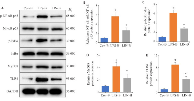
Results: Linarin treatment significantly improved locomotor function (P < 0.05), reduced spinal cord damage area, increased spinal cord myelination, and increased the number of motor neurons in the anterior horn of the SCI mice (P < 0.05). In both SCI mice and cultured BV2 cells, linarin effectively inhibited glial cell activation and suppressed the release of iNOS, COX-2, TNF-α, IL-6, and IL-1β, resulting also in reduced neuronal apoptosis in SCI mice (P < 0.05). Western blotting suggested that linarin-induced microglial activation inhibition was mediated by inhibition of the TLR4/NF- κB signaling pathway. In the cell co-culture experiments, linarin treatment significantly decreased inflammation-mediated apoptosis of HT22 cells (P < 0.05).
Conclusion: The neuroprotective effect of linarin is medicated by inhibition of microglia activation via suppressing the TLR4/NF‑κB signaling pathway, which mitigates neural inflammation and reduce neuronal apoptosis to enhance motor function of the SCI mice.
目的: 探讨蒙花苷(LIN)在脊髓损伤(SCI)后对激活小胶质细胞介导的炎症和神经元凋亡的神经保护作用和潜在机制。
方法: 将50只8~10周龄C57BL/6J小鼠随机分为假手术组(Sham组)、脊髓损伤模型组(SCI组)和LIN药物治疗组(12.5、25、50 mg/kg),10只/组。使用巴索小鼠评分量表(BMS)、斜板实验和脚印分析评估SCI小鼠的运动功能恢复情况;通过HE染色和LFB染色分别评估脊髓组织损伤面积和髓鞘化面积。采用Nissl染色、免疫荧光及Western blotting观察损伤脊髓组织前角运动神经元的保留情况。体外实验通过LPS诱导BV2细胞建立体外炎症模型,同时给予蒙花苷进行干预,将BV2细胞分为对照组(Con-B)、脂多糖组(LPS-B)及蒙花苷组(LIN-B)。通过免疫荧光、Western blotting、RT-qPCR及ELISA检测体内外小胶质细胞活化及炎症因子的释放情况,进一步采用Western blotting验证信号通路的改变。构建体外BV2细胞和HT22细胞共培养模型,分为Con-H组、LPS-H组以及LIN-H组。采用Western blotting检测激活小胶质细胞介导的HT22细胞凋亡情况。
结果: 行为学检测发现,LIN组小鼠BMS评分显著提高,承受的斜板高度更高,后肢行走协调且足迹清晰(P<0.05)。组织学染色结果显示,LIN组小鼠脊髓组织损伤面积减少,髓鞘化面积增加,并且小鼠脊髓前角运动神经元的数量增加(P<0.05)。体内外实验证实,LIN显著抑制小胶质细胞的活化和炎症因子(iNOS、COX-2、TNF-α、IL-6、IL-1β)的释放,并减少了体内神经元的凋亡(P<0.05)。Western blotting显示LIN抑制小胶质细胞活化可能与抑制TLR4/NF-κB信号通路有关。体外共培养实验结果显示,LIN的干预能够减少炎症介导的神经元凋亡数量(P<0.05)。
结论: LIN可能通过抑制TLR4/NF-κB信号通路抑制小胶质细胞的激活,减轻神经炎症,改善SCI后的神经元凋亡,提高SCI小鼠运动能力,发挥其神经保护作用。
Keywords: TLR4/NF-κB; apoptosis; linarin; neuroinflammation; spinal cord injury.
Figures
Similar articles
-
Effect of Sakuranetin on Microglia-Mediated Neuroinflammation After Spinal Cord Injury.Zhongguo Yi Xue Ke Xue Yuan Xue Bao. 2024 Dec;46(6):836-848. doi: 10.3881/j.issn.1000-503X.16087. Zhongguo Yi Xue Ke Xue Yuan Xue Bao. 2024. PMID: 39212067
-
Wogonoside alleviates microglia-mediated neuroinflammation via TLR4/MyD88/NF-κB signaling axis after spinal cord injury.Eur J Pharmacol. 2024 Jun 15;973:176566. doi: 10.1016/j.ejphar.2024.176566. Epub 2024 Apr 16. Eur J Pharmacol. 2024. PMID: 38636801
-
circ-Ncam2 (mmu_circ_0006413) Participates in LPS-Induced Microglia Activation and Neuronal Apoptosis via the TLR4/NF-κB Pathway.J Mol Neurosci. 2022 Aug;72(8):1738-1748. doi: 10.1007/s12031-022-02018-6. Epub 2022 Jun 10. J Mol Neurosci. 2022. PMID: 35687299
-
The Impact of Nuclear Factor Kappa B on the Response of Microglia in Spinal Cord Injuries.Cureus. 2025 Feb 20;17(2):e79367. doi: 10.7759/cureus.79367. eCollection 2025 Feb. Cureus. 2025. PMID: 40125122 Free PMC article. Review.
-
The Inhibition of Inflammatory Signaling Pathway by Secretory Leukocyte Protease Inhibitor can Improve Spinal Cord Injury.Cell Mol Neurobiol. 2020 Oct;40(7):1067-1073. doi: 10.1007/s10571-020-00799-1. Epub 2020 Jan 28. Cell Mol Neurobiol. 2020. PMID: 31993863 Free PMC article. Review.
Cited by
-
[Monotropein improves motor function of mice with spinal cord injury by inhibiting the PI3K/AKT signaling pathway to suppress neuronal apoptosis].Nan Fang Yi Ke Da Xue Xue Bao. 2025 Apr 20;45(4):774-784. doi: 10.12122/j.issn.1673-4254.2025.04.13. Nan Fang Yi Ke Da Xue Xue Bao. 2025. PMID: 40294928 Free PMC article. Chinese.
References
-
- Lin FX, Pan QL, Gu HY, et al. . The role of resveratrol on spinal cord injury: from bench to bedside[J]. Mol Neurobiol, 2024, 61(1): 104-19. - PubMed
Publication types
MeSH terms
Substances
LinkOut - more resources
Full Text Sources
Medical
Research Materials