. 2024 Oct 17;187(21):5967-5980.e17.
doi: 10.1016/j.cell.2024.08.024. Epub 2024 Sep 13.

Co-opting templated aggregation to degrade pathogenic tau assemblies and improve motor function

Affiliations

Co-opting templated aggregation to degrade pathogenic tau assemblies and improve motor function

Lauren V C Miller et al. Cell. .

Abstract

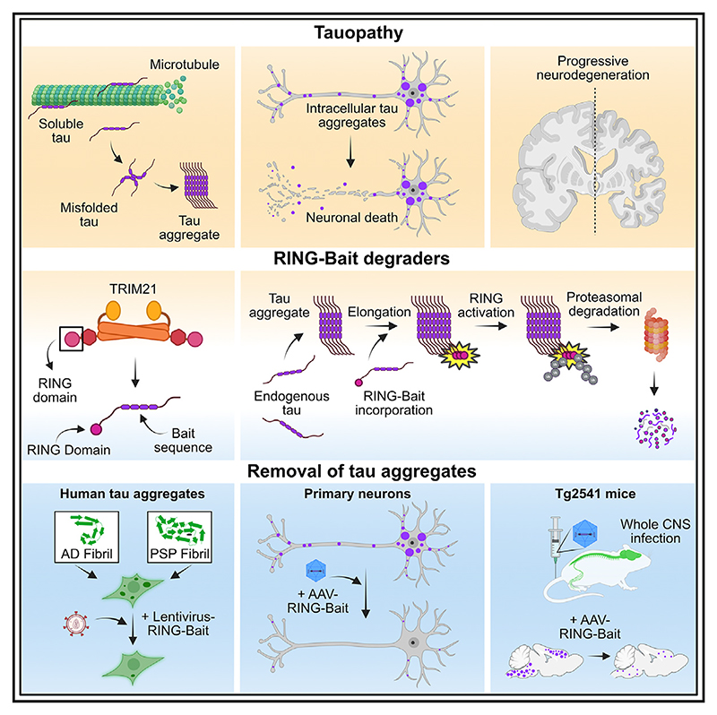
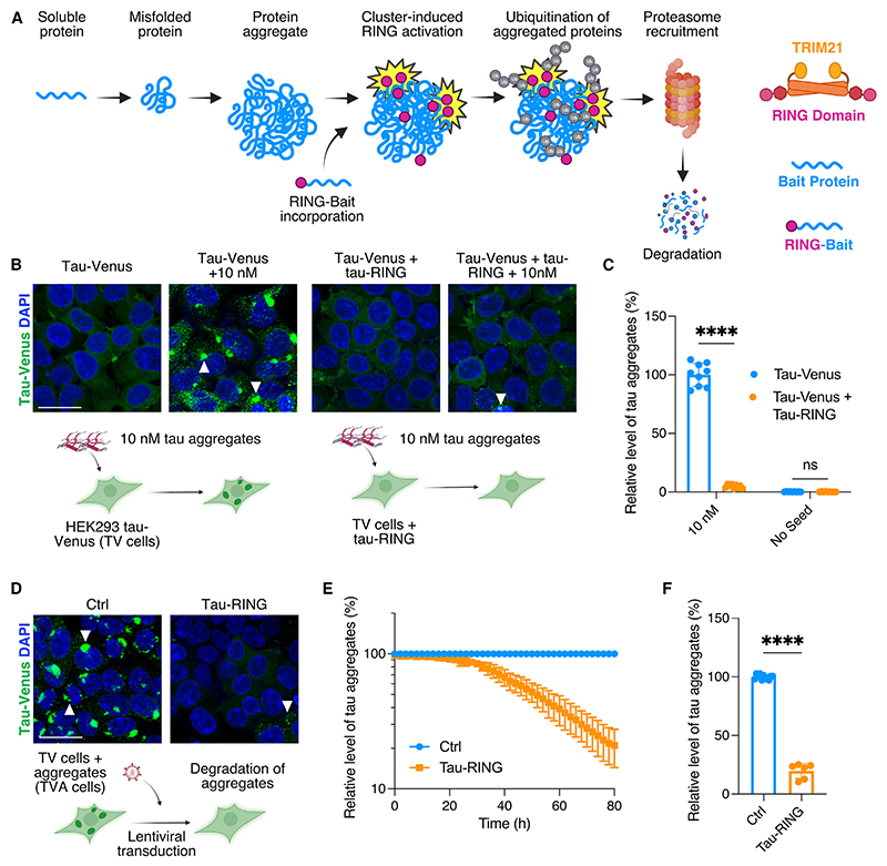
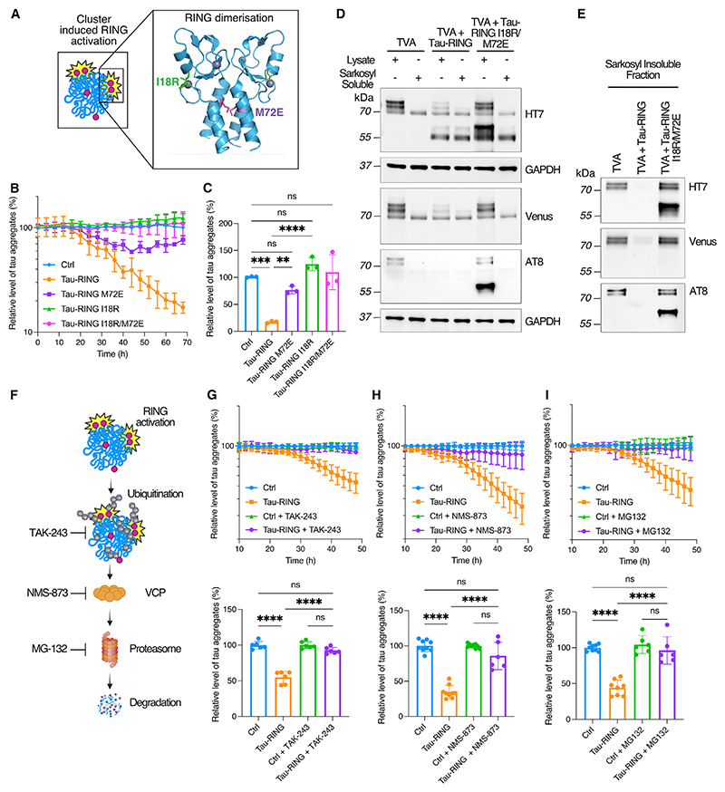
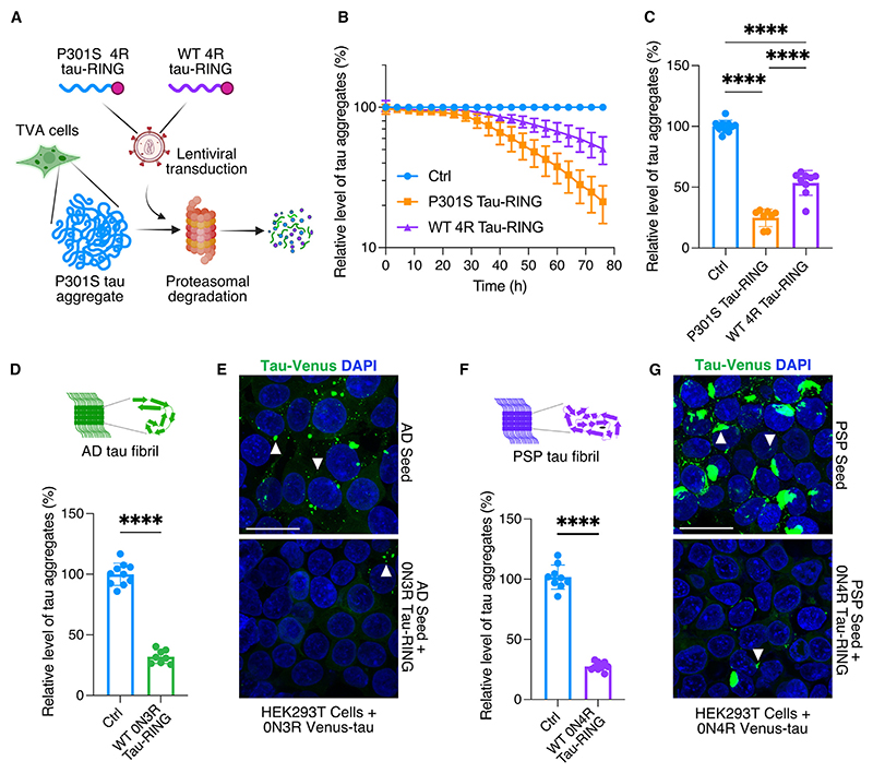
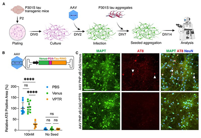
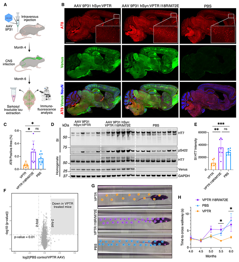
Protein aggregation causes a wide range of neurodegenerative diseases. Targeting and removing aggregates, but not the functional protein, is a considerable therapeutic challenge. Here, we describe a therapeutic strategy called "RING-Bait," which employs an aggregating protein sequence combined with an E3 ubiquitin ligase. RING-Bait is recruited into aggregates, whereupon clustering dimerizes the RING domain and activates its E3 function, resulting in the degradation of the aggregate complex. We exemplify this concept by demonstrating the specific degradation of tau aggregates while sparing soluble tau. Unlike immunotherapy, RING-Bait is effective against both seeded and cell-autonomous aggregation. RING-Bait removed tau aggregates seeded from Alzheimer's disease (AD) and progressive supranuclear palsy (PSP) brain extracts and was also effective in primary neurons. We used a brain-penetrant adeno-associated virus (AAV) to treat P301S tau transgenic mice, reducing tau pathology and improving motor function. A RING-Bait strategy could be applied to other neurodegenerative proteinopathies by replacing the Bait sequence to match the target aggregate.

Keywords: Alzheimer’s disease; TRIM21; antibodies; gene therapy; neurobiology; neurodegeneration; protein engineering; targeted protein degradation; tauopathy; ubiquitination.


Conflict of interest statement

Declaration of interests L.V.C.M., G.P., W.A.M., and L.C.J. are listed as inventors on a patent containing data published in this paper.

Figures

Graphical abstract
Graphical abstract
Figure 1
Figure 1. Tau-RING prevents seeded aggregation and removes existing aggregates
(A) Schematic of RING-Bait technology. (B) Representative confocal images of HEK293 reporter cell line expressing tau-venus (TV cells) ± tau-RING, seeded ±10 nM tau aggregates with Lipofectamine 2000. White arrows denote examples of aggregates. (C) Quantification of live-cell images from cells treated as in (B). n = 3. (D) Representative confocal images of HEK293 reporter cell line expressing tau-venus, constitutively bearing tau aggregates (TVA cells), infected with lentivirus containing tau-RING and evaluated after 72 h. (E) Time course of tau aggregates in TVA reporter cells with and without tau-RING. n = 3. (F) Quantification of the number of aggregates in TVA cells 72 h post transduction with tau-RING lentivirus from live-cell images. N = 3. Scale bars, 25 μm. Statistical significance for (C) determined by two-way ANOVA and Sidak’s multiple comparison test. Statistical significance for (F) determined by unpaired t test. ****p < 0.0001. ns, non-significant. See also Figures S1 and S2.
Figure 2
Figure 2. Tau-RING requires canonical TRIM21 pathway components for degradation
(A) Schematic of RING domain activation via dimerization. The I18 and M72 residues are highlighted in green and purple, respectively. (B) Time course of tau-RING ± I18R, M72E, I18R/M72E RING mutations in TVA cells. (C) Quantification of the number of aggregates in TVA cells 72 h post transduction with tau-RING lentivirus. N = 3. (D and E) TVA cells treated ±tau-RING or tau-RING I18R/M72E lentivirus for 72 h and then fractionated into sarkosyl-soluble and sarkosyl-insoluble tau by ultracentrifugation. Western blots probed for total tau (HT7), venus protein, hyperphorphorylated tau (AT8), and loading control GAPDH. N = 3 experiments pooled. (F) Schematic of inhibitors of degradation pathway components utilized by TRIM21. TAK-243 is an E1 inhibitor, NMS-873 is a VCP inhibitor, and MG-132 is a proteasome inhibitor. (G) Time course of TVA cells treated with tau-RING ± TAK-243 (100 nM) and quantification of end point at 48 h post transduction. N = 3. (H) Time course of TVA cells treated with tau-RING ± NMS-873 (2 μM) and quantification at 48 h post transduction. N = 3. (I) Time course of TVA cells treated with tau-RING ± MG-132 (5 μM) and quantification at 48 h post transduction. N = 3. Statistical significance for (C) and (G)–(I) determined by one-way ANOVA and Tukey’s multiple comparisons test. **p < 0.01, ***p < 0.001, ****p < 0.0001; ns, non-significant. See also Figure S3.
Figure 3
Figure 3. RING-Bait is effective against human brain-derived tau aggregates
(A) Schematic of the TVA cell assay, with a lentivirus used to express different isoforms of tau-RING as examples of different “Baits.” Incorporation of the Bait into the aggregate leads to proteasomal degradation and a reduction in the number of puncta by high-content microscopy. (B) Time course of TVA assay, where lentivirus carrying P301S 0N4R tau-RING or WT 0N4R tau-RING is applied to cells. N = 3. (C) Quantification of the number of aggregates in TVA cells treated as in (B) 72 h post transduction. N = 3. (D) Quantification of the number of aggregates in HEK293T cells expressing venus-3R tau, seeded with AD-derived tau aggregates, ±0N3R tau-RING. N = 3. (E) Representative images from cells treated as in (D). (F) Quantification of the number of aggregates in HEK293T cells expressing venus-4R tau, seeded with PSP-derived tau aggregates, ±0N4R tau-RING. N = 3. (G) Representative images from cells treated as in (F). White arrows denote tau aggregates. Scale bars, 25 μm. Statistical significance for (C) determined by one-way ANOVA and Tukey’s multiple comparisons test. Statistical significance for (D) and (F) determined by unpaired t test. ****p < 0.0001.
Figure 4
Figure 4. Tau-RING prevents seeded aggregation in primary neurons
(A) Schematic of the primary neuron seeding assay, using P301S tau transgenic mice at P2. Cultures were infected with AAV PHP.eB carrying venus-P2A-tau-RING (VPTR) or venus only at day in vitro 2 (DIV2), and P301S tau aggregates were added to the media at DIV7. Cultures were evaluated at DIV14 for the number of AT8-positive tau aggregates. (B) Quantification of primary neurons treated ±100 nM tau aggregates, ±venus, or venus-P2A-tau-RING (VPTR) AAV at DIV14. N = 3. (C) Representative immunofluorescence images of primary neuron cultures at DIV14, treated with venus or VPTR AAV, +100 nM P301S tau aggregates. Scale bar, 100 μm. Statistical significance for (B) determined by two-way ANOVA and Sidak’s multiple comparisons test. ****p < 0.0001. ns, non-significant. See also Figure S3.
Figure 5
Figure 5. Tau-RING reduces tau pathology in vivo and improves motor function
(A) Schematic of intravenous injection of P301S mice at 4 months with AAV 9P31. At 6 months, one half of the brain was homogenized in order to extract sarkosyl insoluble (SI) tau assemblies, and the other half was fixed and analyzed for AT8-positive tau aggregates by immunofluorescent staining. (B) Representative immunoflourescence images of mice infected with AAV 9P31 hSyn:VPTR (active tau-RING) or hSyn:VPTR I18R/M72E (inactive tau-RING) at 4 months, or injected with PBS, and evaluated at 6 months for tau aggregates via AT8 staining. Venus fluorescence detected from virally expressed protein. Neuronal nuclei were probed for using an antibody against NeuN. Enlarged cortical region shown to exemplify tau aggregate levels. Scale bar, 2 mm. (C) Quantification of AT8-positive tau aggregates in frontal cortex, as shown in (B). (D) Western blot of the sarkosyl insoluble (SI) fraction of mouse brains treated as in (B), stained for total human tau (HT7) and hyperphosphorylated tau at serine 422 (pS422). Mouse brain homogenate was also probed with HT7 and venus protein, in addition to GAPDH as a loading control. (E) Quantification of SI HT7 in (D). (F) Mass spectrometry analysis of whole brain homogenate from mice treated with PBS compared with hSyn:VPTR AAV. (G) Footsteps of the median mouse from each condition (VPTR, VPTR I18R/M72E, PBS) on the MouseWalker apparatus. (H) Quantification of the time to traverse the walkway for mice from each condition (VPTR, VPTR I18R/M72E, PBS) from 4 to 6 months. N = 6–8 mice per group. Statistical significance for (C) and (E) determined by Brown-Forsythe and Welch ANOVA. Statistical significance for (F) determined by unpaired t test with group-wise correction. Statistical significance for (H) determined by Kruskal-Wallis test. *p < 0.05. **p < 0.01. ****p < 0.0001. ns, non-significant. See also Figures S4 and S5.

References

    1. Shi Y, Zhang W, Yang Y, Murzin AG, Falcon B, Kotecha A, van Beers M, Tarutani A, Kametani F, Garringer HJ, et al. Structure-based classification of tauopathies. Nature. 2021;598:359–363. doi: 10.1038/s41586-021-03911-7. - DOI - PMC - PubMed
    1. Goedert M. NEURODEGENERATION. Alzheimer’s and Parkinson’s diseases: The prion concept in relation to assembled Aβ, tau, and α-synuclein. Science. 2015;349:1255555. - PubMed
    1. Yang Y, Shi Y, Schweighauser M, Zhang X, Kotecha A, Murzin AG, Garringer HJ, Cullinane PW, Saito Y, Foroud T, et al. Structures of α-synuclein filaments from human brains with Lewy pathology. Nature. 2022;610:791–795. doi: 10.1038/s41586-022-05319-3. - DOI - PMC - PubMed
    1. Arseni D, Chen R, Murzin AG, Peak-Chew SY, Garringer HJ, Newell KL, Kametani F, Robinson AC, Vidal R, Ghetti B, et al. TDP-43 forms amyloid filaments with a distinct fold in type A FTLD-TDP. Nature. 2023;620:898–903. doi: 10.1038/s41586-023-06405-w. - DOI - PMC - PubMed
    1. Ji C, Sigurdsson EM. Current Status of Clinical Trials on Tau Immunotherapies. Drugs. 2021;81:1135–1152. doi: 10.1007/s40265-021-01546-6. - DOI - PMC - PubMed

MeSH terms