. 2024 Oct 15:300:120845.
doi: 10.1016/j.neuroimage.2024.120845. Epub 2024 Sep 12.

High-quality lipid suppression and B0 shimming for human brain 1H MRSI

Affiliations

High-quality lipid suppression and B0 shimming for human brain 1H MRSI

Chathura Kumaragamage et al. Neuroimage. .

Abstract

Magnetic Resonance Spectroscopic Imaging (MRSI) is a powerful technique that can map the metabolic profile in the brain non-invasively. Extracranial lipid contamination and insufficient B0 homogeneity however hampers robustness, and as a result has hindered widespread use of MRSI in clinical and research settings. Over the last six years we have developed highly effective extracranial lipid suppression methods with a second order gradient insert (ECLIPSE) utilizing inner volume selection (IVS) and outer volume suppression (OVS) methods. While ECLIPSE provides > 100-fold in lipid suppression with modest radio frequency (RF) power requirements and immunity to B1+ field variations, axial coverage is reduced for non-elliptical head shapes. In this work we detail the design, construction, and utility of MC-ECLIPSE, a pulsed second order gradient coil with Z2 and X2Y2 fields, combined with a 54-channel multi-coil (MC) array. The MC-ECLIPSE platform allows arbitrary region of interest (ROI) shaped OVS for full-axial slice coverage, in addition to MC-based B0 field shimming, for robust human brain proton MRSI. In vivo experiments demonstrate that MC-ECLIPSE allows axial brain coverage of 92-95 % is achieved following arbitrary ROI shaped OVS for various head shapes. The standard deviation (SD) of the residual B0 field following SH2 and MC shimming were 25 ± 9 Hz and 18 ± 8 Hz over a 5 cm slab, and 18 ± 5 Hz and 14 ± 6 Hz over a 1.5 cm slab, respectively. These results demonstrate that B0 magnetic field shimming with the MC array supersedes second order harmonic capabilities available on standard MRI systems for both restricted and large ROIs. Furthermore, MC based B0 shimming provides comparable shimming performance to an unrestricted SH5 shim set for both restricted, and 5-cm slab shim challenges. Phantom experiments demonstrate the high level of localization performance achievable with MC-ECLIPSE, with ROI edge chemical shift displacements ranging from 1-3 mm with a median value of 2 mm, and transition width metrics ranging from 1-2.5 mm throughout the ROI edge. Furthermore, MC based B0 shimming is comparable to performance following a full set of unrestricted spherical harmonic fields up to order 5. Short echo time MRSI and GABA-edited MRSI acquisitions in the human brain following MC-shimming and arbitrary ROI shaping demonstrate full-axial slice coverage and extracranial lipid artifact free spectra. MC-ECLIPSE allows full-axial coverage and robust MRSI acquisitions, while allowing interrogation of cortical tissue proximal to the skull, which has significant value in a wide range of neurological and psychiatric conditions.

Keywords: B0 field shimming; ECLIPSE; GABA-MRSI, GOIA; Human brain; Lipid suppression; MRSI; Multicoil; Proton MRSI, OVS.


Conflict of interest statement

Declaration of competing interest The authors have no competing interests (financial or personal) to disclose.

Figures

Fig. 1.
Fig. 1.
Components and construction of the MC-ECLIPSE. (A-C) illustrates the modular components of a MC element. 3D printed with ABS plastic. An assembled MC element is illustrated (B). Illustration of the overall MC-ECLISE system (C-D), which include the Z2, and X2Y2 gradient coil wire patterns and the 54 channel MC-array. The 54 channel MC array populated on the inner surface of the nylon cylinder (D).
Fig. 2.
Fig. 2.
Segmented X2Y2 gradient coil. The X2Y2 coil was segmented into two parts illustrated in bronze (X2Y2–1) and black (X2Y2–2) illustrated in A, driven by two independent current amplifiers. The simulated B0 field patterns in the x-y plane generated by the X2Y2–1, X2Y2–2 coil patterns, and combined contribution with equal amplitude X2Y2T = X2Y2–1 + X2Y2–2 are illustrated in B, C, and D, respectively. (E) An example axial brain slice with elliptical selection for outer volume suppression with Z2 (F), X2Y2–1 (G), X2Y2–2 (H), and X2Y2T (I) fields. Here the amplitude ratio of X2Y2–1:X2Y2–2 is 1:1 providing a pure X2Y2 field, and as a result produces an elliptical field patten (Total) when Z2 and necessary linear gradients (X, Y, Z) are imposed (J). (K) The resulting elliptical ROI provides 90.7 % axial coverage. Alternatively, X2Y2–1 and X2Y2–2 coils can be independently driven with a −1:3.5 ratio (M-N), that produces an asymmetric ROI around the y-axis (O-P) that provides a substantially improved 98.5 % axial coverage (Q) relative to elliptical localization for an asymmetrical axial head shape that deviates from an ellipse.
Fig. 3.
Fig. 3.
Pulse sequence illustrating ROI shaping and B0 shimming for 1H MRSI acquisitions. A 4-cycle asymmetric-GOIA pulse-based OVS method with an interleaved VAPOR water suppression method was utilized. OVS pulses with relative amplitudes of 0.74, 0.78, 0.87 and 1.00 are placed 1179, 558, 216 and 48 ms before the slice-selective excitation pulse, respectively. Water suppression pulses have relative flip angles of α, α, 1.78α, α, 1.78α, α and 1.78α, which are placed 280, 265, 250, 170, 98, 28 and 13 ms before the MRSI component, respectively. To attain arbitrary ROI shaping, gradient modulation was incorporated in both ECLIPSE and MC as illustrated. A static DC shim optimized over the MRSI slab of interest is superimposed using the MC-array. For brevity, only MC 1,27, and 54 are illustrated for ROI shaping and DC shimming. The MRSI method precedes the OVS and VAPOR water suppression components, where short TE unedited, and J-difference edited MRSI methods were employed.
Fig. 4.
Fig. 4.
Process of arbitrary ROI shaping with MC-ECLIPSE. (A) Axial anatomical image of a highly asymmetrical head shape. (B) skull stripped anatomical in (A), which is used to generate a brain mask illustrated in (C). Using (C), three layers of contours around the ROI edge is generated, where the three contours are assigned B0 values that represent a desired gradient along the ROI edge to achieve high selectivity and low chemical shift displacements (CSD) (D). In this example a 600 Hz/mm gradient is set, at the ROI-edge, which results in a target chemical shift displacement of ~ 1 mm (fat-water separation at 4 T). A fit of the contour field in (D) is performed with ECLIPSE and MC B0 basis fields, which results in the spatial-B0 field illustrated in (E). The resulting ROI selected in simulation is illustrated in (F), where the red and green ROI’s illustrate lipid and water ROIs respectively. Notably, the simulated CSD around the ROI ranged from 1–3 mm, with a median of 2 mm. (G) illustrates experimentally acquired MR images following MC-ECLIPSE based OVS, where excellent agreement is present between the theoretical ROI and experimental acquisition, with the acquired image closely following the theoretical ROI shape. Transition width metrics (5–95 %) were evaluated in a Silicone oil phantom for ROI shaping parameters for the illustrated volunteer (H). Locations with worst-case and best-case TW metrics observed are illustrated in the zoomed cut outs (I), (J) respectively, where the ROI edge TW spanned from 1–2.5 mm throughout the entire ROI.
Fig. 5.
Fig. 5.
Combined results of ROI shaping and B0 shimming with the MC-ECLIPSE on five volunteers. The 1-cm thick axial slice location is illustrated in the sagittal image (I), anatomical ROI’s with superimposed simulated OVS ROI is illustrated in (II). (III) Experimental axial images following OVS, with attained axial slice coverage marked, which spans over the 1 cm slab ranged from 92 % to 95 % for the different head shapes investigated. (IV-VI) are B0 maps of the lowest slice within the slab illustrated in (I) following theoretical SH2, theoretical MC, and experimental MC for the five volunteers, respectively. (VII) and (VIII) illustrate water linewidth maps calculated over 1 × 1cm2 grid following SH2 and MC shimming from water unsuppressed MRSI acquisitions, respectively. The pooled histogram demonstrates the overall reduction in water linewidth following MC shimming with nearly 70 % of voxels with < 12 Hz water linewidth, in comparison to 52 % with SH2 shimming.B0 Magnetic Field Shimming with MC-ECLIPSE.
Fig. 6.
Fig. 6.
The utility of MC-ECLIPSE for B0 shimming of the human brain. Illustrated are static B0 shimming comparisons in vivo over the illustrated region spanning 5.1 cm. The rows in order are: B0 map before shimming, B0 map following theoretical 2nd order shimming, theoretical B0 map following MC shimming, and experimentally acquired B0 map following MC shimming. The improved shimming performance with the MC is evident by the histogram illustrated compared to best-case SH2 shimming, where std, 80 %, 90 %, and 95 % frequency spans for theoretical SH2 were 28 Hz, 37 Hz, 59 Hz, and 17 Hz, 24 Hz, 37 Hz, and 53 Hz, following experimental MC shimming, respectively.
Fig. 7.
Fig. 7.
Overall ROI shaping and shimming with MC-ECLIPSE over a 20 mm axial slab for GABA edited MRSI (TE/TR = 68/2000 ms, 1.5 × 1.5 cm2 in-plane resolution, 1.5 cm axial slab, 4 averages, leading to a 26-minute acquisition). (A) ROI shaping and attained axial coverage over 3 mm axial slices that span the entire axial slab of interest providing 91 % of overall axial coverage. (B) A comparison of theoretical SH2 and experimentally attained B0 shimming over the slab of interest. Inhomogeneities present following SH2 are virtually eliminated with MC shimming, as illustrated. (C) LCModel fitting of an example unedited and GABA edited spectra. MC-ECLISPE based extracranial lipid suppression allows low amplitude and a stable lipid footprint, even at ROI edge voxels as illustrated, providing artifact-free J-difference edited MR spectra. (D) Water referenced metabolic maps over the entire axial slab following quantification of the unedited spectra (NAA, tCr, tCho, and mI), and edited spectra (Glx, and GABA+). CRLB maps associated with each metabolic map is illustrated below each metabolic map.
Fig. 8.
Fig. 8.
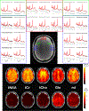
MRSI acquisitions (TE/TR = 30/2000 ms, 1 × 1 cm2 in-plane resolution, 1 cm axial slab, 8.2-minute acquisition) following MC-ECLISPE based OVS and B0 shimming over a 1.5 cm slab. Example spectra (black) and LCModel fits (red) for voxels corresponding to locations marked in the anatomical MRI. The lower panel illustrates metabolic maps for tNAA, tCr, tCho, Glx (Glutamate + Glutamine), and myo-inositol (mI) covering the near-complete axial slab.

References

    1. Aghaeifar A, Zhou J, Heule R, et al., 2020. A 32-channel multi-coil setup optimized for human brain shimming at 9.4T. Magn. Reson. Med. 83 (2), 749–764. - PubMed
    1. Bai X, Edden RA, Gao F, et al., 2015. Decreased gamma-aminobutyric acid levels in the parietal region of patients with Alzheimer’s disease. J. Magn. Reson. Imaging 41 (5), 1326–1331. - PMC - PubMed
    1. Balchandani P, Spielman D, 2008. Fat suppression for 1H MRSI at 7T using spectrally selective adiabatic inversion recovery. Magn. Reson. Med. 59 (5), 980–988. - PMC - PubMed
    1. Bilgic B, Chatnuntawech I, Fan AP, et al., 2014. Fast image reconstruction with L2-regularization. J. Magn. Reson. Imaging 40 (1), 181–191. - PMC - PubMed
    1. Bilgic B, Gagoski B, Kok T, 2013. Adalsteinsson E. Lipid suppression in CSI with spatial priors and highly undersampled peripheral k-space. Magn. Reson. Med. 69 (6), 1501–1511. - PMC - PubMed

LinkOut - more resources