. 2024 Sep 14;15(1):8071.
doi: 10.1038/s41467-024-52021-1.

Dual client binding sites in the ATP-independent chaperone SurA

Affiliations

Dual client binding sites in the ATP-independent chaperone SurA

Bob Schiffrin et al. Nat Commun. .

Abstract

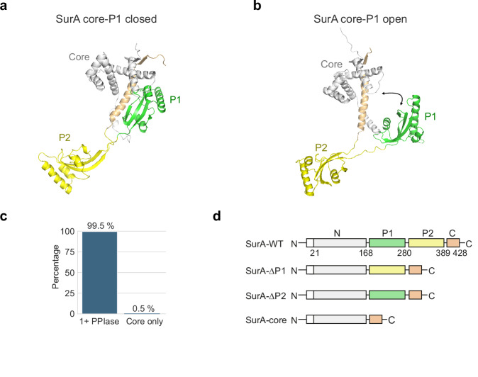
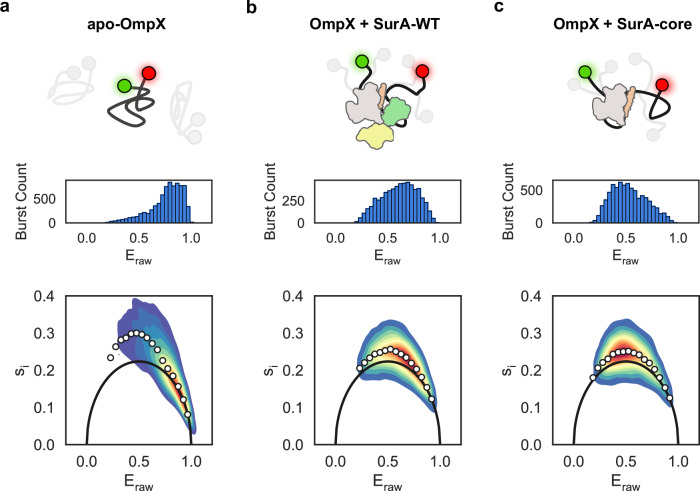
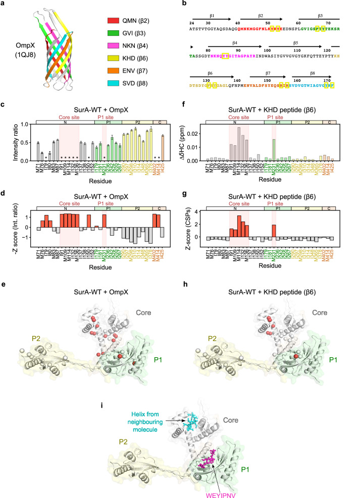
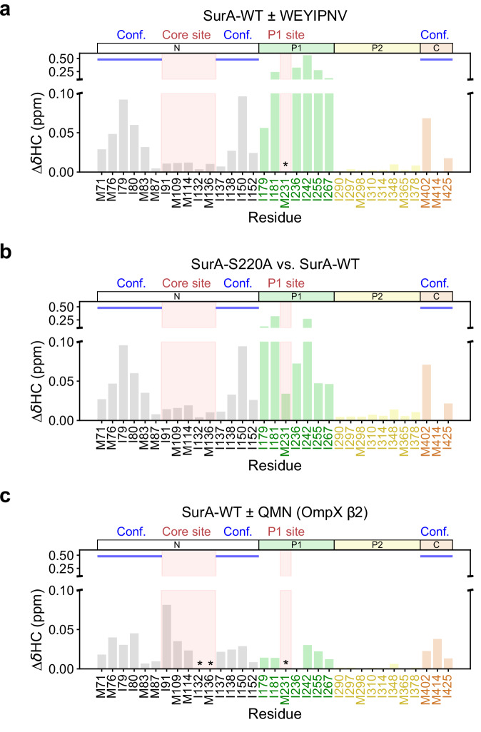
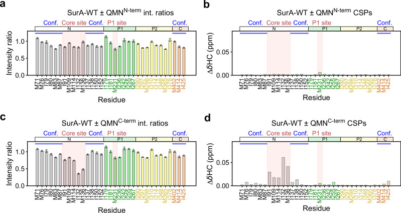
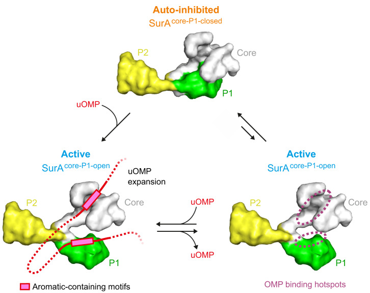
The ATP-independent chaperone SurA protects unfolded outer membrane proteins (OMPs) from aggregation in the periplasm of Gram-negative bacteria, and delivers them to the β-barrel assembly machinery (BAM) for folding into the outer membrane (OM). Precisely how SurA recognises and binds its different OMP clients remains unclear. Escherichia coli SurA comprises three domains: a core and two PPIase domains (P1 and P2). Here, by combining methyl-TROSY NMR, single-molecule Förster resonance energy transfer (smFRET), and bioinformatics analyses we show that SurA client binding is mediated by two binding hotspots in the core and P1 domains. These interactions are driven by aromatic-rich motifs in the client proteins, leading to SurA core/P1 domain rearrangements and expansion of clients from collapsed, non-native states. We demonstrate that the core domain is key to OMP expansion by SurA, and uncover a role for SurA PPIase domains in limiting the extent of expansion. The results reveal insights into SurA-OMP recognition and the mechanism of activation for an ATP-independent chaperone, and suggest a route to targeting the functions of a chaperone key to bacterial virulence and OM integrity.


Conflict of interest statement

The authors declare no competing interests.

Figures

Fig. 1
Fig. 1. Structure, conformations and domain architecture of E. coli SurA.
a Crystal structure of wild-type (WT) E. coli SurA (PDB: 1M5Y) with missing residues added using MODELLER,. In this structure the P1 domain is bound to the core domain (named here, SurA core-P1 closed). Regions are coloured grey (N-terminal region of the core domain), green (P1), yellow (P2) and orange (C-terminal region of the core domain). This colour scheme for SurA domains is used throughout. b Model of WT SurA in which the P1 domain is extended away from the core domain (named here SurA core-P1 open). The model was built using MODELLER, and the crystal structures of full-length SurA (PDB: 1M5Y) and SurA-ΔP2 (PDB: 2PV3). c Bar chart showing the percentage of SurA homologues from the InterPro family IPRO15391 (14,422 homologues) which contain at least one PPIase domain (1+PPIase) or are core domain only (Core only) homologues. d Domain architecture of E. coli SurA-WT and the SurA domain deletion variants used in this study. The signal sequence was not present in the constructs used here, but the numbering used reflects the gene numbering and includes the signal peptide (residues 1–22) (white). Source data are provided as a Source Data file.
Fig. 2
Fig. 2. smFRET captures the conformational dynamics of unfolded OmpX in the presence or absence of SurA variants.
a Schematic showing OmpX (black line) with donor and acceptor fluorophores on the N- and C-termini (green and red) with example alternate conformers shaded in grey (top), Eraw histogram (middle) and BVA analysis (lower) for apo-OmpX. The solid black line represents the expected standard deviation for a static (ms timescale) FRET population. The white circles show the mean si for bin widths of 0.05 of Eraw. b OmpX + SurA-WT and (c) OmpX + SurA-core with panels as in (a). The schematic of SurA is coloured according to Fig. 1. Source data are provided as a Source Data file.
Fig. 3
Fig. 3. Dual binding hotspots in SurA mediate affinity for OMP substrates.
a OmpX-derived peptides analysed here mapped onto the crystal structure of natively folded OmpX (PDB: 1QJ8). b OmpX sequence highlighting peptides analysed, coloured as in (a). Ar-X-Ar and Ar-Ar motifs are highlighted with yellow boxes. c Raw intensity change data and (d) Z-score plots of peak intensity ratios for SurA-WT binding to OmpX. Peaks which are broadened below the noise in the intensity ratio plots are indicated by an asterisk. For these peaks, the noise value for the bound spectra was used in the calculation of Z-scores. Signal to noise ratios were used for the calculation of errors in peak intensities (Methods). e Residues showing significant Z-scores for SurA-WT binding to OmpX mapped onto a model of SurA in a core-P1 open conformation. The 13C- labelled δ1 and ε carbon atoms of Ile and Met residues, respectively, are shown as spheres coloured by Z-score (red for Z-scores > 0 and white for Z-scores <= 0). f Raw CSP data and (g) Z-score plots of CSPs for SurA-WT binding to the OmpX-derived KHD (β6) peptide, comprising a 15-residue sequence that forms β-strand 6 in the OmpX native state. h Residues showing different CSP Z-scores for SurA-WT binding to the KHD (β6) peptide mapped onto a SurA core-P1-open model (coloured as in (g)). The effects of other OmpX-derived peptides on the SurA spectrum are shown in Supplementary Figs. 9, 10. i Model of SurA-WT in the core-P1 open state bound with peptides modelled in at the two binding hotspots. A short polypeptide sequence from a neighbouring molecule in the crystal structure (PDB: 1M5Y) (cyan) is located in the core domain binding crevice. A peptide identified by phage-display which binds to the SurA P1 domain (WEYIPNV) (pink) is modelled in from the P1-WEYIPNV peptide crystal structure (PDB: 2PV1). Source data are provided as a Source Data file.
Fig. 4
Fig. 4. Binding of an OmpX-derived peptide promotes core-P1 open ‘activated’ SurA.
Chemical shift differences between (a) SurA-WT + WEYIPNV and SurA-WT alone, (b) SurA-S220A and SurA-WT, and (c) SurA-WT + QMN peptide (OmpX β2) and SurA-WT alone. Peaks which were broadened beyond detection in peptide-containing samples are indicated with an asterisk. Conf.: residues indicative of conformational change (Supplementary Fig. 14). Samples contained 5 µM SurA or SurA-S220A, in 5 mM EDTA, 20 mM Tris-HCl, pH 8, at 25 °C. In samples containing the WEYIPNV or QMN peptides, these were present at 50 µM or 200 µM, respectively. Source data are provided as a Source Data file.
Fig. 5
Fig. 5. Effects of truncation of the OmpX-derived QMN (b2) peptide on the methyl-TROSY NMR spectrum of SurA-WT.
Intensity ratio (a, c) and CSP (b, d) plots are shown for SurA-WT bound to (a, b) the N-terminal 8 residues of QMN (QMNN-term - QMNKMGGF), and (c, d) the C-terminal 7 residues of QMN (QMNC-term - NLKYRYE). Peaks which were broadened beyond detection in peptide-containing samples are indicated with an asterisk. Conf.: residues indicative of conformational change (Supplementary Fig. 14). Samples contained 5 µM SurA ± 200 µM peptide, 5 mM EDTA, 20 mM Tris-HCl, pH 8, at 25 °C. int: intensity. Signal to noise ratios were used for the calculation of errors in peak intensities in (a, c) (Methods). Source data are provided as a Source Data file.
Fig. 6
Fig. 6. Proposed activation mechanism for OMP binding to SurA.
In the absence of substrate SurA is predominantly in an auto-inhibited core-P1 closed conformation (top) with a minor population (~25%) of an active core-P1 open conformation (right). Binding of OMP substrates (red) to the two identified binding hotspots in the core and P1 domains enables activation of the chaperone and leads to OMP expansion (left). These dual binding sites recognise aromatic containing motifs (e.g., Ar-X-Ar) that are enriched in OMP sequences. Whether it is OMP binding to the core site, P1 site, or both, that leads to chaperone activation is currently not clear, though our results implicate P1 binding in this process (Fig. 4, Supplementary Fig. 13). Binding sites of both hotspots are accessible in the core-P1 closed state suggesting that auto-inhibition is due to repression of conformational dynamics of the core domain, rather than steric blocking, and that sequences linking binding motifs in intact unfolded OMP clients are important for chaperone activation. Expansion of the substrate and prevention of locally collapsed regions in the unfolded chain by activated SurA suggests a mechanism for delivery of the ‘linearised’ client to BAM for its vectorial folding into the OM,.

Similar articles

Cited by

References

    1. Nikaido, H. Molecular basis of bacterial outer membrane permeability revisited. Microbiol. Mol. Biol. Rev.67, 593–656 (2003). 10.1128/MMBR.67.4.593-656.2003 - DOI - PMC - PubMed
    1. Rojas, E. R. et al. The outer membrane is an essential load-bearing element in Gram-negative bacteria. Nature559, 617–621 (2018). 10.1038/s41586-018-0344-3 - DOI - PMC - PubMed
    1. Jarosławski, S., Duquesne, K., Sturgis, J. N. & Scheuring, S. High‐resolution architecture of the outer membrane of the Gram‐negative bacteria Roseobacter denitrificans. Mol. Microbiol.74, 1211–1222 (2009). 10.1111/j.1365-2958.2009.06926.x - DOI - PubMed
    1. Benn, G. et al. Phase separation in the outer membrane of Escherichia coli. Proc. Natl Acad. Sci. USA118, e2112237118 (2021). 10.1073/pnas.2112237118 - DOI - PMC - PubMed
    1. Webby, M. N. et al. Lipids mediate supramolecular outer membrane protein assembly in bacteria. Sci. Adv.8, eadc9566 (2022). 10.1126/sciadv.adc9566 - DOI - PMC - PubMed

Publication types

MeSH terms

LinkOut - more resources