. 2024 Sep 14;15(9):675.
doi: 10.1038/s41419-024-07061-2.

Inhibiting S-palmitoylation arrests metastasis by relocating Rap2b from plasma membrane in colorectal cancer

Affiliations

Inhibiting S-palmitoylation arrests metastasis by relocating Rap2b from plasma membrane in colorectal cancer

Jiangli Zhu et al. Cell Death Dis. .

Abstract

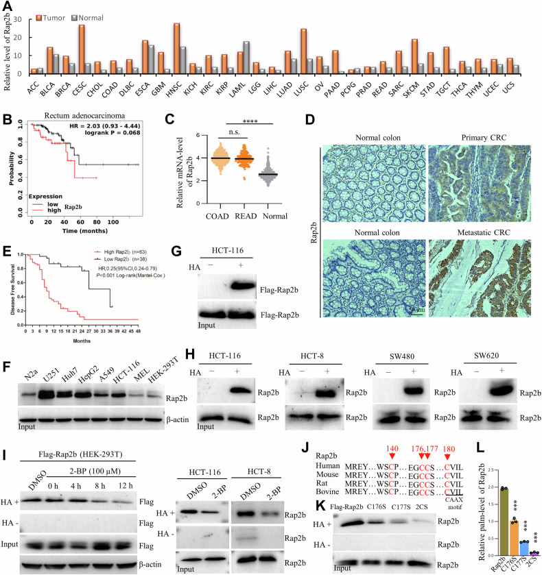
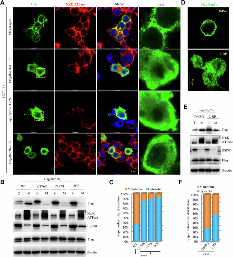
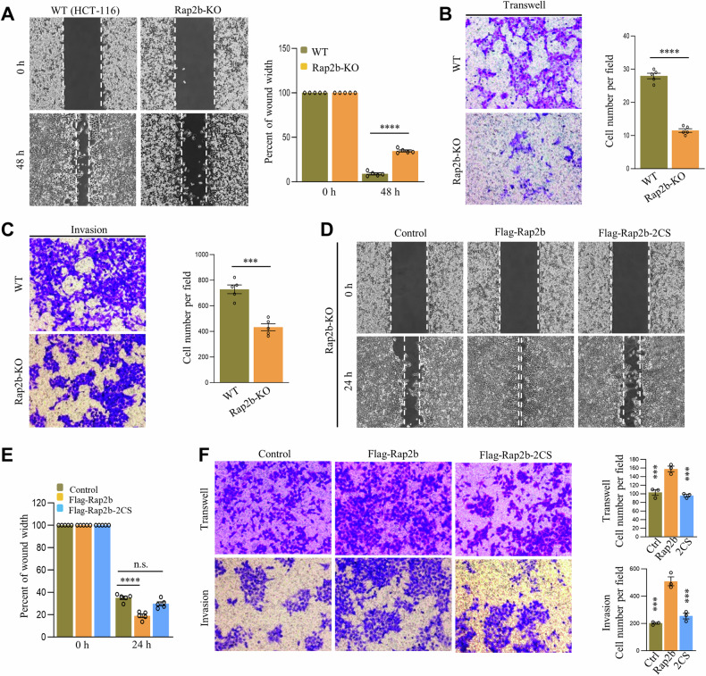
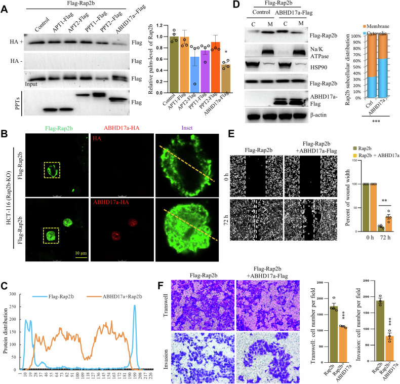
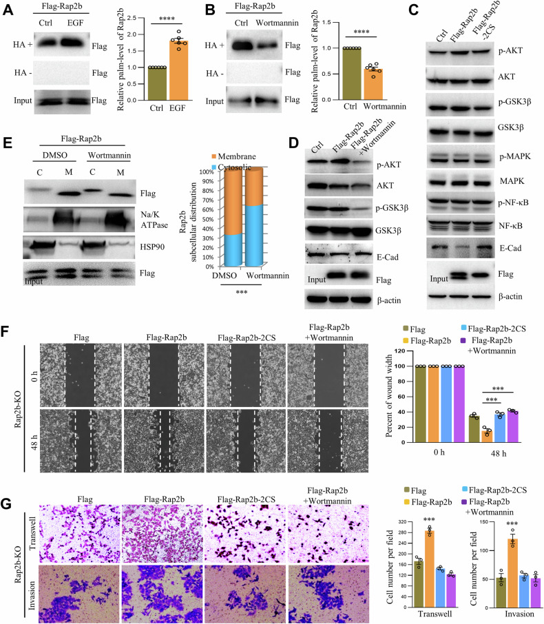
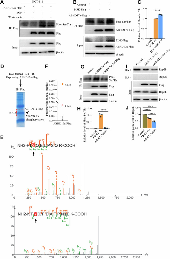
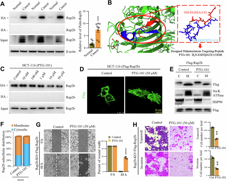
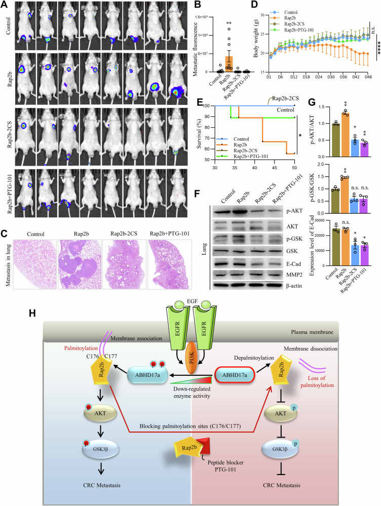
Rap2b, a proto-oncogene upregulated in colorectal cancer (CRC), undergoes protein S-palmitoylation at specific C-terminus sites (C176/C177). These palmitoylation sites are crucial for Rap2b localization on the plasma membrane (PM), as mutation of C176 or C177 results in cytosolic relocation of Rap2b. Our study demonstrates that Rap2b influences cell migration and invasion in CRC cells, independent of proliferation, and this activity relies on its palmitoylation. We identify ABHD17a as the depalmitoylating enzyme for Rap2b, altering PM localization and inhibiting cell migration and invasion. EGFR/PI3K signaling regulates Rap2b palmitoylation, with PI3K phosphorylating ABHD17a to modulate its activity. These findings highlight the potential of targeting Rap2b palmitoylation as an intervention strategy. Blocking the C176/C177 sites using an interacting peptide attenuates Rap2b palmitoylation, disrupting PM localization, and suppressing CRC metastasis. This study offers insights into therapeutic approaches targeting Rap2b palmitoylation for the treatment of metastatic CRC, presenting opportunities to improve patient outcomes.


Conflict of interest statement

JLZ and EYK are listed as inventors on pending patent covering the targeting of C176 and C177 in Rap2b as intervention strategy for treating cancer.

Figures

Fig. 1
Fig. 1. Rap2b is associated with a poor prognosis in CRC and is palmitoylated at cysteine-176 and cysteine-177.
A Upregulation of Rap2b in various types of cancers (data from TCGA). B Negative correlation between high Rap2b expression and survival in rectum adenocarcinoma (TCGA). C Comparison of Rap2b mRNA levels between Colorectal cancer and normal tissues (TCGA). D Immunostaining of Rap2b in paraffin sections of colon cancer to evaluate protein expression. E Kaplan–Meier analysis of Rap2B expression and disease-free survival (DFS) in CRC patients. ***P < 0.001, n = 101. F Evaluation of endogenous Rap2b levels in various cancer cell lines by WB. G, H RAC assay to examine protein palmitoylation of ectopically expressed Flag-Rap2b or endogenously expressed Rap2b. HA− without hydroxylamine; HA+ with hydroxylamine. I RAC assay to examine protein palmitoylation of ectopically expressed Flag-Rap2b or endogenously expressed Rap2b treated with DMSO or 2-BP. J Protein sequence alignment of Rap2b for conservation analysis. K, L RAC assay and quantification of Rap2b and its mutants for protein palmitoylation. n = 3 biological replicates, one-way ANOVA followed by Tukey’s post hoc test, ****P < 0.0001. Data are presented as mean ± S.E.M.
Fig. 2
Fig. 2. Palmitoylation is required for the membrane localization of Rap2b.
A Immunofluorescence microscopy of HCT-116 cells expressing Rap2b or its mutants. Na/K-ATPase was used as a marker for the plasma membrane. B, C Cytosol/membrane fractionation and western blot analysis of HCT-116 cells expressing Rap2b or its mutants. Quantification of the results is shown. Statistical analysis was performed using one-way ANOVA followed by Tukey’s post hoc test. ****P < 0.0001. HSP90 was used as a marker for the cytosol. D Immunofluorescence microscopy of HCT-116 cells expressing Flag-Rap2b treated with either DMSO or 2-BP. E, F Cytosol/membrane fractionation and western blot analysis of HCT-116 cells expressing Flag-Rap2b treated with either DMSO or 2-BP. Quantification of the results is shown. Statistical analysis was performed using a two-tailed t-test. ****P < 0.0001. Data are presented as mean ± S.E.M.
Fig. 3
Fig. 3. Role of Rap2b palmitoylation in migration and invasion of CRC cells.
A Wound-healing analysis of wild-type (WT) and Rap2b knockout (Rap2b-KO) HCT-116 cells, with quantification. Statistical analysis was performed using a two-tailed t-test. ****P < 0.0001. n = 5 biological replicates. B Transwell analysis of WT and Rap2b-KO HCT-116 cells, with quantification. Statistical analysis was performed using a two-tailed t-test. ****P < 0.0001. n = 5 biological replicates. C Matrigel invasion analysis of WT and Rap2b-KO HCT-116 cells, with quantification. Statistical analysis was performed using a two-tailed t-test. ***P < 0.001. n = 5 biological replicates. D, E Wound-healing assay of Rap2b-KO cells rescued with either Rap2b or Rap2b-2CS, with quantification (E). Statistical analysis was performed using one-way ANOVA followed by Tukey’s post hoc test. ****P < 0.0001. n = 5 biological replicates. F Matrigel invasion assay of Rap2b-KO cells rescued with either Rap2b or Rap2b-2CS, with quantification. Statistical analysis was performed using one-way ANOVA followed by Tukey’s post hoc test. ***P < 0.001. n = 3 biological replicates. Data are presented as mean ± S.E.M.
Fig. 4
Fig. 4. ABHD17a-mediated depalmitoylation relocates Rap2b from the plasma membrane and regulates cell migration and invasion.
A Various thioesterases were coexpressed with Rap2b in HCT-116 cells, and the level of palmitoylated Rap2b (palm-Rap2b) was analyzed by RAC assay and quantified. Statistical analysis was performed using one-way ANOVA followed by Tukey’s post hoc test. *P < 0.05. n = 3 biological replicates. Fluorescence microscopy of HCT-116 cells (Rap2b-KO) coexpressing Flag-Rap2b with or without ABHD17a (B), with the distribution of Rap2b profiled (C). D Cytosol/membrane fractionation of HCT-116 cells (Rap2b-KO) coexpressing Flag-Rap2b with or without ABHD17a, analyzed by western blot and quantified. Statistical analysis was performed using a two-tailed t-test. ***P < 0.001. n = 3 biological replicates. Wound-healing (E), Transwell, and Matrigel invasion analysis (F) of HCT-116 cells (Rap2b-KO) coexpressing Flag-Rap2b with or without ABHD17a, with quantification. Statistical analysis was performed using a two-tailed t-test. **P < 0.01, ***P < 0.001. n = 3 biological replicates. Data are presented as mean ± S.E.M.
Fig. 5
Fig. 5. Palm-Rap2b regulates cell migration and invasion via EGFR/PI3K/AKT signaling.
A HCT-116 cells (Rap2b-KO) expressing Flag-Rap2b were incubated with or without EGF for 12 h, and the level of palmitoylated Rap2b (palm-Rap2b) was analyzed by RAC assay and quantified. Statistical analysis was performed using a two-tailed t-test. ****P < 0.0001. n = 6 biological replicates. B HCT-116 cells (Rap2b-KO) expressing Flag-Rap2b were incubated with or without Wortmannin and analyzed by RAC assay for the level of palm-Rap2b, and quantified. Statistical analysis was performed using a two-tailed t-test. ****P < 0.0001. n = 6 biological replicates. C Cytosol/membrane fractionation of HCT-116 cells (Rap2b-KO) expressing Flag-Rap2b and treated with DMSO or Wortmannin, analyzed by western blot and quantified. Statistical analysis was performed using a two-tailed t-test. ***P < 0.001. n = 3 biological replicates. D Western blot analysis of HCT-116 cells (Rap2b-KO) expressing Flag-Rap2b or Flag-Rap2b-2CS. E Western blot analysis of HCT-116 cells (Rap2b-KO) expressing Flag-Rap2b and treated with DMSO or Wortmannin. F Wound-healing assay of HCT-116 cells (Rap2b-KO) expressing Flag-Rap2b or Flag-Rap2b-2CS and treated with DMSO or Wortmannin. Statistical analysis was performed using one-way ANOVA followed by Tukey’s post hoc test. ***P < 0.001. n = 3 biological replicates. G Transwell and matrigel invasion analysis of HCT-116 cells (Rap2b-KO) expressing Flag-Rap2b or Flag-Rap2b-2CS and treated with DMSO or Wortmannin. Statistical analysis was performed using one-way ANOVA followed by Tukey’s post hoc test. ***P < 0.001. n = 3 biological replicates. Data are presented as mean ± S.E.M.
Fig. 6
Fig. 6. PI3K phosphorylates ABHD17a to regulate Rap2b palmitoylation.
A HCT-116 cells expressing ABHD17a-Flag were treated with EGF alone or in combination with Wortmannin, and immunoprecipitated with Flag-agarose beads for western blot analysis. HCT-116 cells expressing Flag-ABHD17a alone or in combination with PI3K-Flag were immunoprecipitated with Flag-agarose beads for western blot analysis (B), and quantified (C). Statistical analysis was performed using one-way ANOVA followed by Tukey’s post hoc test. ****P < 0.0001. n = 3 biological replicates. Purified ABHD17a-Flag (D) was subjected to MS analysis to identify site-specific phosphorylation signals (E). F MS spectrum showing phosphorylation modifications at S302 and Y229. HCT-116 cells expressing ABHD17a-Flag or ABHD17a-2M-Flag were immunoprecipitated with Flag-agarose beads for western blot analysis (G), and quantified (H). Statistical analysis was performed using one-way ANOVA followed by Tukey’s post hoc test. ****P < 0.0001. n = 3 biological replicates. HCT-116 cells expressing ABHD17a-Flag or ABHD17a-2M-Flag were subjected to ABE analysis (I), and the level of Rap2b palmitoylation was quantified (J). Statistical analysis was performed using one-way ANOVA followed by Tukey’s post hoc test. ****P < 0.0001. n = 4 biological replicates. Data are presented as mean ± S.E.M.
Fig. 7
Fig. 7. Targeting palmitoylation inhibits migration and invasion by relocating Rap2b from plasma membrane.
A Colorectal cancer/normal tissue samples were collected for western blot analysis, and the level of Rap2b was quantified. Statistical analysis was performed using a two-tailed t-test. ****P < 0.0001. n = 3 biological replicates. B Design strategy of the peptide PTG-101, which binds to the palmitoylation peptide of Rap2b. PDB code: 1X0C. C HCT-116 cells were incubated with different concentrations of PTG-101, and the level of palmitoylated Rap2b (palm-Rap2b) was evaluated by RAC assay. D HCT-116 cells expressing Flag-Rap2b were treated with or without PTG-101 and analyzed by immunofluorescence microscopy. E, F HCT-116 cells expressing Flag-Rap2b were treated with or without PTG-101 and subjected to cytosol/membrane fractionation. The fractions were analyzed by western blot and quantified. Statistical analysis was performed using a two-tailed t-test. ****P < 0.0001. n = 3 biological replicates. G Rap2b-KO cells expressing Flag-Rap2b were treated with or without PTG-101 and subjected to a wound-healing assay. Statistical analysis was performed using a two-tailed t-test. ****P < 0.0001. n = 3 biological replicates. H Rap2b-KO cells expressing Flag-Rap2b were treated with or without PTG-101 and subjected to transwell and matrigel invasion assays. Statistical analysis was performed using a two-tailed t-test. **P < 0.01. n = 3 biological replicates. Data are presented as mean ± S.E.M.
Fig. 8
Fig. 8. Inhibiting Rap2b palmitoylation by PTG-101 suppresses metastasis in xenograft mouse model of CRC.
A, B HCT-116 cells expressing Rap2b or Rap2b-2CS were injected into the tail vein to establish a xenograft mouse model. One group was treated with PTG-101 and imaged for luciferase fluorescence at day 45. C Mouse lungs from different groups were paraffin-sectioned and subjected to HE staining. D Mouse body weight was recorded every other day throughout the establishment of the xenograft mouse model. Statistical analysis was performed using one-way ANOVA followed by Tukey’s post hoc test. ****P < 0.0001. n = 8 for each group. E Kaplan–Meier plot comparing lifespans between groups. P values were calculated using the log-rank test. *P < 0.05. n = 8 for each group. F, G Mouse lung homogenates from different groups were analyzed by western blot and quantified. Statistical analysis was performed using one-way ANOVA followed by Tukey’s post hoc test. **P < 0.01. n = 3 biological replicates. Data are presented as mean ± S.E.M. H Schematic representation of the potential pathological mechanism mediated by Rap2b palmitoylation to promote CRC metastasis. EGF/EGFR activates PI3K, leading to the phosphorylation of ABHD17a. This deactivates ABHD17a, resulting in elevated levels of palmitoylated Rap2b. Palmitoylated Rap2b enhances downstream signaling of AKT and GSK3β, promoting CRC metastasis. PTG-101, a peptide blocker of the palmitoylation sites (C176/C177) of Rap2b, inhibits Rap2b palmitoylation, causing Rap2b to be relocated from the plasma membrane. This disrupts signaling transmission, suppresses AKT and GSK3β phosphorylation, and ultimately inhibits CRC metastasis.

Similar articles

Cited by

References

    1. Bos JL. Linking Rap to cell adhesion. Curr Opin Cell Biol. 2005;17:123–8. 10.1016/j.ceb.2005.02.009 - DOI - PubMed
    1. Qu D, Huang H, Di J, Gao K, Lu Z, Zheng J. Structure, functional regulation and signaling properties of Rap2B. Oncol Lett. 2016;11:2339–46. 10.3892/ol.2016.4261 - DOI - PMC - PubMed
    1. Ohmstede CA, Farrell FX, Reep BR, Clemetson KJ, Lapetina EG. RAP2B: a RAS-related GTP-binding protein from platelets. Proc Natl Acad Sci USA. 1990;87:6527–31. 10.1073/pnas.87.17.6527 - DOI - PMC - PubMed
    1. Canobbio I, Trionfini P, Guidetti GF, Balduini C, Torti M. Targeting of the small GTPase Rap2b, but not Rap1b, to lipid rafts is promoted by palmitoylation at Cys176 and Cys177 and is required for efficient protein activation in human platelets. Cell Signal. 2008;20:1662–70. 10.1016/j.cellsig.2008.05.016 - DOI - PubMed
    1. Farrell FX, Ohmstede CA, Reep BR, Lapetina EG. cDNA sequence of a new ras-related gene (rap2b) isolated from human platelets with sequence homology to rap2. Nucleic Acids Res. 1990;18:4281. 10.1093/nar/18.14.4281 - DOI - PMC - PubMed