Review
. 2024 Sep 14;13(1):252.
doi: 10.1038/s41377-024-01547-6.

Lanthanide ion-doped upconversion nanoparticles for low-energy super-resolution applications

Affiliations
Review

Lanthanide ion-doped upconversion nanoparticles for low-energy super-resolution applications

Simone Lamon et al. Light Sci Appl. .

Abstract

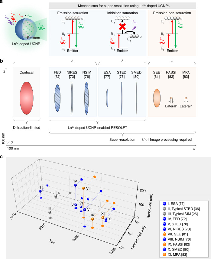
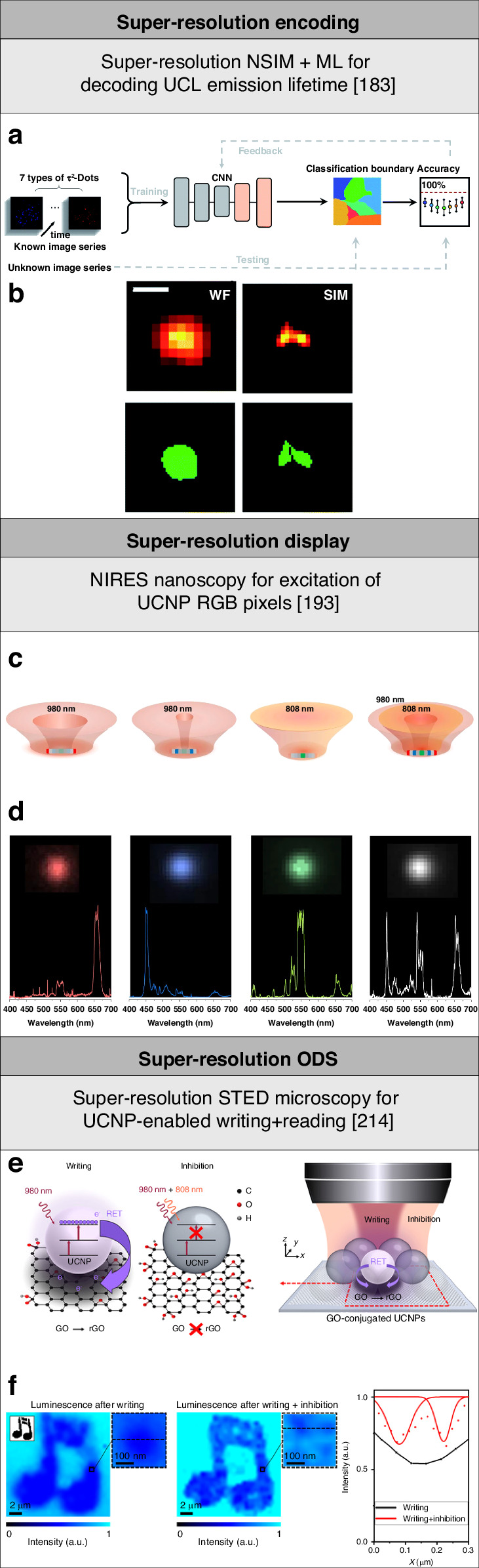
Energy-intensive technologies and high-precision research require energy-efficient techniques and materials. Lens-based optical microscopy technology is useful for low-energy applications in the life sciences and other fields of technology, but standard techniques cannot achieve applications at the nanoscale because of light diffraction. Far-field super-resolution techniques have broken beyond the light diffraction limit, enabling 3D applications down to the molecular scale and striving to reduce energy use. Typically targeted super-resolution techniques have achieved high resolution, but the high light intensity needed to outperform competing optical transitions in nanomaterials may result in photo-damage and high energy consumption. Great efforts have been made in the development of nanomaterials to improve the resolution and efficiency of these techniques toward low-energy super-resolution applications. Lanthanide ion-doped upconversion nanoparticles that exhibit multiple long-lived excited energy states and emit upconversion luminescence have enabled the development of targeted super-resolution techniques that need low-intensity light. The use of lanthanide ion-doped upconversion nanoparticles in these techniques for emerging low-energy super-resolution applications will have a significant impact on life sciences and other areas of technology. In this review, we describe the dynamics of lanthanide ion-doped upconversion nanoparticles for super-resolution under low-intensity light and their use in targeted super-resolution techniques. We highlight low-energy super-resolution applications of lanthanide ion-doped upconversion nanoparticles, as well as the related research directions and challenges. Our aim is to analyze targeted super-resolution techniques using lanthanide ion-doped upconversion nanoparticles, emphasizing fundamental mechanisms governing transitions in lanthanide ions to surpass the diffraction limit with low-intensity light, and exploring their implications for low-energy nanoscale applications.


Conflict of interest statement

The authors declare no competing interests.

Figures

Fig. 1
Fig. 1. Overview of the working principles and features of targeted super-resolution techniques using lanthanide ion-doped UCNPs.
a Schematic of the use of excitation and inhibition light to control optical transitions of lanthanide ion emitters of UCNPs for targeted super-resolution techniques based on UCL emission saturation, upconversion inhibition saturation, and UCL emission non-saturation. b Comparison of the 3D resolution achieved using different techniques using lanthanide ion-doped UCNPs. The ellipsoids represent the lateral (x,y) and axial (z) resolution of the stated techniques. Diffraction-limited confocal microscopy is shown in red, assuming the case of typical Yb3+/Tm3+-doped UCNPs that show UCL emission at 450 nm excited at 980 nm. Lanthanide ion-doped UCNP-enabled targeted super-resolution techniques based on UCL emission saturation, such as super-resolution FED microscopy, NIRES nanoscopy, and super-resolution NSIM, or upconversion inhibition saturation, such as super-resolution ESA microscopy, super-resolution STED microscopy, and super-resolution SMED microscopy, are shown in blue. Techniques based on UCL emission non-saturation, such as super-resolution SEE microscopy, PASSI nanoscopy, and MPA nanoscopy, are shown in orange. c Timeline showing a comparison of the resolution and light intensity needed by different targeted super-resolution techniques including super-resolution STED microscopy and super-resolution SIM using typical fluorophores and luminescent nanomaterials, and techniques using lanthanide ion-doped UCNPs,,–,–. *For PASSI nanoscopy and MPA nanoscopy, only the value of the achieved lateral resolution is reported[,. λex. excitation wavelength, λem. emission wavelength, λin. inhibition wavelength, FED fluorescence emission difference, NIRES near-infrared emission saturation, NSIM nonlinear structured illumination microscopy, ESA excited-state absorption, STED stimulated emission depletion, SMED surface-migration emission depletion, SEE super-linear excitation-emission, PASSI photon-avalanche single-beam super-resolution imaging, MPA migrating photon avalanche
Fig. 2
Fig. 2. Analysis of the dynamics of optical transitions for super-resolution using lanthanide ion-doped UCNPs.
a Schematic of the upconversion processes of ESA, ETU, and PA of targeted super-resolution techniques using lanthanide ion-doped UCNPs. b Summary of optical transitions of lanthanide ions of UCNPs, λex. (colored in red), λin. (colored in purple), and λem. (colored in green) of lanthanide ion-doped UCNPs used in the demonstrations of targeted super-resolution techniques. c Schematic of a simplified four-state system used to describe the dynamics of optical transitions for super-resolution using lanthanide ion-doped UCNPs under excitation and inhibition light. d Schematic of the intensity saturation curve of lanthanide ion-doped UCNPs that shows the relationship between IUCL and P under non-saturating and saturating light excitation. Lanthanide ion-doped UCNPs with highly super-linear and PA, UCL emission show n»1 under non-saturating light excitation
Fig. 3
Fig. 3. Summary of targeted super-resolution techniques based on UCL emission saturation of lanthanide ion-doped UCNPs.
a Simulated solid, donut, and super-resolution FED PSFs, and corresponding intensity profiles of saturated UCL emission of lanthanide ion-doped UCNPs. Adapted with permission from ref. Copyright 2017 Optical Society of America. b Solid, donut, and super-resolution FED microscopy imaging, and corresponding intensity profiles of saturated UCL emission of lanthanide ion-doped UCNPs. Image size: 1.56 μm. Adapted with permission from ref. Copyright 2017 Optical Society of America. c Simulated ‘negative’ contrast images of cross-section profiles of the saturated UCL emission of a lanthanide ion-doped UCNP used in NIRES nanoscopy at different excitation intensity. Scale bar: 500 nm. Reproduced with permission from ref. , CC BY 4.0. Copyright 2018 Springer Nature Limited. d NIRES nanoscopy imaging and corresponding intensity profiles of lanthanide ion-doped UCNPs with different doping. Scale bar: 500 nm. Adapted with permission from ref. , CC BY 4.0. Copyright 2018 Springer Nature Limited. e Schematic of the super-resolution NSIM setup using lanthanide ion-doped UCNPs. Reproduced with permission from ref. . Copyright 2020 American Chemical Society. f UCL emission imaging of lanthanide ion-doped UCNPs with 80 μm thick brain tissues under sinusoidal structured excitation and line profile of Fourier spectrum on a logarithmic scale of the diagonal cross-section profiles. Adapted with permission from ref. . Copyright 2020 American Chemical Society. g Wide-field and super-resolution NSIM imaging of lanthanide ion-doped UCNPs. Scale bar: 2 μm. Reproduced with permission from ref. . Copyright 2020 American Chemical Society. h Comparison imaging results of the green framed area in (g), and corresponding intensity profiles of lanthanide ion-doped UCNPs. Scale bar: 1 μm. Reproduced with permission from ref. . Copyright 2020 American Chemical Society
Fig. 4
Fig. 4. Summary of targeted super-resolution techniques based on upconversion inhibition saturation of lanthanide ion-doped UCNPs.
a Schematic of the energy level diagram of Pr3+-doped UCNPs for super-resolution ESA microscopy. Reproduced with permission from ref. . Copyright 2011 American Physical Society. b Schematic of the sequence of laser pulses for super-resolution ESA microscopy imaging of Pr3+-doped UCNPs. Reproduced with permission from ref. . Copyright 2011 American Physical Society. c Confocal and super-resolution ESA microscopy imaging of Pr3+-doped UCNPs. Reproduced with permission from ref. . Copyright 2011 American Physical Society. d Schematic of the energy level diagram of lanthanide ion-doped UCNPs with high Tm3+ doping for UCL emission excitation and inhibition through STED. Reproduced with permission from ref. Copyright 2017 Springer Nature Limited. e Schematic of the super-resolution STED microscopy imaging system based on spatially overlapped Gaussian-shaped excitation and donut-shaped depletion laser beams and lanthanide ion-doped UCNPs with high Tm3+ doping. Reproduced with permission from ref. . Copyright 2017 Springer Nature Limited. f Confocal and super-resolution STED microscopy imaging, and corresponding intensity profiles of lanthanide ion-doped UCNPs with high Tm3+ doping. Scale bars: 500 nm (main images) and 200 nm (insets). Reproduced with permission from ref. . Copyright 2017 Springer Nature Limited. g Dual-color confocal and super-resolution STED microscopy imaging, and corresponding intensity profiles of lanthanide ion-doped UCNPs with high Tm3+ doping. Scale bar: 1 μm. Reproduced with permission from ref. , CC BY 4.0. Copyright 2017 Springer Nature Limited. h Schematic of the SMED mechanism for UCL emission depletion through surface migration. Reproduced with permission from ref. , CC BY 4.0. Copyright 2022 Springer Nature Limited. i Schematic of the energy level diagram of lanthanide ion-doped UCNPs with surface quenchers for super-resolution SMED microscopy. Reproduced with permission from ref. , CC BY 4.0. Copyright 2022 Springer Nature Limited. j Confocal and super-resolution SMED microscopy imaging, and corresponding intensity profiles of lanthanide ion-doped UCNPs with surface quenchers. Reproduced with permission from ref. , CC BY 4.0. Copyright 2022 Springer Nature Limited
Fig. 5
Fig. 5. Summary of targeted super-resolution techniques based on UCL emission non-saturation of lanthanide ion-doped UCNPs.
a Schematic of the experimental setup based on a standard confocal optical microscopy system for super-resolution SEE microscopy using lanthanide ion-doped UCNPs. Reproduced with permission from ref. , CC BY 4.0. Copyright 2019 Springer Nature Limited. b Intensity saturation curve of lanthanide ion-doped UCNPs with high Tm3+ doping. Reproduced with permission from ref. , CC BY 4.0 Copyright 2019 Springer Nature Limited. c 3D confocal and super-resolution SEE microscopy imaging, and corresponding intensity profiles of lanthanide ion-doped UCNPs with high Tm3+ doping. Reproduced with permission from ref. , CC BY 4.0. Copyright 2019 Springer Nature Limited. d Schematic of the PA effect in Tm3+-doped UCNPs. Reproduced with permission from ref. , 2021. Springer Nature Limited. e Model plot of the intensity saturation curve of Tm3+-doped UCNPs with PA UCL emission with a non-linearity of more than 15. Reproduced with permission from ref. . Copyright 2021 Springer Nature Limited. f Confocal and PASSI nanoscopy imaging, and corresponding intensity profile of Tm3+-doped UCNPs. Reproduced with permission from ref. Copyright 2021 Springer Nature Limited. g Schematic of the PA mechanism in Yb3+/Pr3+-doped UCNPs. Reproduced with permission from ref. . Copyright 2022 Springer Nature Limited. h Confocal and MPA nanoscopy imaging, and corresponding intensity profiles of Yb3+/Pr3+-doped UCNPs. Reproduced with permission from ref. . Copyright 2022 Springer Nature Limited
Fig. 6
Fig. 6. Summary of low-energy super-resolution bio-imaging and tracking applications using lanthanide ion-doped UCNPs.
a Confocal and super-resolution STED microscopy imaging of the cytoskeleton of HeLa cancer cells using Yb3+/Tm3+-doped UCNPs. Scale bar: 2 μm. Reproduced with permission from ref. , CC BY 4.0. Copyright 2017 Springer Nature Limited. b Schematic of a mouse liver tissue slice and deep-tissue confocal and NIRES nanoscopy imaging using Yb3+/Tm3+ UCNPs. Scale bar: 500 nm. Reproduced with permission from ref. , CC BY 4.0. Copyright 2018 Springer Nature Limited. c 3D NIRB nanoscopy imaging of deep cancer spheroid using Yb3+/Tm3+-doped UCNPs. Reproduced with permission from ref. . Copyright 2020 John Wiley & Sons, Inc.. d Confocal and super-resolution SMED microscopy imaging of the cytoskeleton actin filaments of HeLa cancer cells using Yb3+/Tm3+-doped UCNPs. Scale bar: 800 nm. Reproduced with permission from ref. , CC BY 4.0. Copyright 2022 Springer Nature Limited. e Wide-field and super-resolution LSIM imaging of deep mouse liver tissue using Yb3+/Tm3+-doped UCNPs. Scale bar: 1 μm. Reproduced with permission from ref. . Copyright 2020 American Chemical Society. f 3D confocal and super-resolution SEE microscopy imaging of neuronal cells using Yb3+/Tm3+-doped UCNPs. Reproduced with permission from ref. , CC BY 4.0, Copyright 2019 Springer Nature Limited. g Confocal and MPA nanoscopy imaging of actin protein filaments of HeLa cancer cells using Yb3+/Pr3+-doped UCNPs. Reproduced with permission from ref. . Copyright 2022 Springer Nature Limited
Fig. 7
Fig. 7. Summary of low-energy super-resolution encoding, display and data storage applications using lanthanide ion-doped UCNPs.
a Schematic of deep learning assisted super-resolution decoding UCL emission lifetime of lanthanide ion-doped UCNPs. Reproduced with permission from ref. . Copyright 2022 Royal Society of Chemistry. b Time-domain wide-field and super-resolution SIM imaging of lanthanide ion-doped UCNPs decoded using deep learning. Scale bar: 500 nm. Adapted with permission from ref. . Copyright 2022 Royal Society of Chemistry. c Schematic of a nanorod-shaped lanthanide ion-doped UCNP and light excitation patterns for a super-resolution RGB pixel. Reproduced with permission from ref. , CC BY 4.0. Copyright 2020 Springer Nature Limited. d Wide-field microscopy imaging and corresponding UCL emission spectra of a super-resolution pixel emitting RGB and white light using nanorod-shaped lanthanide ion-doped UCNPs. Reproduced with permission from ref. , CC BY 4.0. Copyright 2020 Springer Nature Limited. e Schematic of super-resolution optical data writing using lanthanide ion-doped UCNPs to reduce GO flakes. Reproduced with permission from ref. , CC BY-NC 4.0. Copyright 2021 American Association for the Advancement of Science. f Diffraction-limited and super-resolution optical data readout based on UCL emission quenching of lanthanide ion-doped UCNPs by reduced GO flakes. Reproduced with permission from ref. , CC BY-NC 4.0. Copyright 2021 American Association for the Advancement of Science
Fig. 8
Fig. 8. Overview of research directions and challenges of low-energy super-resolution applications using lanthanide ion-doped UCNPs.
a Ultrasmall lanthanide ion-doped UCNPs with high emitter doping (blue circles) are brighter than nanoparticles with low emitter doping and same size (red circles). Scale bar: 1 μm. Reproduced with permission from ref. . Copyright 2014 Springer Nature Limited. b Ligand coordination induced energy-level reconstruction in ultrasmall lanthanide ion-doped UCNPs for higher UCL emission intensity. Reproduced with permission from ref. . Copyright 2021 Springer Nature Limited. c Lanthanide ion-doped UCNPs with a core-shell-shell structure (top) show higher UCL emission intensity than lanthanide ion-doped UCNPs with a core structure (bottom) under low excitation light intensity of 8 W cm−2. Scale bars: 500 nm (main panel) and 30 nm (inset). Reproduced with permission from ref. . Copyright 2018 Springer Nature Limited. d Cascade amplified depletion lowers laser beam intensity for inhibition of UCL emission of lanthanide ion-doped UCNPs. Reproduced with permission from ref. , CC BY 4.0. Copyright 2022 Springer Nature Limited. e The MPA mechanism enables extremely high order of non-linearity of up to 46 in Tm3+-doped UCNPs through cascading multiplicative effects of the PA behavior. Reproduced with permission from ref. . Copyright 2022 Springer Nature Limited. f Developing a multi-modal super-resolution technique based on 3D super-resolution SEE microscopy with super-resolution STED microscopy using lanthanide ion-doped UCNPs. Adapted with permission from ref. . Copyright 2020 Optical Society of America
Fig. 9
Fig. 9. Overview of the working principles of diffraction-limited (standard) and targeted super-resolution techniques based on fluorescence and UCL emission.
a Schematic of SPE fluorescence emission of a fluorophore through absorption of one photon at λex. and emission at λem., and resolution of fluorescence microscopy based on SPE fluorescence emission. b Schematic of MPE fluorescence emission of a fluorophore through simultaneous absorption of multiple photons mediated by a virtual state and resolution of fluorescence microscopy based on MPE fluorescence emission. c Schematic of UCL emission of lanthanide ion-doped UCNPs through sequential absorption of multiple photons mediated by a real state and resolution of UCL microscopy based on saturated UCL emission. d Schematic of RESOLFT based on the STED mechanism through saturation of bright (ON) and dark (OFF) states under irradiation at λex. and λin., respectively, and resolution of RESOLFT techniques. e Schematic of UCL emission of lanthanide ion-doped UCNPs and resolution of UCL microscopy based on non-saturated UCL emission. Real and saturated states are represented by solid lines, while virtual and non-saturated states are represented by dotted and dashed lines, respectively. S0=ground energy state; S1=excited energy state; S2=higher-lying excited energy state; λex.=excitation wavelength; λem.=emission wavelength; λin.=inhibition wavelength

Similar articles

Cited by

References

    1. Chu, S., Cui, Y. & Liu, N. The path towards sustainable energy. Nat. Mater.16, 16–22 (2017).10.1038/nmat4834 - DOI - PubMed
    1. Davis, S. J. et al. Net-zero emissions energy systems. Science360, eaas9793 (2018). 10.1126/science.aas9793 - DOI - PubMed
    1. Tabor, D. P. et al. Accelerating the discovery of materials for clean energy in the era of smart automation. Nat. Rev. Mater.3, 5–20 (2018).10.1038/s41578-018-0005-z - DOI
    1. Masanet, E. et al. Recalibrating global data center energy-use estimates. Science367, 984–986 (2020). 10.1126/science.aba3758 - DOI - PubMed
    1. Cullen, D. A. et al. New roads and challenges for fuel cells in heavy-duty transportation. Nat. Energy6, 462–474 (2021).10.1038/s41560-021-00775-z - DOI

LinkOut - more resources