. 2024 Sep 14;14(1):21519.
doi: 10.1038/s41598-024-70060-y.

Assessing the toxicity of green Agaricus bisporus-based Cadmium Sulfide nanoparticles on Musca domestica as a biological model

Affiliations

Assessing the toxicity of green Agaricus bisporus-based Cadmium Sulfide nanoparticles on Musca domestica as a biological model

Hanan I Elhenawy et al. Sci Rep. .

Abstract

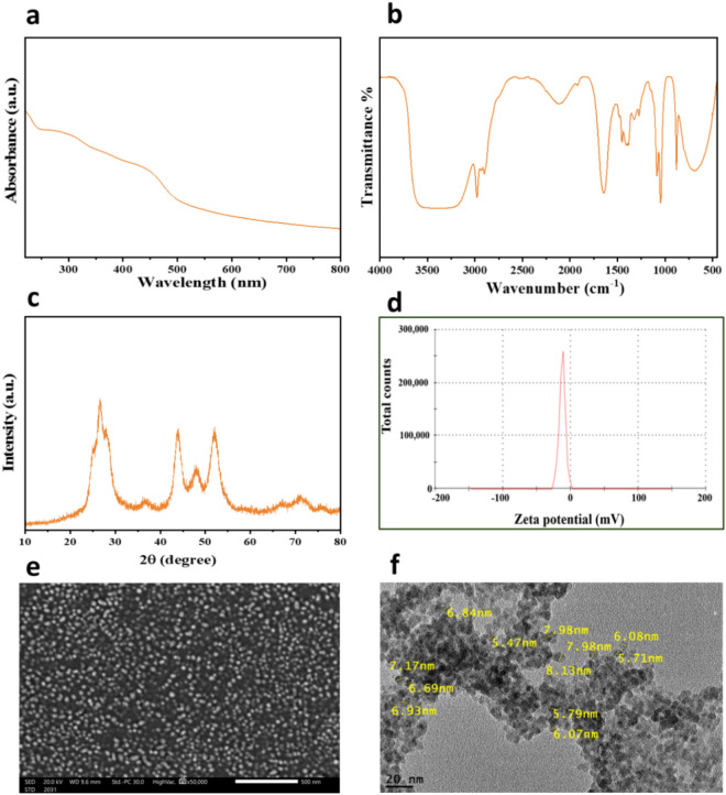
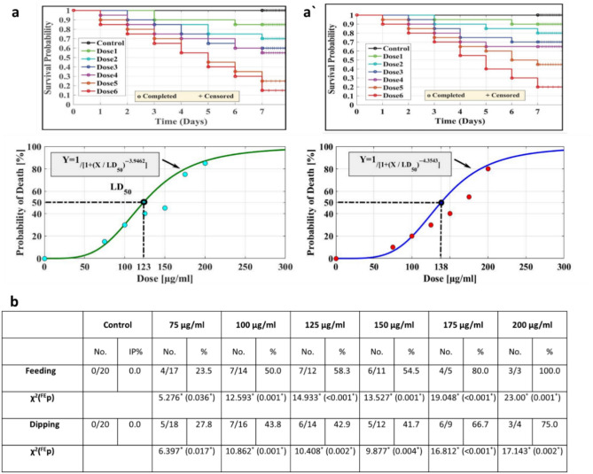
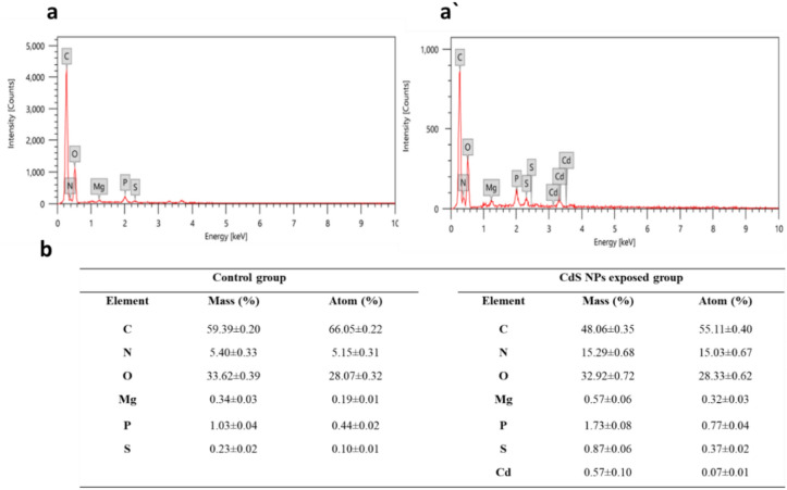
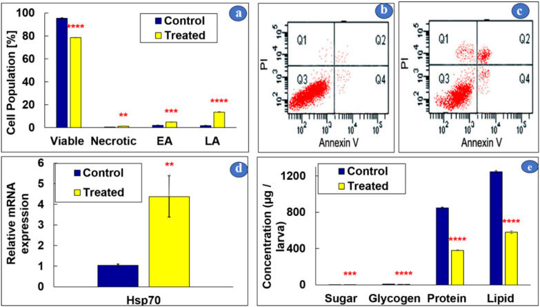
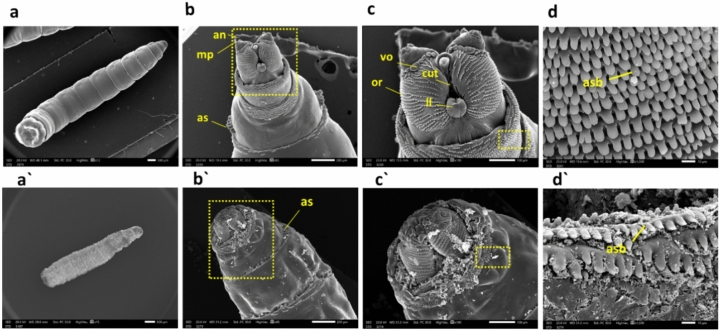
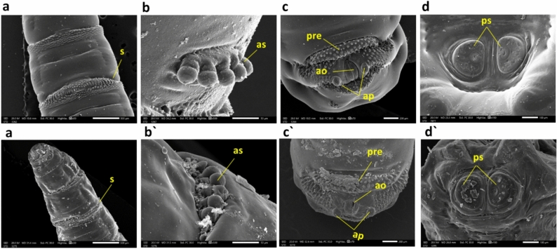
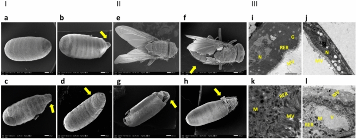
The common housefly, Musca domestica, known for transmitting over 100 infections, was studied using green-synthesized Cadmium Sulfide nanoparticles (CdS NPs) from Agaricus bisporus. These CdS NPs were tested on third-instar larvae under laboratory conditions using dipping and feeding methods with concentrations (75, 100, 125, 150, 175, and 200 µg/mL). The toxicity, measured by LC50, was found to be 138 µg/mL for dipping treatment and 123 µg/mL for feeding treatment. Analysis with an energy-dispersive X-ray microanalyzer confirmed Cd accumulation in the larval midgut, indicating penetration of CdS NPs into the organism, which may potentially increase their toxicity. CdS NPs caused disruptions in Heat Shock Protein 70, cell apoptosis, and various biochemical components. Scanning electron microscopy revealed morphological abnormalities in larvae, pupae, and adults exposed to CdS NPs. Ultrastructural examination showed significant midgut tissue abnormalities in larvae treated with 123 µg/mL of CdS NPs. Our study demonstrated that green-synthesized CdS NPs from A. bisporus can effectively control the development of M. domestica larvae.

Keywords: Agaricus bisporus; Musca domestica; Biochemical components; Cadmium Sulfide; Cell apoptosis; HSP70 expression.


Conflict of interest statement

The authors declare no competing interests.

Figures

Figure 1
Figure 1
CdS NPs: UV–Vis full spectrum (a), FTIR spectrum (b), XRD pattern (c), zeta potential analysis (d), SEM image (e), and TEM image (f) of CdS NPs.
Figure 2
Figure 2
Kaplan–Meier survival analysis for larvae treated with CdS NPs at the following doses 1 ÷ 6: 75, 100, 125, 150, 175, and 200 µg/mL, respectively, by (a) feeding method with LC50 123 μg/mL. (a') the dipping method with LC50 138 μg/mL. Pupal inhibition percentages (b) of M. domestica resulting from CdS NP treatment using feeding and dipping methods. (Abbreviations: χ2—Chi-square test, FE—Fisher Exact, p—p value for comparing the studied groups, and *—Statistically significant at p ≤ 0.05).
Figure 3
Figure 3
Illustrating the EDX analysis of both control (a) and treated larvae (a') display EDX spectra of larval tissues from typical larvae and the CdS NPs-treated larvae, respectively, showcasing the presence of Cd and S in the CdS NPs-treated larvae. The EDX analysis (b) of larval tissues of M. domestica in response to CdS NP exposure, with results displayed as mean ± SEM.
Figure 4
Figure 4
Flow cytometric analyses employing Annexin-V-FITC, conducted on M. domestica midgut larval tissues (a), with plots for the control (b) and CdS NPs-treated larvae at LC50 (123 μg/mL) through feeding (c). Relative expressions of HSP70 mRNA are presented in (d), and (e) illustrates the effect of CdS NPs on biochemical reserves. One-way ANOVA was used to evaluate the data, and Tukey's test was used for multiple comparisons. The stars above the bars indicate significant differences between groups (**p < 0.01, ***p < 0.001, ****p < 0.0001). Q1, Q2, Q3, and Q4 represent necrotic cells, late apoptotic cells (LA), viable cells, and early apoptotic cells (EA), respectively.
Figure 5
Figure 5
M. domestica L3 scanning electron photomicrographs: (a) Displays the control larva, while (a') illustrates a shrunken larva from the CdS NPs-treated group. Moving to (b), it shows the anterior end of the 3rd larval instar with standard body details, including anterior spiracles (as), maxillary palpus (mp), and antennal complex (an). In contrast, (b') depicts the anterior end of a larva treated with CdS NPs, indicating the degeneration of the antennal complex, maxillary palpus, and anterior spiracle. (c) Presents a higher magnification of the inset in (b), showing the control facial mask with details of cutaneous teeth (cut), oral ridges (or), labial lobe (ll), and ventral organ (vo). Meanwhile, (c') shows a higher magnification of the inset in (b'), displaying the anterior end of the treated larva with extreme deformation of the antenna, maxillary palpus, and labial lobe. Finally, (d) illustrates a higher magnification of the inset in (c) of the control larva with an organized anterior spinose band (asb), and (d') depicts a higher magnification of the inset in (c') of the treated group with an unorganized, damaged anterior spinose band. Abbreviations: as = anterior spiracles; mp = maxillary palpus; an = antennal complex; cut = cutaneous teeth; or = oral ridges; ll = labial lobe; vo = ventral organ; s = spinules.
Figure 6
Figure 6
M. domestica L3 scanning electron photomicrographs: (a) depict the control group with normal intersegmental spinules, while (a') illustrates the degeneration of intersegmental spinules in the treated group. In (b), a typical anterior spiracle is presented, and (b') shows the anterior end of a larva treated with degeneration of the anterior spiracle. Moving to (c), the control larva's anal division is observed, noting the anal opening (ao), pre-anal welt (pre), and anal papillae (ap). In contrast, (c') illustrates the posterior end of a larva treated with CdS NPs, indicating severe degeneration in the anal division. Finally, (d) displays the posterior end of the control larva with typical posterior spiracles (ps), while (d') shows the posterior end of the larva treated with CdS NPs, revealing severe damage to the posterior spiracles. Abbreviations: s = spinules; as = Anterior spiracle; ao = anal opening; ap = anal papillae; pre = pre-anal welt; ps = posterior spiracles.
Figure 7
Figure 7
Part(I) arrow shows M. domestica pupae, with (a) representing the normal control appearance compared to treated pupae with an LC50 of CdS NPs through the feeding method, exhibiting aberrant anomalies demonstrated as (b, c) abnormal appendages of pupae, or another curved pupa (d). Part (II) arrow presents the adult housefly with (e) representing a typical appearance and (f) a distorted adult treated with the LC50 of CdS NPs through the feeding method, resulting in the adult being unable to fly. It also illustrates emergence with only the head region from the capsule (g) and emergence with only the head and thorax region from the capsule (h). Part (III) displays the transmission electron micrograph of midgut tissues of M. domestica L3: (i) Control L3 midgut details, including the normal nucleus (N), microvilli (MV), rough endoplasmic complex (RER), and Golgi complex (G). (j) Shows L3 midgut treated with the LC50 of CdS NPs through the feeding method. Note the extreme distortion of the nucleus and microvilli. (k) Presents the control L3 with well-organized microvilli, mitochondria (M), and rough endoplasmic complex. (l) Presents the treated group with damaged microvilli, abnormal rough endoplasmic complex combined with vacuolation (V) in the cytoplasm, and malformed mitochondria. Key: N = nucleus; MV = microvilli; RER = rough endoplasmic complex; G = Golgi complex; V = vacuolated cytoplasm.

Similar articles

References

    1. Olagunju, E. A. Housefly: Common zoonotic diseases transmitted and control. J. Zoonotic Dis.6, 1–10. 10.22034/jzd.2022.14378 (2022).10.22034/jzd.2022.14378 - DOI
    1. Chintalchere, J., Dar, M., Raut, K. & Pandit, R. Bioefficacy of lemongrass and tea tree essential oils against house fly, Musca domestica. Proc. Natl. Acad. Sci. India Sect. B Biol. Sci.10.1007/s40011-020-01220-z (2021). 10.1007/s40011-020-01220-z - DOI - PMC - PubMed
    1. Norris, R. H. et al. Selection for, and characterization of, fluralaner resistance in the house fly, Musca domestica. Pestic. Biochem. Physiol.191, 105355. 10.1016/j.pestbp.2023.105355 (2023). 10.1016/j.pestbp.2023.105355 - DOI - PubMed
    1. Raele, D. et al. Study on the role of the common house fly, Musca domestica, in the spread of ORF virus (Poxviridae) DNA under laboratory conditions. Microorganisms10.3390/microorganisms9112185 (2021). 10.3390/microorganisms9112185 - DOI - PMC - PubMed
    1. Zahn, L. & Gerry, A. Diurnal flight activity of house flies (Musca domestica) is influenced by sex, time of day, and environmental conditions. Insects11, 391. 10.3390/insects11060391 (2020). 10.3390/insects11060391 - DOI - PMC - PubMed

Supplementary concepts

LinkOut - more resources