. 2024 Sep 14;22(1):567.
doi: 10.1186/s12951-024-02773-1.

Umbilical cord blood-derived exosomes attenuate dopaminergic neuron damage of Parkinson's disease mouse model

Affiliations

Umbilical cord blood-derived exosomes attenuate dopaminergic neuron damage of Parkinson's disease mouse model

Junjie Ye et al. J Nanobiotechnology. .

Abstract

Background: Umbilical cord blood (UCB) is a rich source of multifunctional stem cells characterized by low immunogenicity. Recent research in the fields of aging and regenerative medicine has revealed the potential of human umbilical cord blood-derived exosomes (UCB-Exos) in promoting wound healing, anti-aging, and regeneration. However, their role in neurodegenerative diseases, specifically Parkinson's disease (PD), remains unexplored. This study investigates the potential therapeutic effects and underlying mechanisms of UCB-Exos on PD.

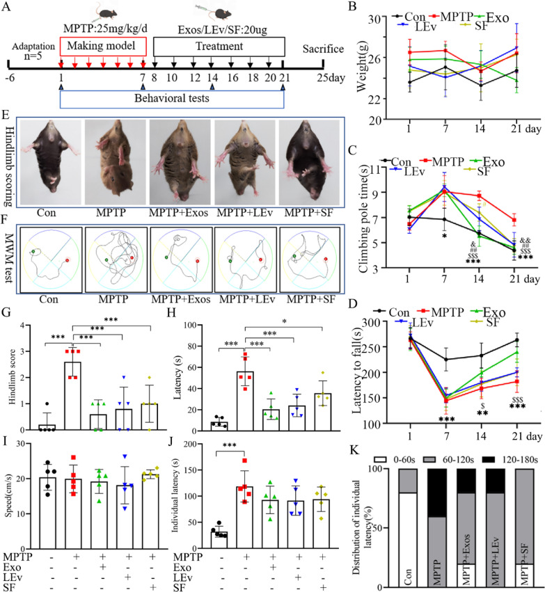
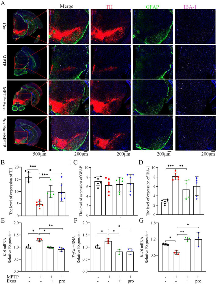
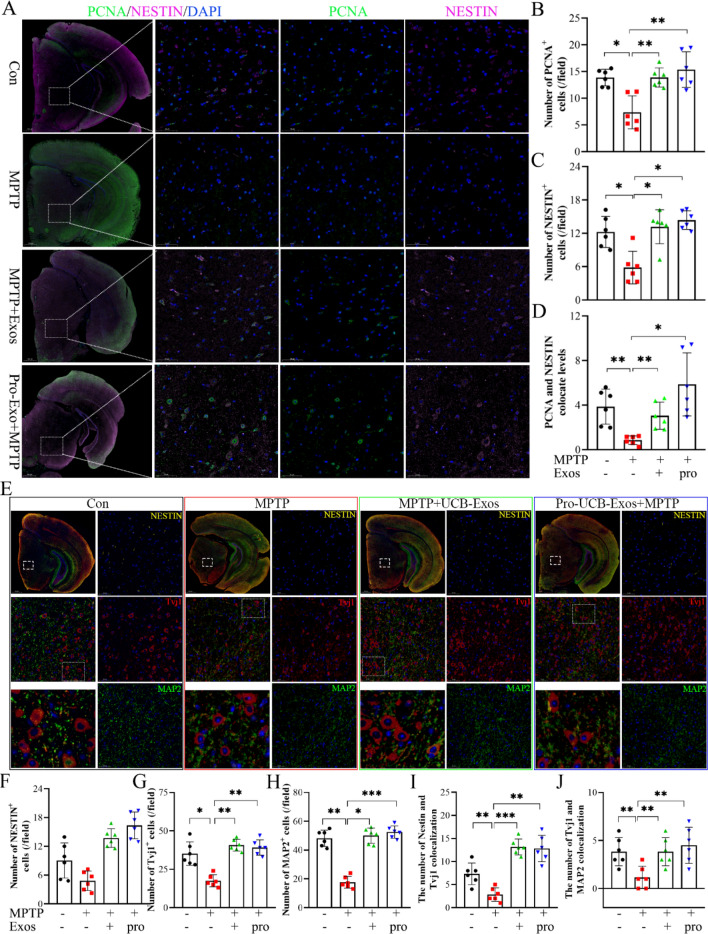
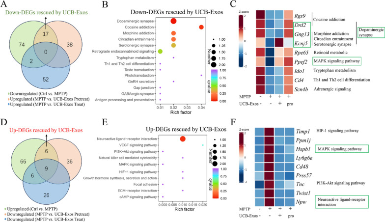
Methods: Large extracellular vesicles (LEv), Exos, and soluble fractions (SF) of human UCB plasma were extracted to investigate their effects on motor dysfunction of the MPTP-induced PD mouse model and identify the key components that improve PD symptoms. UCB-Exos were administered by the caudal vein to prevent or treat the PD mouse model. The motor function and pathological markers were detected. Differentially expressed gene and KEGG enrichment pathways were screened by transcriptome sequence. MN9D and SH-SY5Y cells were cultured and evaluated for cell viability, oxidative stress, cell cycle, and aging-related indexes by qRT-PCR, western blot, immunofluorescence, and flow cytometry. The protein expression level of the MAPK p38 and ERK1/2 signaling pathway was detected by western blot.

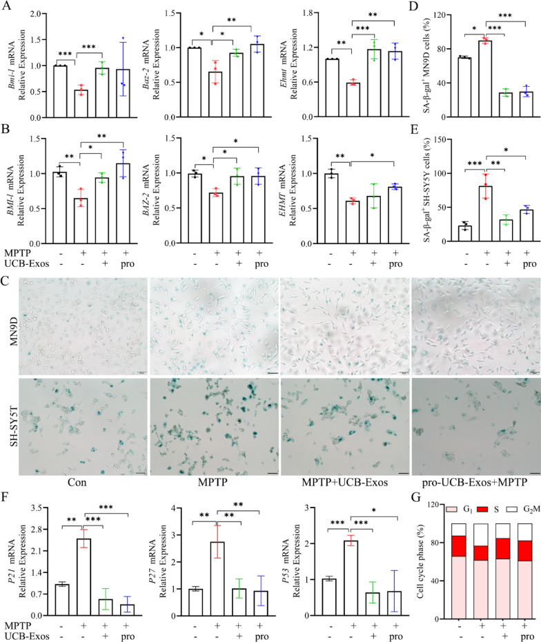
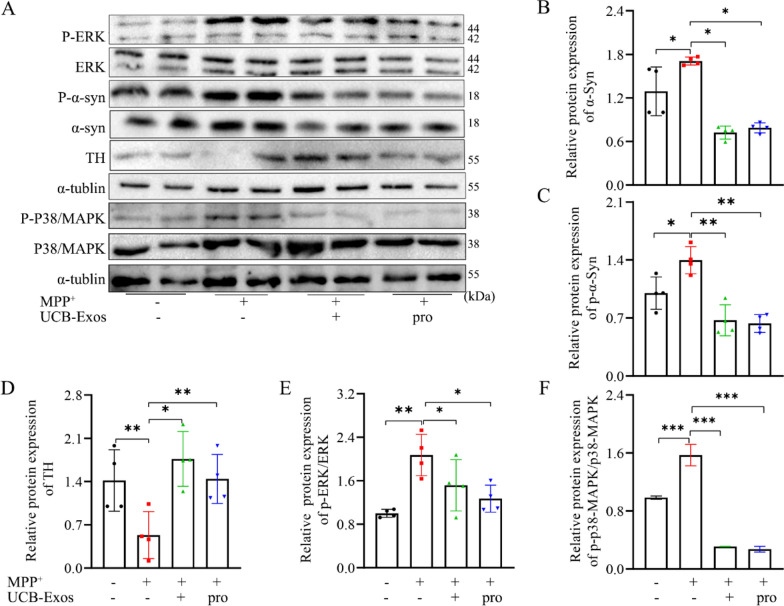
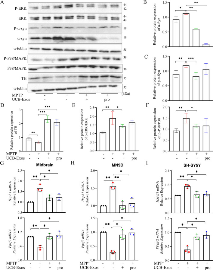
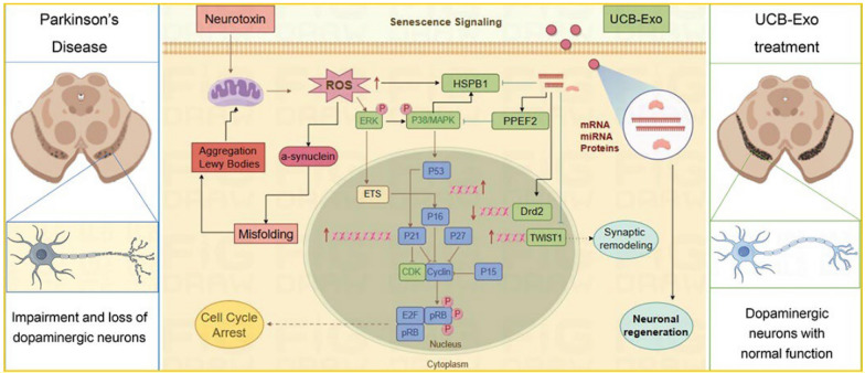
Results: We observed that LEv, Exos, and SF all exhibited potential in ameliorating motor dysfunction in MPTP-induced PD model mice, with UCB-Exos demonstrating the most significant effect. UCB-Exos showed comparable efficacy in preventing and treating motor dysfunction, cognitive decline, and substantia nigra pathological damage in PD mice. Further investigations revealed that UCB-Exos could potentially alleviate oxidative damage, aging and degeneration, and energy metabolism disorders in neurons. Transcriptome sequencing results corroborated that genes differentially expressed due to UCB-Exos were primarily enriched in the neuroactive ligand-receptor interaction, Dopaminergic synapse, and MAPK signaling pathway. We also observed that UCB-Exos significantly inhibited the hyperphosphorylation of the MAPK p38 and ERK1/2 signaling pathways both in vitro and in vivo.

Conclusions: Our study provides a comprehensive evaluation of UCB-Exos on the neuroprotective effects and suggests that inhibition of hyperphosphorylation of MAPK p38 and ERK 1/2 signaling pathways by regulating transcription levels of HspB1 and Ppef2 may be the key mechanism for UCB-Exos to improve PD-related pathological features.

Keywords: Exosome; MAPK signaling pathway; Neuroprotection; Parkinson’s disease; Senescence; Umbilical cord blood.


Conflict of interest statement

The authors declare that they have no competing interests.

Figures

Fig. 1
Fig. 1
Effects of LEv, UCB-Exos, and SF derived from human cord blood plasma on motor function of PD model mice. A Diagram of the experimental design. MPTP (25 mg/kg/d) or control agent (PBS) was injected intrabitoneally for 7 days, and UCB-Exos (20 μg), LEv (20 μg), SF (20 μg) or PBS was injected intravenously every other day for 2 weeks. B Weight of mice in each group. C The climbing pole time (***P < 0.005, Con vs MPTP; $$$P < 0.005, MPTP + UCB-Exos vs MPTP; ##P < 0.01, MPTP + LEv vs MPTP; &P < 0.05, &&P < 0.005, MPTP + SF vs MPTP). D Rotating rod detection (**P < 0.01, ***P < 0.005, Con vs MPTP; $P < 0.05, $$$P < 0.005, MPTP + UCB-Exos vs MPTP). E The representative picture of the hindlimb score. F The representative picture of Morris water maze. G Hindlimb score. H Latency. I Average swimming velocity. J, K Olfactory detection. n = 5 for each group, data are presented as mean ± SD, *P < 0.05, **P < 0.01, ***P < 0.005
Fig. 2
Fig. 2
UCB-Exos improved motor dysfunction in MPTP-induced PD mice. A Diagram of the experimental design. MPTP (25 mg/kg/d) or vehicle (PBS) was intraperitoneally injected for 7 consecutive days, and UCB-Exos (20 μg) or PBS was injected intravenously every other day for 2 weeks. The pro-UCB-Exos + MPTP group was given UCB-Exos before MPTP modeling. B Weight of mice in each group. C Feed consumption. D Pole test, E Rota-Rod test Rotating rod detection, and F Grip strength test (**P < 0.01, ***P < 0.005, Con vs MPTP; #P < 0.05, ##P < 0.01, MPTP + UCB-Exos vs MPTP; $$P < 0.01, $$$P < 0.005, pro-UCB-Exos + MPTP vs MPTP). G Hindlimb score. H The percentage of time covered in the target quadrant. I The percentage of distance covered in the target quadrant. J Stride length variance. K The standard deviation of stride length. n = 6 for each group, data are presented as mean ± SD, *P < 0.05, **P < 0.01, ***P < 0.005
Fig. 3
Fig. 3
UCB-Exos attenuated PD-associated histological features in the substantia nigra region of the MPTP-induced PD mice. A Representative capture of immunofluorescence in the substantia nigra of nuclei (DAPI, blue), astrocytes (GFAP, green), total dopaminergic neurons (TH, red), and microglial cells (IBA-1, pink), scale bar: 500 μm and 200 μm. BD Quantitative results of (A). E The Il-6 mRNA relative expression level in substantia nigra. F The Tnf-α mRNA relative expression level in substantia nigra. G The Il-10 mRNA relative expression level in substantia nigra. n = 3 for each group, data are presented as mean ± SD, *P < 0.05, **P < 0.01, ***P < 0.005
Fig. 4
Fig. 4
UCB-Exos promoted neuronal regeneration in the substantia nigra region of MPTP-induced PD mice. A Representative capture of immunofluorescence in the substantia nigra of nuclei (DAPI, blue), NSCs (PCNA, green, and NESTIN, pink), scale bar: 500 μm and 50 μm. B, C Quantitative results of (A). D Quantitative results of NESTIN and PCNA double positive. E Representative capture of immunofluorescence in the substantia nigra of nuclei (DAPI, blue), stem cell (NESTIN, pink), immature neuron (Tvj1, red), and neuron (MAP2, green), scale bar: 500 μm and 50 μm. F, G, H Quantitative results of (E). I Quantitative results of NESTIN and Tvj1 double positive. J Quantitative results of Tvj1 and MAP2 double positive. n = 3 for each group, data are presented as mean ± SD, *P < 0.05, **P < 0.01, ***P < 0.005
Fig. 5
Fig. 5
UCB-Exos reversed the expression abundance of MPTP-induced differential genes. A Venn diagrams showing down-DEGs rescued by UCB-Exos. B KEGG pathway of down-DEGs rescued by UCB-Exos. C Heatmap analysis of down-DEGs rescued by UCB-Exos. D Venn diagrams showing up-DEGs rescued by UCB-Exos. E KEGG pathway of up-DEGs rescued by UCB-Exos. F Heatmap analysis of up-DEGs rescued by UCB-Exos. n = 4 for each group
Fig. 6
Fig. 6
UCB-Exos regulated the neuronal cell cycle and alleviated senescence in vitro. A The Bmi-l, Baz-2, and Ehmt mRNA relative expression level in MN9D cells. B The Bmi-l, Baz-2, and Ehmt mRNA relative expression level in SH-SY5Y cells. C Representative pictures of senescence-associated β-galactosidase(SA-β-Gal) activity in MN9D cells and SH-SY5Y cells. D, E Quantitative results of SA-β-Gal+ cells in MN9D cells and SH-SY5Y cells. F The P21, P27, and P53 mRNA relative expression levels in MN9D cells. G The cell cycle of MN9D cells was determined by flow cytometry. n = 3 for each group, data are presented as mean ± SD, *P < 0.05, **P < 0.01, ***P < 0.005
Fig. 7
Fig. 7
UCB-Exos inhibited hyperphosphorylation of the MAPK p38 and ERK1/2 signaling pathway in vitro. A TH, α-Syn, p-α-Syn, ERK, p-ERK, p38/MAPK, and p-p38/MAPK protein levels in MN9D cells treated with UCB-Exos or the vehicle for 24 h were determined by western blot analysis. Full-length blots are presented in Supplementary Fig. 9. BF Quantitative results of (A). n = 4 for each group, data are presented as mean ± SD, *P < 0.05, **P < 0.01, ***P < 0.005
Fig. 8
Fig. 8
UCB-Exos inhibited hyperphosphorylation of the MAPK p38 and ERK1/2 signaling pathway in vivo. A TH, α-Syn, p-α-Syn, ERK, p-ERK, p38/MAPK, p-p38/MAPK protein levels in mouse midbrain tissue by western blot analysis. Full-length blots are presented in Supplementary Fig. 10. BF Quantitative results of (A). n = 2 for each group, data are presented as mean ± SD,*P < 0.05, **P < 0.01, ***P < 0.005. mRNA expression levels of Hspb1 and Ppef2 in mouse midbrain tissue (G), MN9D cells (H), and SH-SY5Y cells (I) were detected by Q-PCR analysis
Fig. 9
Fig. 9
The schematic illustration demonstrates the potential neuroprotective effects of UCB-Exos on dopaminergic neurons. The effects are primarily manifested through the significant inhibition of neurotoxin-induced cellular stress responses and the subsequent hyperphosphorylation of MAPK p38 and ERK1/2 signaling pathways. UCB-Exos may modulate the expression of various genes, including Hspb1, Ppef2, Twist1, and Drd2, via the cargo they carry, such as miRNA, mRNA, and proteins. This modulation could potentially inhibit neuronal senescence signaling and safeguard synaptic functions, thereby mitigating Parkinson's Disease (PD)-related motor dysfunction and pathological progression. Image created with Figdraw (Home for Researchers)

Similar articles

Cited by

References

    1. Iba M, McDevitt RA, Kim C, Roy R, Sarantopoulou D, Tommer E, Siegars B, Sallin M, Kwon S, Sen JM, et al. Aging exacerbates the brain inflammatory micro-environment contributing to alpha-synuclein pathology and functional deficits in a mouse model of DLB/PD. Mol Neurodegener. 2022;17:60. 10.1186/s13024-022-00564-6 - DOI - PMC - PubMed
    1. Sano T, Umemoto G, Fujioka S, Iwashita Y, Dotsu Y, Wada N, Tsuboi Y. Relationship between motor dysfunction and chewing movement in patients with Parkinson’s disease: a transversal study. Front Neurol. 2022;13:1062134. 10.3389/fneur.2022.1062134 - DOI - PMC - PubMed
    1. Schaffner SL, Wassouf Z, Lazaro DF, Xylaki M, Gladish N, Lin DTS, MacIsaac J, Ramadori K, Hentrich T, Schulze-Hentrich JM, et al. Alpha-synuclein overexpression induces epigenomic dysregulation of glutamate signaling and locomotor pathways. Hum Mol Genet. 2022;31:3694–714. 10.1093/hmg/ddac104 - DOI - PMC - PubMed
    1. Verma DK, Seo BA, Ghosh A, Ma SX, Hernandez-Quijada K, Andersen JK, Ko HS, Kim YH. Alpha-synuclein preformed fibrils induce cellular senescence in Parkinson’s disease models. Cells. 2021;10(7):1694. 10.3390/cells10071694 - DOI - PMC - PubMed
    1. Kritsilis M, Rizou SV, Koutsoudaki PN, Evangelou K, Gorgoulis VG, Papadopoulos D. Ageing, cellular senescence and neurodegenerative disease. Int J Mol Sci. 2018;19(10):2937. 10.3390/ijms19102937 - DOI - PMC - PubMed