. 2024 Nov 20;33(23):2003-2023.
doi: 10.1093/hmg/ddae136.

Pathomechanisms of Monoallelic variants in TTN causing skeletal muscle disease

Affiliations

Pathomechanisms of Monoallelic variants in TTN causing skeletal muscle disease

Jochen Gohlke et al. Hum Mol Genet. .

Erratum in

Abstract

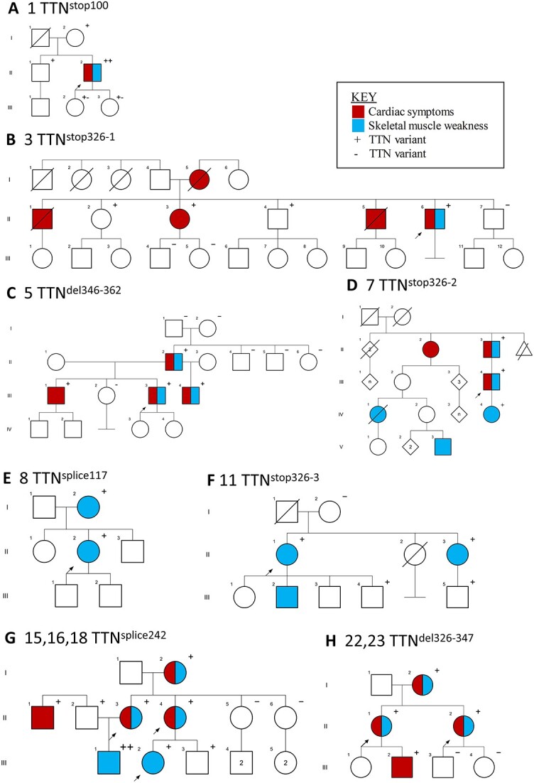
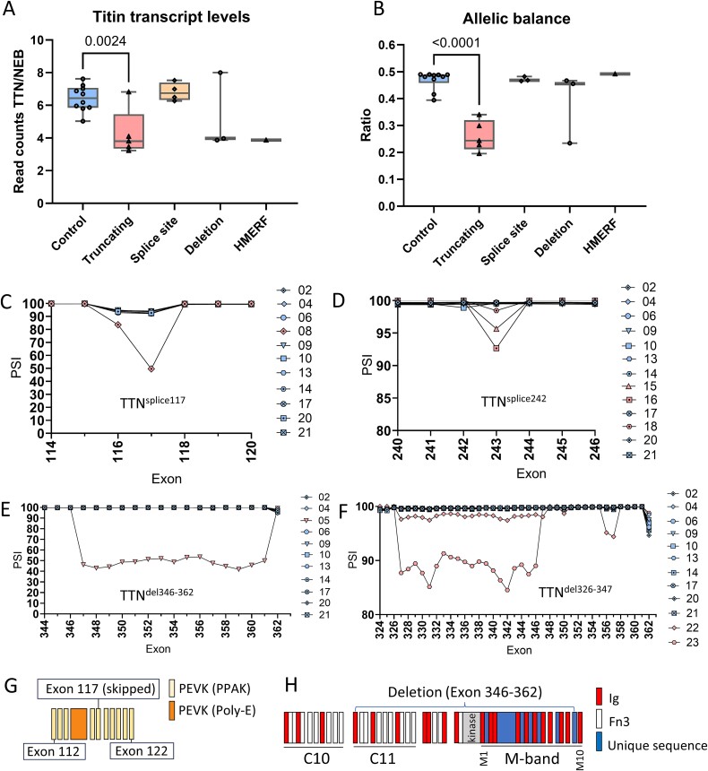
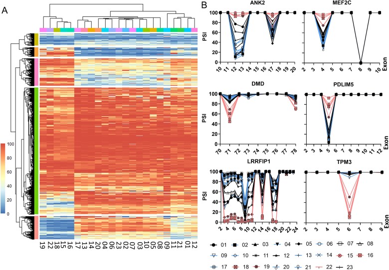
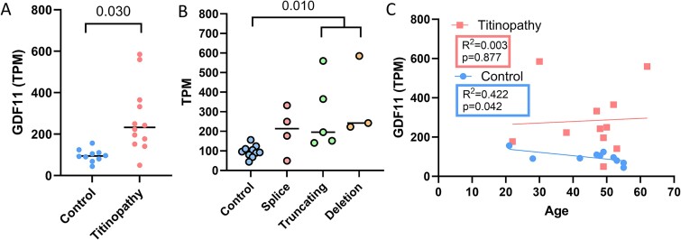
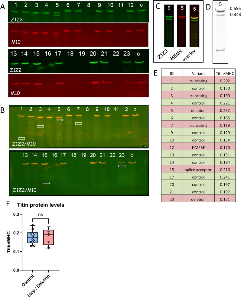
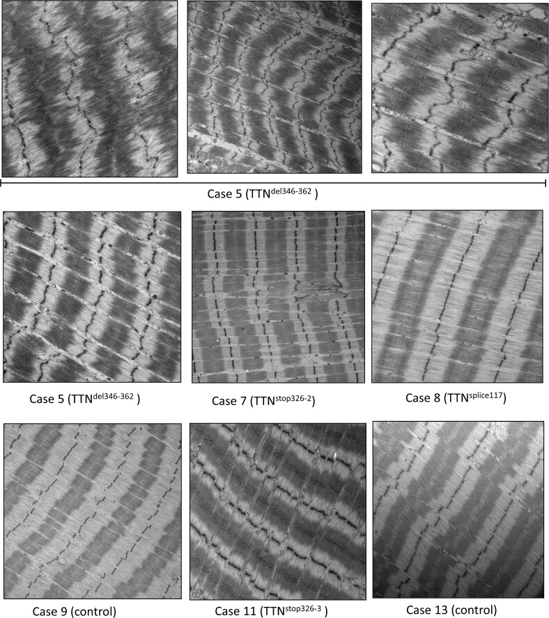
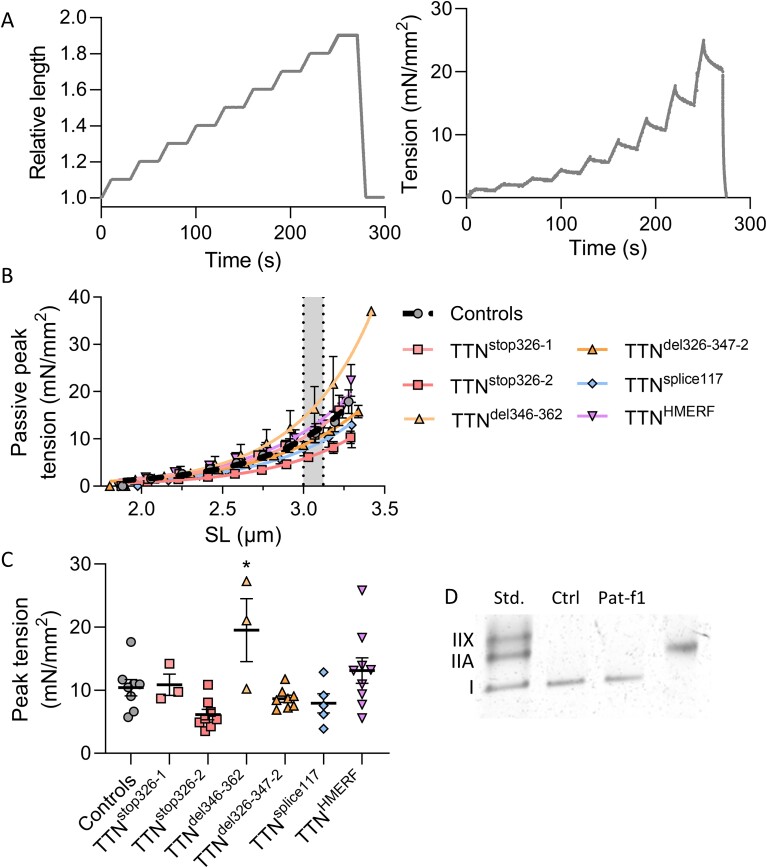
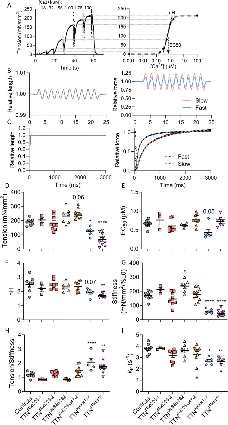
Pathogenic variants in the titin gene (TTN) are known to cause a wide range of cardiac and musculoskeletal disorders, with skeletal myopathy mostly attributed to biallelic variants. We identified monoallelic truncating variants (TTNtv), splice site or internal deletions in TTN in probands with mild, progressive axial and proximal weakness, with dilated cardiomyopathy frequently developing with age. These variants segregated in an autosomal dominant pattern in 7 out of 8 studied families. We investigated the impact of these variants on mRNA, protein levels, and skeletal muscle structure and function. Results reveal that nonsense-mediated decay likely prevents accumulation of harmful truncated protein in skeletal muscle in patients with TTNtvs. Splice variants and an out-of-frame deletion induce aberrant exon skipping, while an in-frame deletion produces shortened titin with intact N- and C-termini, resulting in disrupted sarcomeric structure. All variant types were associated with genome-wide changes in splicing patterns, which represent a hallmark of disease progression. Lastly, RNA-seq studies revealed that GDF11, a member of the TGF-β superfamily, is upregulated in diseased tissue, indicating that it might be a useful therapeutic target in skeletal muscle titinopathies.

Keywords: TTN deletion; TTNtv; Dominant titinopathy; Titin; titinopathy pathomechanisms.


Figures

Figure 1
Figure 1
Pedigrees of titinopathy cases included in this study. Studied cases are indicated by arrows. Individuals with known symptoms are shaded blue (for skeletal muscle) or red (for cardiac muscle). Individuals who underwent genetic testing are denoted with a ‘+’ to indicate the TTN variant was found, or a ‘-’ to indicate the TTN variant was not found. Individuals not marked with ‘+’ or ‘-’ were not available for testing. Cases 12 and 19 are not listed because they were included as disease controls.
Figure 2
Figure 2
Titin transcript levels, allelic balance and exon inclusion levels. Titin transcript levels, determined by RNA sequencing analysis and normalized to nebulin levels (A). Truncating variants displayed reduced transcript levels, while splice site variants were similar to controls. Analysis of biallelic sites confirmed a shift in the allelic balance ratio for this group of variants (B). Numbers represent p-values from unpaired t-tests. Exon 117 was skipped in almost half TTNsplice117 transcripts (C), while only low percentages of exon skipping (1.5%–7.5%) were observed in TTNsplice242 (patients 15, 16 & 18, D). In-frame deletion TTNdel346–362 had the affected exons missing in half of the transcripts (E). Two patients with a large out-of-frame deletion (TTNdel326–347) showed low degrees if exon skipping over the deletion site (F). One PEVK-domain is absent in TTNsplice117 due to exon skipping (G) while the last C-zone super-repeat and most of the M-band (except for M10) are deleted in TTNdel346–362 (H).
Figure 3
Figure 3
A subset of titinopathy samples shows differential splicing patterns. A: An initial analysis of differential splicing between three patient samples with an intron 242 splice acceptor variant [&, 15, 16, 18] and controls revealed a total of 1333 differentially spliced exons (p < 0.05 & PSI change > 5%). Together with two other patient samples [19 & 22], they form a distinct group with splicing patterns that are different from all other control & patient samples during column-wide clustering. Shown are inclusion levels (PSI) of differentially spliced exons, as determined during the initial analysis. B: One or more exons of genes known to be important for muscle development and function are differentially spliced in titinopathy patients (red). Controls are plotted in blue and titinopathy patients without genome-wide splicing changes in black. PSI values are plotted against exon numbers.
Figure 4
Figure 4
GDF11 levels are increased Titinopathy patients. Transcript-specific expression revealed increased levels of GDF11 in titinopathy patients (A). Sample grouping based on variant type revealed a high level of significance in truncating and deletion variants versus controls (B). Significance was calculated from transcript abundances and represents multiple-testing corrected p-values. GDF11 levels did not increase with age of the patient population and correlated negatively in controls (C). Average age did not differ between patients and controls (patients:45.45, controls: 45).
Figure 5
Figure 5
Titin protein analysis. Titin protein was separated on agarose gels and detected with antibodies against its N-termini (Z1Z2, (A), top panels) and C-termini (M10, bottom panels). (A) Shortened TTN protein could only be detected in-frame deletion TTNdel346–362 (patient 5). Overlays of Z1Z2 and M10 images (B) demonstrate that no C-terminally truncated proteins are present at expected protein sizes (indicated by white boxes). Domains M8 and M9 are internally deleted in sample 5 and not detected in shortened titin with an M8M9-specific antibody (C) in contrast to domain M10 (A & B). Shortened titin is less abundant in sample 5 than full-length protein when quantified on agarose gels (D, values represent intensity). Protein levels of truncating and deletion variants (E) were not significantly changed from controls when normalized to MHC (F). Five biopsy samples had protein levels that were too low to detect by gels or Western blots (samples 8, 11, 16, 19 & 22).
Figure 6
Figure 6
TTNdel346–362 muscle shows abnormalities in muscle structure. Analysis of tibialis anterior muscle by electron microscopy revealed irregular Z-disks and M-lines from in-frame deletion TTNdel346–362 (case 5, first 4 panels), which produces shortened titin protein. Normal muscle structure was observed for two patients with truncating variants (TTNstop326–2 and TTNstop326–3), a patient with a splice donor variant (8,TTNsplice117) and controls [9 and 13].
Figure 7
Figure 7
Passive mechanics in slow fibers from titinopathy patients. (A) Left panel: Ramp-stretch protocol for determining passive tension (force divided by fiber cross-sectional area) in skeletal muscle fibers. Fibers were set at slack sarcomere length and stretched in steps of 10% of fiber length (FL) at a speed of 1% FL/s and held for 20 s until the next step. Right panel: Passive force response in a control fiber from the ramp-stretch protocol. (B) Peak tensions plotted against sarcomere length for each step in the stretch protocol. (C) Peak tension at the sarcomere length range 3.0–3.12 μm (shaded area in D). (D) Cut out gel image of silver-stained 8% SDS-PAGE used to determine fiber types. Std; standard, Ctrl; control fiber, Pat-f#; patient fiber. Individual patient fibers are displayed. Controls are the average of 3–11 fibers per individual. *p < 0.05, and **P < 0.01 vs controls.
Figure 8
Figure 8
Membrane-permeabilized muscle fiber experiments reveal myofilament dysfunction in slow fibers from some individual titinopathy patients. (A) Left panel: Recording of tension developed by a slow skeletal muscle fiber at incremental [Ca2+]. Right panel: Tension-Ca2 + −curve for the fiber in A with EC50 ([Ca2+] for 50% tension) and Hill coefficient (nH) of curve shown. (B) Active stiffness protocol. Left panel: Relative length change; 0.3% of fiber length (FL) 500 Hz sinusoid for 20 ms was used to determine active stiffness. Right panel: Force trace for a fast and a slow fiber. Stiffness is calculated by dividing the force change with the length change. (C) Rate of force redevelopment protocol. Left panel: Relative length change; the fiber is shortened to 75% of FL in a ~ 1 ms step, held for 20 ms, stretched 103% of FL and immediately returned to 100% of FL. Right panel: Force trace for a fast and a slow fiber fitted with double exponential function. (D) Active tension (force normalized to fiber cross-sectional area). (E) Calcium sensitivity as EC50. (F) Hill coefficient (nH) is slope of the steep rise segment (black line in A, left panel) in tension-Ca2+-curve. (G) Active stiffness, an indicator of strongly-bound crossbridges. (H) Tension-stiffness ratio. (I) Rate of force redevelopment (ktr). Control values are average of 5–21 fibers from 8 subjects without known neuromuscular disease. Individual fibers are shown for patients. One-way ANOVA with Dunnett’s multiple comparisons post hoc test was used for statistical testing. *P < 0.05, **P < 0.01, ***P < 0.001 and ****P < 0.0001 vs control.

Similar articles

  • Novel heterozygous truncating titin variants affecting the A-band are associated with cardiomyopathy and myopathy/muscular dystrophy.
    Rich KA, Moscarello T, Siskind C, Brock G, Tan CA, Vatta M, Winder TL, Elsheikh B, Vicini L, Tucker B, Palettas M, Hershberger RE, Kissel JT, Morales A, Roggenbuck J. Rich KA, et al. Mol Genet Genomic Med. 2020 Oct;8(10):e1460. doi: 10.1002/mgg3.1460. Epub 2020 Aug 20. Mol Genet Genomic Med. 2020. PMID: 32815318 Free PMC article.
  • Interpreting Genetic Variants in Titin in Patients With Muscle Disorders.
    Savarese M, Maggi L, Vihola A, Jonson PH, Tasca G, Ruggiero L, Bello L, Magri F, Giugliano T, Torella A, Evilä A, Di Fruscio G, Vanakker O, Gibertini S, Vercelli L, Ruggieri A, Antozzi C, Luque H, Janssens S, Pasanisi MB, Fiorillo C, Raimondi M, Ergoli M, Politano L, Bruno C, Rubegni A, Pane M, Santorelli FM, Minetti C, Angelini C, De Bleecker J, Moggio M, Mongini T, Comi GP, Santoro L, Mercuri E, Pegoraro E, Mora M, Hackman P, Udd B, Nigro V. Savarese M, et al. JAMA Neurol. 2018 May 1;75(5):557-565. doi: 10.1001/jamaneurol.2017.4899. JAMA Neurol. 2018. PMID: 29435569 Free PMC article.
  • Reading Frame Repair of TTN Truncation Variants Restores Titin Quantity and Functions.
    Romano R, Ghahremani S, Zimmerman T, Legere N, Thakar K, Ladha FA, Pettinato AM, Hinson JT. Romano R, et al. Circulation. 2022 Jan 18;145(3):194-205. doi: 10.1161/CIRCULATIONAHA.120.049997. Epub 2021 Dec 14. Circulation. 2022. PMID: 34905694 Free PMC article.
  • Titin mutations and muscle disease.
    Kellermayer D, Smith JE 3rd, Granzier H. Kellermayer D, et al. Pflugers Arch. 2019 May;471(5):673-682. doi: 10.1007/s00424-019-02272-5. Epub 2019 Mar 27. Pflugers Arch. 2019. PMID: 30919088 Free PMC article. Review.
  • Titin-related Cardiomyopathy: Is it a Distinct Disease?
    Santiago CF, Huttner IG, Fatkin D. Santiago CF, et al. Curr Cardiol Rep. 2022 Sep;24(9):1069-1075. doi: 10.1007/s11886-022-01726-0. Epub 2022 Jun 27. Curr Cardiol Rep. 2022. PMID: 35759169 Review.

Cited by

References

    1. Granzier H, Wu Y, Siegfried L. et al. Titin: physiological function and role in cardiomyopathy and failure. Heart Fail Rev 2005;10:211–223. - PubMed
    1. Bang ML, Centner T, Fornoff F. et al. The complete gene sequence of titin, expression of an unusual approximately 700-kDa titin isoform, and its interaction with obscurin identify a novel Z-line to I-band linking system. Circ Res 2001;89:1065–1072. - PubMed
    1. Savarese M, Maggi L, Vihola A. et al. Interpreting genetic variants in Titin in patients with muscle disorders. JAMA Neurol 2018;75:557–565. - PMC - PubMed
    1. Hackman P, Vihola A, Haravuori H. et al. Tibial muscular dystrophy is a Titinopathy caused by mutations in TTN, the gene encoding the Giant skeletal-muscle protein Titin. Am J Hum Genet 2002;71:492–500. - PMC - PubMed
    1. Palmio J, Leonard-Louis S, Sacconi S. et al. Expanding the importance of HMERF titinopathy: new mutations and clinical aspects. J Neurol 2019;266:680–690. - PMC - PubMed

MeSH terms