. 2024 Aug 27:28:101213.
doi: 10.1016/j.mtbio.2024.101213. eCollection 2024 Oct.

Magnetic stirring with iron oxide nanospinners accretes neurotoxic Aβ42 oligomers into phagocytic clearable plaques for Alzheimer's disease treatment

Affiliations

Magnetic stirring with iron oxide nanospinners accretes neurotoxic Aβ42 oligomers into phagocytic clearable plaques for Alzheimer's disease treatment

Arjun Sabu et al. Mater Today Bio. .

Abstract

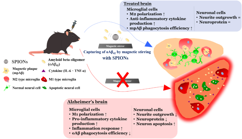
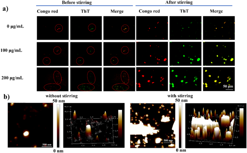
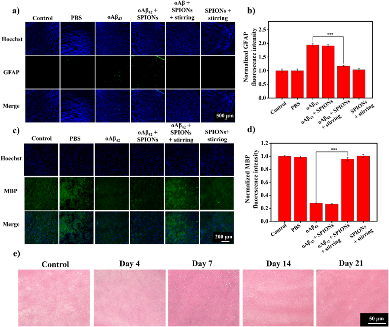
An increasing number of medications have been explored to treat the progressive and irreversible Alzheimer's disease (AD) that stands as the predominant form of dementia among neurodegenerative ailments. However, assertions about toxic side effects of these drugs are a significant hurdle to overcome, calling for drug-free nanotherapeutics. Herein, a new therapeutic strategy devoid of conventional drugs or other cytotoxic species was developed. The constructed superparamagnetic iron oxide nanoparticles (SPIONs) nanospinners can accrete neurotoxic β-amyloid 42 oligomers (oAβ42) into aggregated magnetic plaques (mpAβ) by mechanical rotating force via remote interaction between nanoparticles and the applied magnetic field. While the cellular uptake of mpAβ attained from the magnetic stirring treatment by neuronal cells is severely limited, the facile phagocytic uptake of mpAβ by microglial cells leads to the polarization of the brain macrophages to M2 phenotype and thus the increased anti-inflammatory responses to the treatment. The SPION stirring treatment protects the AD mice from memory deterioration and maintain cognitive ability as evidenced from both nesting and Barnes maze tests. The examination of the oAβ42 injected brain tissues with the stirring treatment showed significant amelioration of functional impairment of neurons, microglia, astrocytes and oligodendrocytes alongside no obvious tissue damage caused by stirring meanwhile complete degradation of SPION was observed at day 7 after the treatment. The in vitro and animal data of this work strongly corroborate that this new modality of undruggable stirring treatment with SPIONs provides a new feasible strategy for developing novel AD treatments.

Keywords: Alzheimer's disease; Microglial cell polarization; Nanoscaled stirring treatment; Phagocytic clearance; Superparamagnetic iron oxide nanoparticles; β-Amyloid 42 oligomers.


Conflict of interest statement

The authors whose names are listed immediately below certify that they have declare no conflict of interest in the subject matter or materials discussed in this manuscript.

Figures

Image 1
Graphical abstract
Fig. 1
Fig. 1
Schematic illustration of effective therapeutic action of SPIONs by accreting cytotoxic oAβ42 species into magnetic plaques and enhancing cellular clearance by microglial cells for the treatment of AD.
Fig. 2
Fig. 2
(a) ThT fluorescence intensity of Aβ42 (20 μM) in PBS with different incubation times. (b) Dot blot analysis of the formation of Aβ42 oligomers and fibrils using anti-oligomer and anti-fibril antibodies with different incubation times. (c) TEM images of various Aβ42 species (50 μM) after incubation in PBS at 37 °C for different times.
Fig. 3
Fig. 3
(a) Fluorescence images of accretion of oAβ42 (25 μM) by SPIONs stirring treatment with different SPIONs concentrations (0, 100 and 200 μg/mL) at 2500 rpm for 30 min. The Aβ species were stained with CR (red) and ThT (green), respectively. (b) AFM 2-D and 3-D images of oAβ42 (25 μM) with and without the SPIONs (200 μg/mL) stirring treatment at 2500 rpm for 30 min. (For interpretation of the references to color in this figure legend, the reader is referred to the Web version of this article.)
Fig. 4
Fig. 4
(a) Cell viability of N2a cells after 24 h incubation with Aβ42 (25 μM) pre-treated over different time intervals. The cell viability was determined by the MTT assay. (b) Cell viability of N2a cells after the incubation with oAβ42 of varying concentrations for 24 h. The oAβ42 was attained by preincubation of Aβ42 in PBS for 20 h. (c) Cell viability of N2a cells treated together with oAβ42 (25 μM) and SPION magnetic stirring with different SPION concentrations at 2500 rpm for 30 min. (d) LSCM images of N2a cells incubated with οΑβ42 (25 μM) and SPIONs (200 μg/mL) without and with magnetic stirring at 2500 rpm for 30 min to evaluate the cellular uptake of oAβ42. Cell nuclei (blue) and oAβ42 (green) were stained with Hoechst and ThT, respectively. Scale bar 50 μm. (e) Morphology of N2a cells receiving various treatments. Scale bar 20 μm. (f) Graphical representation of the SPION stirring treatment safeguarding the neuronal cells from the uptake of oAβ42. (g) Images of N2a cells receiving various treatments highlighting the intracellular MAP2 fluorescence signals (in red). Scale bar 50 μm. (h) Images of N2a cells receiving various treatments highlighting the intracellular β3-tubulin fluorescence signals (in red). Scale bar 50 μm. (i) Fluorescence images of intracellular ROS stained with DCF-DA in N2a cells receiving various treatments. Scale bar 50 μm. (For interpretation of the references to color in this figure legend, the reader is referred to the Web version of this article.)
Fig. 5
Fig. 5
Concentrations of IL-6 (a), IL-10 (b) and TNF-α (c) secretion from BV-2 cells receiving various treatments as determined by ELISA assay (n = 6). *p < 0.05, ***p < 0.001. d) Schematic of macrophage polarization with and without SPION magnetic stirring treatment. The naïve macrophages when are exposed to oAβ42 uptake undergo the M1 polarization. On the other hand, the magnetic stirring treatment with SPIONs causes the aggregation of oAβ42 into mpAβ. This aggregated form is then phagocytosed by microglial cells, leading to the polarization of brain macrophages into the M2 phenotype, thereby enhancing anti-inflammatory responses.
Fig. 6
Fig. 6
(a) Nest construction behaviour of mice injected with oAβ42 and with or without SPION stirring treatments. Nesting materials were 2 pieces of cotton lint (5*5*0.5 cm*cm*cm) (n = 3 in each group). (b) Representative moving path of mice from different groups in the Barnes maze test (n = 3 in each group). (c) Time spans for mice receiving various treatments to find the hidden chamber in Barnes maze test.
Fig. 7
Fig. 7
(a) IHC staining (anti-fibril Aβ42 as the primary antibody) of Aβ42 aggregates in brain tissues harvested from mice receiving various treatments. Cell nuclei were stained with Hoechst 33342. (b) Images of the Nissl-stained brain hippocampus region of the mice receiving various treatments. (c) Images identifying IHC staining of caspase 3 in brain tissues with various treatments. (d) Images of brain tissues after various treatments with IHC staining on Iba1 as a marker to signify the microglia level. (e) Images of brain tissues receiving various treatments with fluorescence staining of CD68 to represent the level of activated microglia.
Fig. 8
Fig. 8
(a) LSCM images of brain tissue sections with IHC staining (in green) of GFAP as the astrocyte activation marker from mice receiving different treatments. Cell nuclei were stained with Hoechst 33342 (blue). Scale bar 500 μm. (b) Quantified data also included. (n = 5) (c) LSCM images of brain tissue sections with IHC staining (in green) of MPB as a marker for oligodendrocyte differentiation and myelination from mice receiving different treatments. Scale bar 200 μm (d) Quantified data are also included, (n = 5). e). H&E staining of brain tissues over various time intervals after being subjected to the SPION stirring treatment at 2500 rpm for 30 min. Scale bar 50 μm. Samples were analyzed with one-way ANOVA. *p < 0.05, **p < 0.01, ***p < 0.001. Error bars represent mean ± s.d. (n = 5 for all groups). (For interpretation of the references to colour in this figure legend, the reader is referred to the Web version of this article.)

Similar articles

Cited by

References

    1. Gustavsson A., Norton N., Fast T., Frölich L., Georges J., Holzapfel D., Kirabali T., Krolak-Salmon P., Rossini P.M., Ferretti M.T., Lanman L., Chadha A.S., van der Flier W.M. Global estimates on the number of persons across the Alzheimer's disease continuum. Alzheimer’s and dementia, Alzheimer’s Dement. 2023;19:658–670. - PubMed
    1. Rudnicka-Drożak E., Drożak P., Mizerski G., Zaborowski T., Ślusarska B., Nowicki G., Drożak M. Links between COVID-19 and Alzheimer's disease—what do we already know? Int. J. Environ. Res. Publ. Health. 2023;20:2146. - PMC - PubMed
    1. Nichols E., Steinmetz J.D., Vollset S.E., Fukutaki K., et al. Estimation of the global prevalence of dementia in 2019 and forecasted prevalence in 2050: an analysis for the global burden of disease study 2019. Lancet Public Health. 2022;7:105–125. - PMC - PubMed
    1. Madhu P., Mukhopadhyay S. Distinct types of amyloid-β oligomers displaying diverse neurotoxicity mechanisms in Alzheimer's disease. J. Cell. Biochem. 2021;122:1594–1608. - PubMed
    1. Panza F., Lozupone M., Logroscino G., Imbimbo B.P. A critical appraisal of amyloid-β-targeting therapies for Alzheimer disease. Nat. Rev. Neurol. 2019;15:73–88. - PubMed

LinkOut - more resources