. 2024 May 6;8(5):1010-1015.
doi: 10.1016/j.jseint.2024.04.013. eCollection 2024 Sep.

Biologic augmentation of rotator cuff repair with microfragmented autologous subacromial bursal tissue enveloped in a patch of compressed autologous long head of biceps tendon tissue: the Bio-Ravioli technique

Affiliations

Biologic augmentation of rotator cuff repair with microfragmented autologous subacromial bursal tissue enveloped in a patch of compressed autologous long head of biceps tendon tissue: the Bio-Ravioli technique

Giuseppe Milano et al. JSES Int. .

Abstract

Background: Rotator cuff repair is one of the most frequently performed procedures in orthopedic surgery. However, considering the limited healing potential of rotator cuff tendons, several augmentation strategies have evolved to enhance tendon healing. The purpose of this article was to present a new surgical technique called Bio-Ravioli.

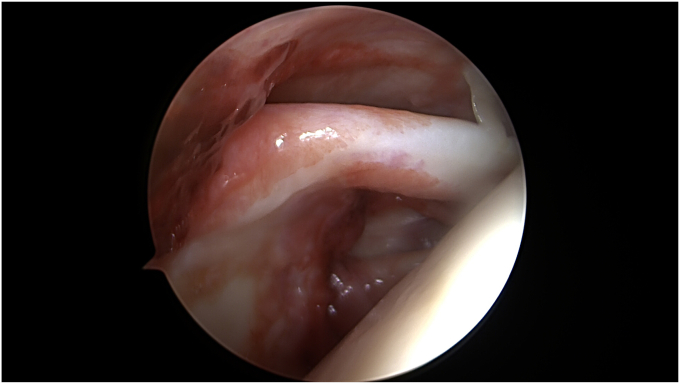
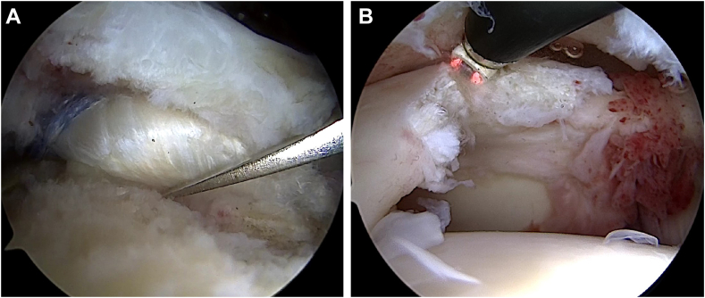
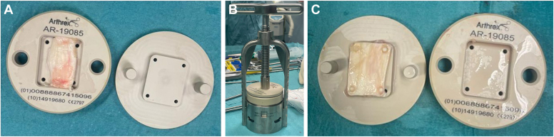
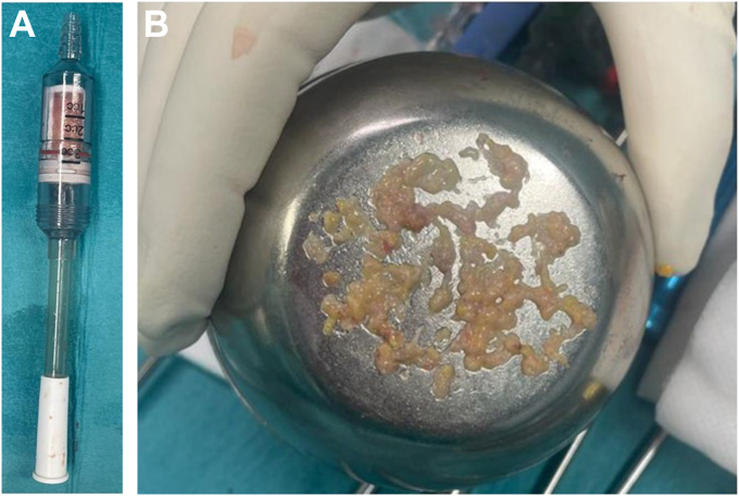
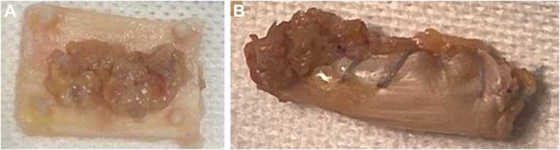
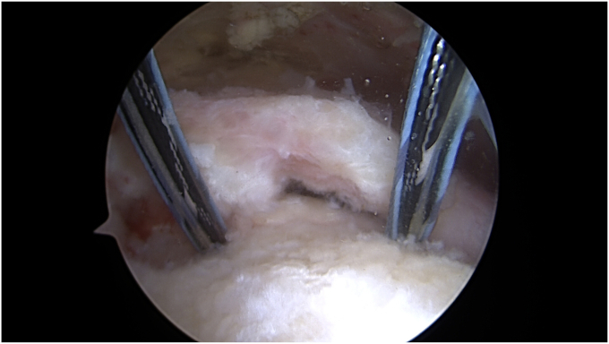
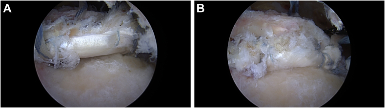
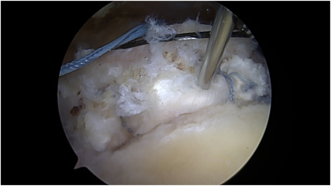
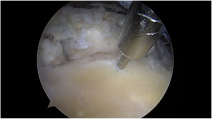
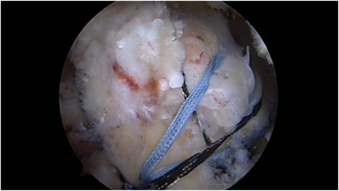
Methods: Patients with repairable full-thickness posterosuperior rotator cuff tear and a moderate-to-high risk of healing failure were chosen as candidates for the Bio-Ravioli procedure. It is a biologic augmentation strategy to increase healing potential of arthroscopic rotator cuff repair by use of a biologic graft fixed at the bone-tendon interface. The Bio-Ravioli consists of microfragmented autologous subacromial bursal tissue enveloped in a patch of compressed autologous long head of biceps tendon tissue. The rotator cuff is then repaired to the bone and over the graft using a transosseus equivalent configuration.

Conclusion: The Bio-Ravioli technique represents an easy and reliable way to increase the healing potential at the bone-tendon interface by using autologous mesenchymal stem cells from different sources: subacromial bursa and long head of the biceps tendon.

Keywords: Biologic enhancement; Long head of the biceps tendon; Mesenchymal stem cells; Rotator cuff repair; Subacromial bursa; Tendon healing.


Figures

Figure 1
Figure 1
Right shoulder. Scope in the posterior portal: diagnostic arthroscopy. Long head of the biceps inspection.
Figure 2
Figure 2
Right shoulder. Scope in the lateral portal: Long head of the biceps harvesting. Biceps tendon is freed from its groove and tenotomized (A); the intraarticular part of the tendon is released from its proximal attachment on the superior pole of the glenoid (B) and a proximal stump approximately 3 cm in length is harvested.
Figure 3
Figure 3
The tendon is cut to a length of approximately 25 mm by removing proximal and distal ends, then it was placed into a tray (Biceps Autograft Tissue Compression Plate; Arthrex) consisting of 2 plates that provide a space for the biceps (A). The tray plates with the tendon tissue in between are then placed into a taper assembly press (Arthrex), and compression is applied (B). The compression force is then released, and the biceps patch is ready (C).
Figure 4
Figure 4
Subacromia bursa is collected in a specific device (GraftNet Autologous Tissue Collector; Arthrex) attached to the suction on the shaver (A). After bursectomy, the bursal tissue is extracted (B).
Figure 5
Figure 5
The extracted microfragmented subacromial bursa is placed over the biceps patch (A), which is then rolled up and closed like an envelope around the bursal tissue with 2-0 permanent braided sutures: the so called Bio-Ravioli (B).
Figure 6
Figure 6
Right shoulder. Scope in the lateral portal. Two triple-loaded anchors are placed at the medial edge of the tendon footprint.
Figure 7
Figure 7
Right shoulder. Scope in the lateral portal. The graft is introduced through the anterior-superior portal and shuttled over the greater tuberosity at the insertion site of the suture anchors (A). From each of the suture anchors, 1 of the 3 suture tapes is used to fix the graft at the medial edge of the tendon footprint (B).
Figure 8
Figure 8
Right shoulder. Scope in the lateral portal. Small vents on the graft are performed by using a needle.
Figure 9
Figure 9
Right shoulder. Scope in the lateral portal. Nanofractures of the greater tuberosity.
Figure 10
Figure 10
Right shoulder. Scope in the lateral portal. Final view: a speed bridge configuration is performed to repair the cuff over the Bio-Ravioli.

Similar articles

Cited by

References

    1. Aicale R., Poeta N., Savarese E., Bernardini G., Oliva F., Maffulli N. The use of long head biceps tendon autograft for massive rotator cuff tears: a PRISMA compliant systematic review. Br Med Bull. 2022;144:76–89. doi: 10.1093/bmb/ldac021. - DOI - PubMed
    1. Baldino J.B., Muench L.N., Kia C., Johnson J., Morikawa D., Tamburini L., et al. Intraoperative and in vitro classification of subacromial bursal tissue. Arthroscopy. 2020;36:2057–2068. doi: 10.1016/j.arthro.2020.03.039. - DOI - PubMed
    1. Berthold D.P., Garvin P., Mancini M.R., Uyeki C.L., LeVasseur M.R., Mazzocca A.D., et al. Arthroscopic rotator cuff repair with biologically enhanced patch augmentation. Oper Orthopadie Traumatol. 2022;34:4–12. doi: 10.1007/s00064-021-00754-3. - DOI - PubMed
    1. Bhatia D.N. Arthroscopic biological augmentation for massive rotator cuff tears: the biceps-cuff-bursa composite repair. Arthrosc Tech. 2021;10:e2279–e2285. doi: 10.1016/j.eats.2021.07.003. - DOI - PMC - PubMed
    1. Bhatia D.N. Arthroscopic bursa-augmented rotator cuff repair: a vasculature-preserving technique for subacromial bursal harvest and tendon augmentation. Arthrosc Tech. 2021;10:e1203–e1209. doi: 10.1016/j.eats.2021.01.013. - DOI - PMC - PubMed

LinkOut - more resources