. 2024 Sep 15;15(9):1979-2001.
doi: 10.4239/wjd.v15.i9.1979.

Bone marrow-derived mesenchymal stem cell-derived exosome-loaded miR-129-5p targets high-mobility group box 1 attenuates neurological-impairment after diabetic cerebral hemorrhage

Affiliations

Bone marrow-derived mesenchymal stem cell-derived exosome-loaded miR-129-5p targets high-mobility group box 1 attenuates neurological-impairment after diabetic cerebral hemorrhage

Yue-Ying Wang et al. World J Diabetes. .

Abstract

Background: Diabetic intracerebral hemorrhage (ICH) is a serious complication of diabetes. The role and mechanism of bone marrow mesenchymal stem cell (BMSC)-derived exosomes (BMSC-exo) in neuroinflammation post-ICH in patients with diabetes are unknown. In this study, we investigated the regulation of BMSC-exo on hyperglycemia-induced neuroinflammation.

Aim: To study the mechanism of BMSC-exo on nerve function damage after diabetes complicated with cerebral hemorrhage.

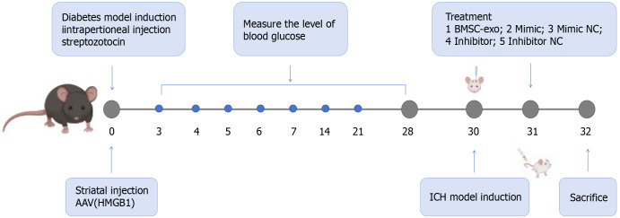
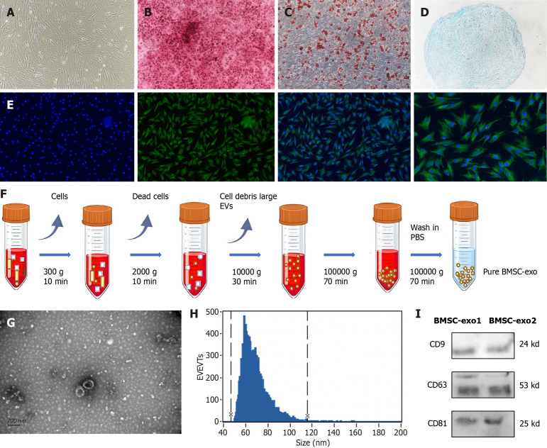
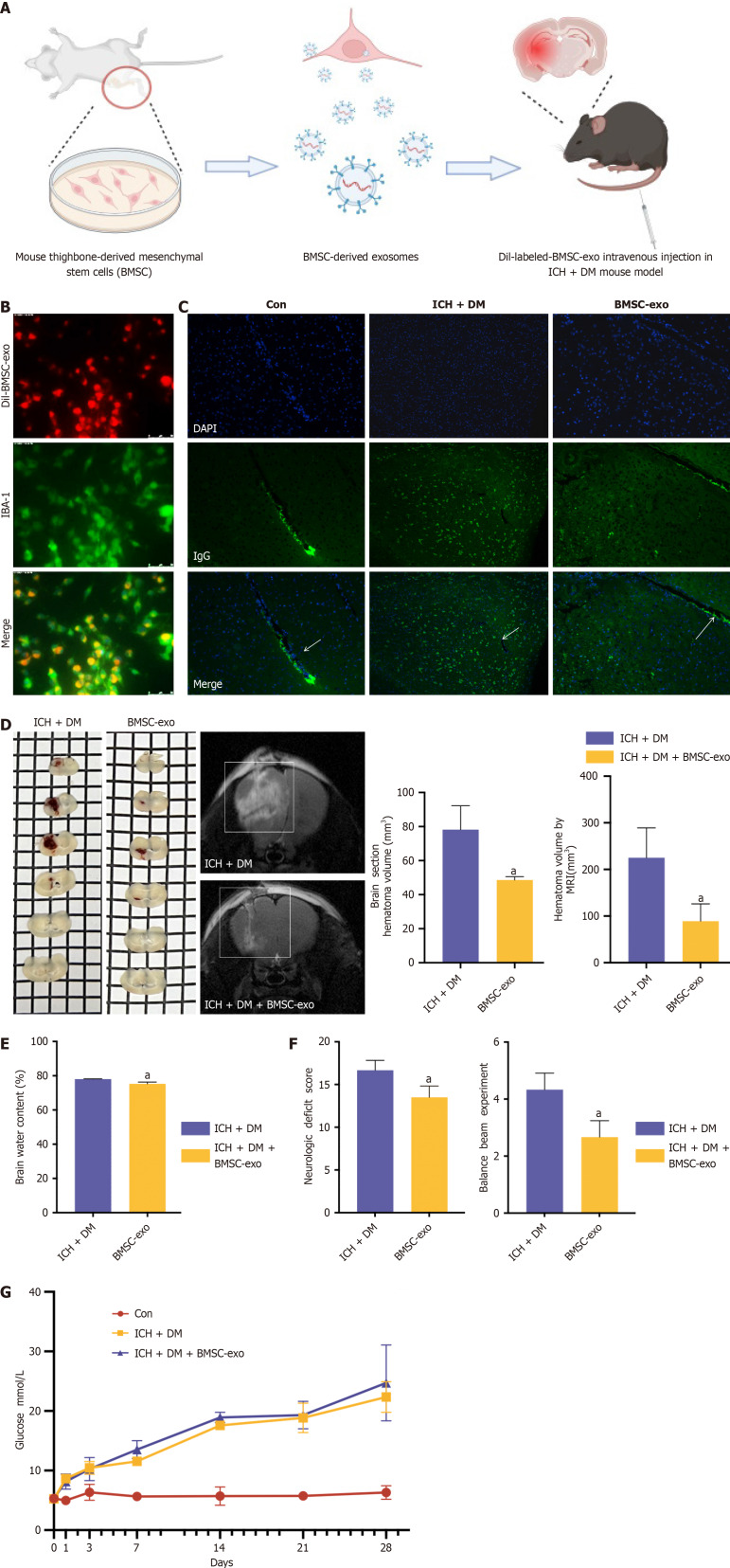
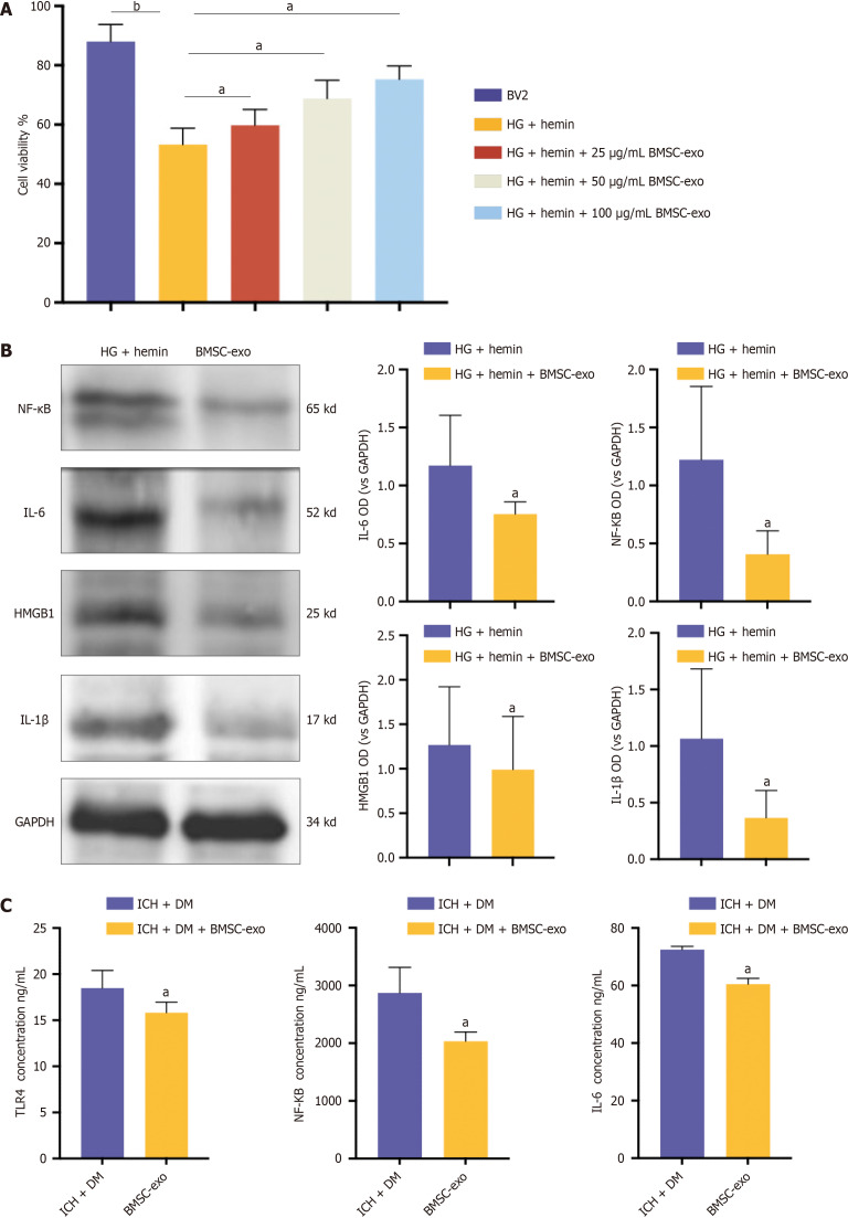
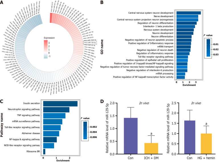
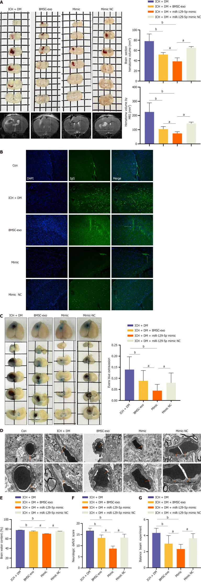
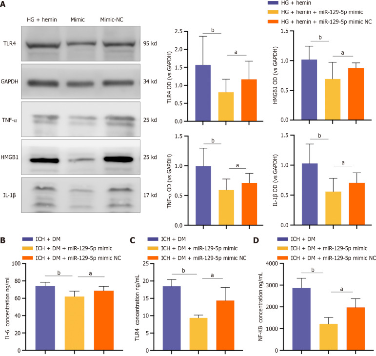
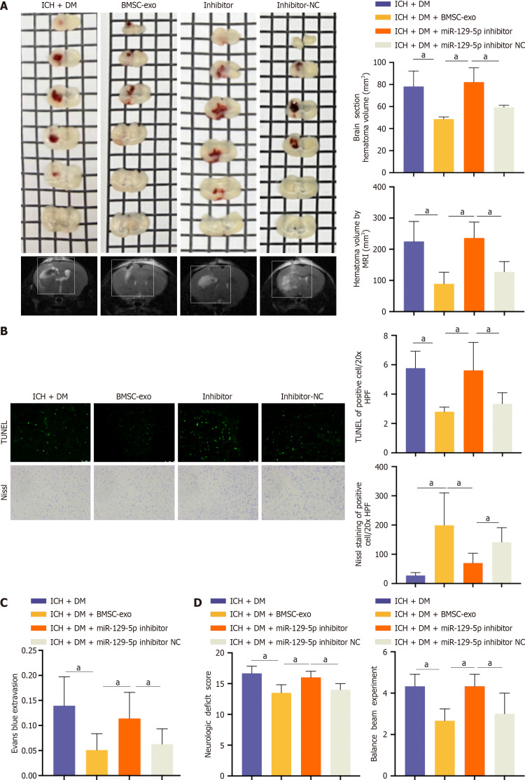
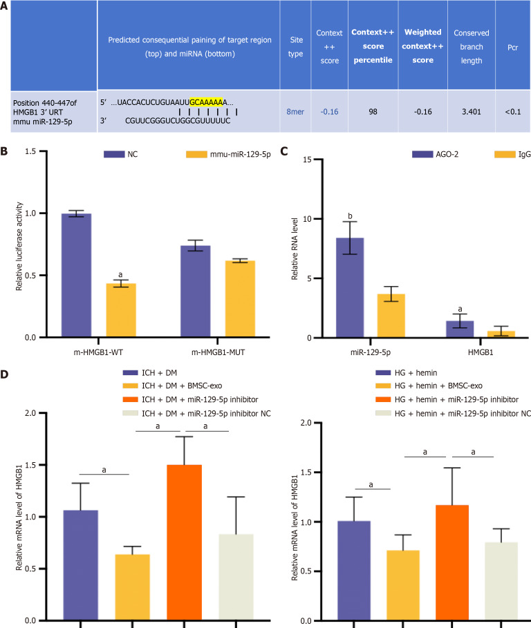
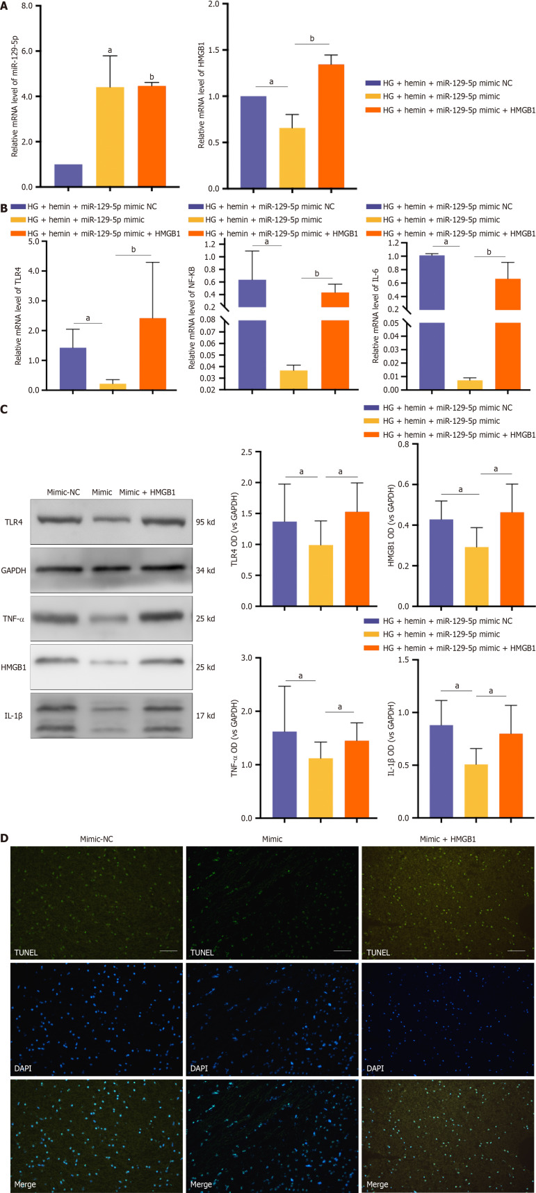
Methods: BMSC-exo were isolated from mouse BMSC media. This was followed by transfection with microRNA-129-5p (miR-129-5p). BMSC-exo or miR-129-5p-overexpressing BMSC-exo were intravitreally injected into a diabetes mouse model with ICH for in vivo analyses and were cocultured with high glucose-affected BV2 cells for in vitro analyses. The dual luciferase test and RNA immunoprecipitation test verified the targeted binding relationship between miR-129-5p and high-mobility group box 1 (HMGB1). Quantitative polymerase chain reaction, western blotting, and enzyme-linked immunosorbent assay were conducted to assess the levels of some inflammation factors, such as HMGB1, interleukin 6, interleukin 1β, toll-like receptor 4, and tumor necrosis factor α. Brain water content, neural function deficit score, and Evans blue were used to measure the neural function of mice.

Results: Our findings indicated that BMSC-exo can promote neuroinflammation and functional recovery. MicroRNA chip analysis of BMSC-exo identified miR-129-5p as the specific microRNA with a protective role in neuroinflammation. Overexpression of miR-129-5p in BMSC-exo reduced the inflammatory response and neurological impairment in comorbid diabetes and ICH cases. Furthermore, we found that miR-129-5p had a targeted binding relationship with HMGB1 mRNA.

Conclusion: We demonstrated that BMSC-exo can reduce the inflammatory response after ICH with diabetes, thereby improving the neurological function of the brain.

Keywords: Bone marrow mesenchymal stem cells; Diabetic cerebral hemorrhage; Exosome; High mobility group box 1; MicroRNA-129-5p; Neuroinflammation.


Conflict of interest statement

Conflict-of-interest statement: All the authors report no relevant conflicts of interest for this article.

Figures

Figure 1
Figure 1
Modeling time point, drug treatment, and grouping of animal models. HMGB1: High-mobility group box 1; BMSC-exo: Bone marrow mesenchymal stem cell derived exosomes; NC: Negative control; ICH: Intracerebral hemorrhage.
Figure 2
Figure 2
Characterization of bone marrow-derived mesenchymal stem cells and bone marrow-derived mesenchymal stem cell-derived exosomes. A: Bone marrow-derived mesenchymal stem cells (BMSC) were fusiform. Scale = 50 μm; B: Osteoblast alizarin red, before and after staining. Scale = 50 μm; C: Lipoblast oil red O, before and after staining. Scale = 50 μm; D: Formation of chondrospheres, stained with alcian blue. Scale = 50 μm; E: Cluster of differentiation (CD) 44 immunofluorescence staining. Scale = 50 μm/25 μm; F: Extraction of BMSC-derived exosomes (BMSC-exo) by ultrafast centrifugation; G: BMSC-exo appeared saucer-shaped under electron microscopy. Scale = 200 nm; H: BMSC-exo particle microscopy with 47 nm and 116 nm diameters; I: Western blot analysis of protein expression of exosome markers: CD9; CD63; and CD81. EVs: Extracellular vesicles; PBS: Phosphate buffered saline; BMSC-exo: Bone marrow mesenchymal stem cell derived exosomes.
Figure 3
Figure 3
Bone marrow-derived mesenchymal stem cell-derived exosome attenuated neurological deficits in vivo. A: Dil-labeled bone marrow-derived mesenchymal stem cell-derived exosomes (BMSC-exo) were injected in the tail vein of mice; B: Microglia (green) were labeled with ionized calcium-binding adaptor molecule 1 to observe the phagocytosis of BMSC-exo (red) by microglia. Scale bar = 50 μm; C: Immunofluorescence staining to observe the location of immunoglobulin G expression. Scale bar = 100 μm; D: Brain slices and hematoma volume were analyzed based on T2 magnetic resonance imaging. Experimental data are expressed as mean ± SD. aP < 0.05, intracerebral hemorrhage (ICH) + diabetes mellitus (DM) group vs ICH + DM + BMSC-exo group, t-test; E: Brain water content analysis. Experimental data are expressed as mean ± SD. aP < 0.01, ICH + DM group vs ICH + DM + BMSC-exo group, t-test; F: Neurological function scores and balance beam scores. Experimental data are expressed as mean ± SD. aP < 0.05, ICH + DM group vs ICH + DM + BMSC-exo group, t-test; G: The blood glucose levels of mice in each group were detected, and it was found that BMSC-exo could not reduce the blood glucose levels after diabetic cerebral hemorrhage in mice. Con: Control; BMSC-exo: Bone marrow mesenchymal stem cell derived exosomes; ICH: Intracerebral hemorrhage; DM: Diabetes mellitus; IgG: Immunoglobulin G.
Figure 4
Figure 4
Bone marrow-derived mesenchymal stem cell-derived exosome attenuated neuroinflammation in mice with diabetes and intracerebral hemorrhage. A: Microglia were treated with different concentrations of bone marrow-derived mesenchymal stem cell-derived exosomes (BMSC-exo), and cell viability was observed using the cell counting kit-8 assay. Experimental data are expressed as mean ± SD. bP < 0.01, BV2 group vs high glucose (HG) + hemin group; aP < 0.05, HG + hemin group vs HG + hemin + 25 μg/mL BMSC-exo group; aP < 0.05, HG + hemin group vs HG + hemin + 50 μg/mL BMSC-exo group; aP < 0.05, HG + hemin group vs HG + hemin + 100 μg/mL BMSC-exo group, t-test; B: Western blot assay was performed to detect the expression levels of nuclear factor kappa B (NF-κB), interleukin (IL) 6, IL-1β, and high-mobility group box 1 (HMGB1) inflammatory factors in the cells. Experimental data are expressed as mean ± SD. aP < 0.05, HG + hemin group vs HG + hemin + BMSC-exo group, t-test; C: Enzyme-linked immunosorbent assay detection of toll-like receptor 4, NF-κB, and IL-6 expression. Experimental data are expressed as mean ± SD. aP < 0.05, Intracerebral hemorrhage (ICH) + diabetes mellitus (DM) group vs ICH + DM + BMSC-exo group, t-test. BMSC-exo: Bone marrow mesenchymal stem cell derived exosomes; HG: High glucose; NF-κB: Nuclear factor kappa B; IL: Interleukin; HMGB1: High-mobility group box 1; TLR: Toll-like receptor; DM: Diabetes mellitus; ICH: Intracerebral hemorrhage.
Figure 5
Figure 5
Screening for microRNA. A: Heat maps showed microRNA content in bone marrow-derived mesenchymal stem cell-derived exosomes; B: Gene Ontology functional enrichment analysis; C: Kyoto Encyclopedia of Genes and Genomes pathway analysis; D: The expression of microRNA-129-5p in the intracerebral hemorrhage + diabetes mellitus mice and high glucose + hemin-treated BV2 was detected by quantitative polymerase chain reaction assay. The experimental data are expressed as mean ± SD. In vivo: aP < 0.05, control group vs intracerebral hemorrhage + diabetes mellitus group, t-test. In vitro: aP < 0.05, control group vs high glucose + hemin group, t-test. GO: Gene Ontology; TNF: Tumor necrosis factor; HG: High glucose; DM: Diabetes mellitus; ICH: Intracerebral hemorrhage.
Figure 6
Figure 6
Overexpression of microRNA-129-5p attenuated neurological deficits after intracerebral hemorrhage in mice with diabetes. A: Brain sections and T2 magnetic resonance imaging to detect intracerebral hematoma volume in mice. Experimental data are expressed as mean ± SD. aP < 0.05, intracerebral hemorrhage (ICH) + diabetes mellitus (DM) + bone marrow-derived mesenchymal stem cell-derived exosomes (BMSC-exo) vs ICH + DM + miR-129-5p mimic group; aP < 0.05, ICH + DM + miR-129-5p mimic group vs ICH + DM + miR-129-5p mimic negative control (NC) group; bP < 0.01, ICH + DM group vs ICH + DM + BMSC-exo group; bP < 0.01, ICH + DM group vs ICH + DM + miR-129-5p mimic group, t-test; B: Immunofluorescence staining to detect the location of immunoglobulin G protein expression; C: Evans blue detection of blood-brain barrier damage. Experimental data are expressed as mean ± SD. aP < 0.05, ICH + DM + BMSC-exo group vs ICH + DM + miR-129-5p mimic group; aP < 0.05, ICH + DM + miR-129-5p mimic group vs ICH + DM + miR-129-5p mimic NC group; bP < 0.01, ICH + DM group vs ICH + DM + BMSC-exo group; bP < 0.01, ICH + DM group vs ICH + DM + miR-129-5p mimic group, t-test; D: Transmission electron microscopic observation of blood-brain barrier damage; E: Dry and wet weight method to measure edema after cerebral hemorrhage in mice. Experimental data are expressed as mean ± SD. aP < 0.05, ICH + DM + BMSC-exo group vs ICH + DM + miR-129-5p mimic group; aP < 0.05, ICH + DM + miR-129-5p mimic group vs ICH + DM + miR-129-5p mimic NC group; bP < 0.01, ICH + DM group vs ICH + DM + BMSC-exo group; bP < 0.01, ICH + DM group vs ICH + DM + miR-129-5p mimic group, t-test; F and G: Neurological score and balance beam test to detect the neurological function of mice after cerebral hemorrhage. Experimental data are expressed as mean ± SD. aP < 0.05, ICH + DM + BMSC-exo group vs ICH + DM + miR-129-5p mimic group; aP < 0.05, ICH + DM + miR-129-5p mimic group vs ICH + DM + miR-129-5p mimic NC group; bP < 0.01, ICH + DM group vs ICH + DM + BMSC-exo group; bP < 0.01, ICH + DM group vs ICH + DM + miR-129-5p mimic group, t-test. DM: Diabetes mellitus; ICH: Intracerebral hemorrhage; BMSC-exo: Bone marrow-derived mesenchymal stem cell-derived exosomes; NC: Negative control.
Figure 7
Figure 7
Bone marrow-derived mesenchymal stem cell-derived exosome overexpression of microRNA-129-5p attenuated neuro-inflammation after intracerebral hemorrhage in mice with diabetes. A: The expression of some inflammatory factors [toll-like receptor (TLR) 4, tumor necrosis factor (TNF)-α, high-mobility group box 1 (HMGB1), and interleukin (IL)-1β] in BV2 cells was detected by western blot. Experimental data are expressed as mean ± SD. aP < 0.05, high glucose (HG) + hemin + miR-129-5p mimic group vs HG + hemin + miR-129-5p mimic negative control (NC) group, t-test; bP < 0.01, HG + hemin group vs HG + hemin + miR-129-5p mimic group; B-D: Levels of inflammatory factors TLR4, IL-6, and nuclear factor kappa B (NF-κB) were detected by enzyme-linked immunosorbent assay. Experimental data are expressed as mean ± SD. aP < 0.05, intracerebral hemorrhage (ICH) + diabetes mellitus (DM) + miR-129-5p mimic group vs ICH + DM + miR-129-5p mimic NC group, t-test; bP < 0.01, ICH + DM group vs ICH + DM + miR-129-5p mimic group. The mimic group in panel A was HG + hemin + microRNA-129-5p mimic; the mimic NC group was HG + hemin + microRNA-129-5p mimic NC. TLR: Toll-like receptor; IL: Interleukin; TNF: Tumor necrosis factor; HMGB1: High-mobility group box 1; DM: Diabetes mellitus; ICH: Intracerebral hemorrhage; NF-κB: Nuclear factor kappa B; NC: Negative control.
Figure 8
Figure 8
Bone marrow-derived mesenchymal stem cell-derived exosome alleviated neuroinflammation and neurological impairment through microRNA-129-5p following intracerebral hemorrhage in mice with diabetes. A: The volume of intracerebral hemorrhage (ICH) detected by magnetic resonance imaging in mice. Experimental data are expressed as mean ± SD. aP < 0.05, ICH + diabetes mellitus (DM) group vs ICH + DM + bone marrow-derived mesenchymal stem cell-derived exosomes (BMSC-exo) group; aP < 0.05, ICH + DM + BMSC-exo group vs ICH + DM + miR-129-5p inhibitor group; aP < 0.05, ICH + DM + miR-129-5p inhibitor group vs ICH + DM + miR-129-5p inhibitor negative control (NC) group, t-test; B: Terminal deoxynucleotidyl transferase dUTP nick end labeling and Nissl staining were used to detect neuronal apoptosis and activity. Scale = 50 μm. Experimental data are expressed as mean ± SD. aP < 0.05, ICH + DM group vs ICH + DM + BMSC-exo group; aP < 0.05, ICH + DM + BMSC-exo group vs ICH + DM + miR-129-5p inhibitor group; aP < 0.05, ICH + DM + miR-129-5p inhibitor group vs ICH + DM + miR-129-5p inhibitor NC group, t-test; C: Blood-brain barrier injury was detected by Evans blue. Experimental data are expressed as mean ± SD. aP < 0.05, ICH + DM group vs ICH + DM + BMSC-exo group; aP < 0.05, ICH + DM + BMSC-exo group vs ICH + DM + miR-129-5p inhibitor group; aP < 0.05, ICH + DM + miR-129-5p inhibitor group vs ICH + DM + miR-129-5p inhibitor NC group, t-test; D: Nervous system score and balance beam test were used to detect the neurological function of the mice after ICH. The experimental data are expressed as mean ± SD. aP < 0.05, ICH + DM group vs ICH + DM + BMSC-exo group; aP < 0.05, ICH + DM + BMSC-exo group vs ICH + DM + miR-129-5p inhibitor group; aP < 0.05, ICH + DM + miR-129-5p inhibitor group vs ICH + DM + miR-129-5p inhibitor NC group, t-test. BMSC-exo: Bone marrow-derived mesenchymal stem cell-derived exosome; NC: Negative control; DM: Diabetes mellitus; ICH: Intracerebral hemorrhage.
Figure 9
Figure 9
MicroRNA-129-5p targeted and regulated high mobility group box 1 expression. A: Bioinformatic analysis of binding sites of microRNA (miR-129-5p) and high mobility group box 1 (HMGB1) 3’ untranslated region; B: A dual luciferase reporter gene assay was used to verify the specific binding of miR-129-5p and HMGB1. Experimental data are expressed as mean ± SD. aP < 0.05, negative control (NC) group vs mmu-miR-129-5p group, t-test; C: RNA binding protein immunoprecipitation assay verified the specific binding of miR-129-5p and HMGB1. Experimental data are expressed as mean ± SD. aP < 0.05, AGO-2 vs immunoglobulin G (IgG) (HMGB1 group); bP < 0.01, AGO-2 vs IgG (miR-129-5p group), t-test; D: The expression of HMGB1 was detected by quantitative polymerase chain reaction in vitro and in vivo, and the experimental data are expressed as mean ± SD. In vivo: aP < 0.05, intracerebral hemorrhage (ICH) + diabetes mellitus (DM) group vs ICH + DM + bone marrow-derived mesenchymal stem cell-derived exosomes (BMSC-exo) group; aP < 0.05, ICH +DM + BMSC-exo group vs ICH + DM + miR-129-5p inhibitor group; aP < 0.05, ICH + DM + miR-129-5p inhibitor group vs ICH +DM + miR-129-5p inhibitor NC group, t-test. In vitro: aP < 0.05, high glucose (HG) + hemin group vs HG + hemin + BMSC-exo group; aP < 0.05, HG + hemin + BMSC-exo group vs HG + hemin + miR-129-5p inhibitor group; aP < 0.05, HG + hemin + miR-129-5p inhibitor group vs HG + hemin + miR-129-5p inhibitor NC group, t-test. BMSC-exo: Bone marrow-derived mesenchymal stem cell-derived exosomes; HG: High glucose; NC: Negative control; ICH: Intracerebral hemorrhage; DM: Diabetes mellitus; HMGB1: High-mobility group box 1; DM: Diabetes mellitus; ICH: Intracerebral hemorrhage; IgG: Immunoglobulin G.
Figure 10
Figure 10
Bone marrow-derived mesenchymal stem cell-derived exosome attenuated neuroinflammation after intracerebral hemorrhage in mice with diabetes by targeting the expression of high-mobility group box 1 in microglia through microRNA-129-5p. A: Quantitative polymerase chain reaction detected the expression levels of miR-129-5p and high-mobility group box 1 (HMGB1). The experimental data are expressed as mean ± SD. aP < 0.05, high glucose (HG) + hemin + miR-129-5p mimic negative control (NC) group vs HG + hemin + miR-129-5p mimic group; bP < 0.01, HG + hemin + miR-129-5p mimic group vs HG + hemin + miR-129-5p mimic + HMGB1 group, t-test; B: Quantitative polymerase chain reaction detected the expression levels of nuclear factor kappa B (NF-κB), interleukin (IL) 6, and toll-like receptor 4 inflammatory factors. The experimental data are expressed as mean ± SD. aP < 0.01, HG + hemin + miR-129-5p mimic NC group vs HG + hemin + miR-129-5p mimic group; bP < 0.001, HG + hemin + miR-129-5p mimic group vs HG + hemin + miR-129-5p mimic + HMGB1 group, t-test; C: Western blot detected the expression level of inflammatory factors, and the experimental data are expressed as mean ± SD. aP < 0.05, HG + hemin + miR-129-5p mimic NC group vs HG + hemin + miR-129-5p mimic group, t-test; D: Terminal deoxynucleotidyl transferase dUTP nick end labeling of neuronal apoptosis. The mimic NC group was HG + hemin + miR-129-5p mimic NC, the mimic group was HG + hemin + miR-129-5p mimic, and the mimic + HMGB1 group was HG + hemin + miR-129-5p mimic + HMGB1 in Panels B and D. HG: High glucose; NC: Negative control; HMGB1: High-mobility group box 1; TLR4: Toll-like receptor 4; IL: Interleukin; NF-κB: Nuclear factor kappa B; TNF: Tumor necrosis factor.

References

    1. Chen S, Wan Y, Guo H, Shen J, Li M, Xia Y, Zhang L, Sun Z, Chen X, Li G, He Q, Hu B. Diabetic and stress-induced hyperglycemia in spontaneous intracerebral hemorrhage: A multicenter prospective cohort (CHEERY) study. CNS Neurosci Ther. 2023;29:979–987. - PMC - PubMed
    1. Ren H, Han R, Chen X, Liu X, Wan J, Wang L, Yang X, Wang J. Potential therapeutic targets for intracerebral hemorrhage-associated inflammation: An update. J Cereb Blood Flow Metab. 2020;40:1752–1768. - PMC - PubMed
    1. Gómez-de Frutos MC, Laso-García F, García-Suárez I, Piniella D, Otero-Ortega L, Alonso-López E, Pozo-Novoa J, Gallego-Ruiz R, Díaz-Gamero N, Fuentes B, Alonso de Leciñana M, Díez-Tejedor E, Ruiz-Ares G, Gutiérrez-Fernández M. The impact of experimental diabetes on intracerebral haemorrhage. A preclinical study. Biomed Pharmacother. 2024;176:116834. - PubMed
    1. Lan X, Han X, Li Q, Yang QW, Wang J. Modulators of microglial activation and polarization after intracerebral haemorrhage. Nat Rev Neurol. 2017;13:420–433. - PMC - PubMed
    1. Hoang DM, Pham PT, Bach TQ, Ngo ATL, Nguyen QT, Phan TTK, Nguyen GH, Le PTT, Hoang VT, Forsyth NR, Heke M, Nguyen LT. Stem cell-based therapy for human diseases. Signal Transduct Target Ther. 2022;7:272. - PMC - PubMed

LinkOut - more resources