Review
. 2024 Aug 14;16(8):e66908.
doi: 10.7759/cureus.66908. eCollection 2024 Aug.

Point-of-Care Ultrasound: A Vital Tool for Anesthesiologists in the Perioperative and Critical Care Settings

Affiliations
Review

Point-of-Care Ultrasound: A Vital Tool for Anesthesiologists in the Perioperative and Critical Care Settings

Ankita Dhir et al. Cureus. .

Abstract

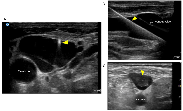
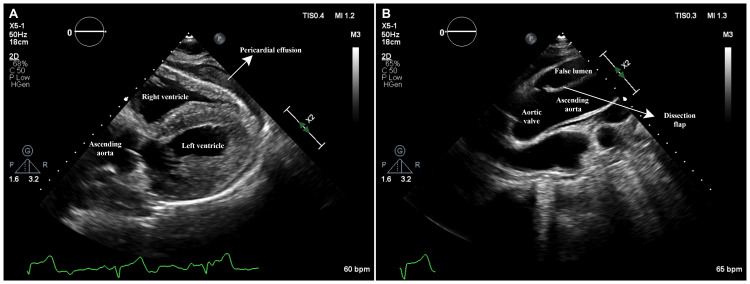
Point-of-care ultrasound (POCUS) is an essential skill in various specialties like anesthesiology, critical care, and emergency medicine. Anesthesiologists utilize POCUS for quick diagnosis and procedural guidance in perioperative and critical care settings. Key applications include vascular ultrasound for challenging venous and arterial catheter placements, gastric ultrasound for aspiration risk assessment, airway ultrasound, diaphragm ultrasound, and lung ultrasound for respiratory assessment. Additional utilities of POCUS can include multi-organ POCUS evaluation for undifferentiated shock or cardiac arrest, ultrasound-guided central neuraxial and peripheral nerve blocks, focused cardiac ultrasound, and novel applications such as venous excess ultrasound. This review highlights these POCUS applications in perioperative and intensive care and summarizes the latest evidence of their accuracy and limitations.

Keywords: airway ultrasound; anaesthesiology; bedside ultrasound; gastric ultrasound; perioperative pocus; pocus (point of care ultrasound).


Conflict of interest statement

Conflicts of interest: In compliance with the ICMJE uniform disclosure form, all authors declare the following: Payment/services info: All authors have declared that no financial support was received from any organization for the submitted work. Financial relationships: Abhilash Koratala declare(s) a grant from KidneyCure and the American Society of Nephrology’s William and Sandra Bennett Clinical Scholars Grant. Other relationships: All authors have declared that there are no other relationships or activities that could appear to have influenced the submitted work.

Figures

Figure 1
Figure 1. Ultrasound of the internal jugular vein for guiding central venous access placement
(A) Needle tip in the vein (arrow). (B) Guidewire in the vein (arrow). (C) Thrombus (arrow) in the internal jugular vein detected prior to catheter placement. Image credit: Abhilash Koratala
Figure 2
Figure 2. Compression ultrasound for evaluating lower extremity DVT
(A) Normal common femoral vein that is fully compressible with transducer pressure (B). (C) Common femoral vein with a hyperechoic thrombus in the lumen (arrow), which is non-compressible (D). DVT: deep vein thrombosis Image credit: Abhilash Koratala
Figure 3
Figure 3. Gastric ultrasound for evaluation of gastric contents
(A, B) Ultrasound probe location and orientation for parasagittal and axial views of the gastric antrum. Parasagittal (C, E, G, I) and axial (D, F, H, J) views of the gastric antrum: empty stomach (C, D); one hour after ingestion of clear liquids (E, F); one minute after drinking approximately 500 mL (16 oz) of clear liquids, showing an expanded lumen with hypoechoic contents (G, H); immediately after a meal with a characteristic “ground-glass” appearance (I, J). A: antrum; Ao: aorta; D: duodenum; IVC: inferior vena cava; L: liver; NPO: nil per os; P: pancreas; Py: pylorus; SMA: superior mesenteric artery Created with BioRender.com. Nguyen et al. (2023) [21]; Creative Commons Attribution (CC BY) license
Figure 4
Figure 4. Airway ultrasound for identifying the CTM
Cricoid cartilage, thyroid cartilage, and CTM in a longitudinal plane. Cc: cricoid cartilage; CTM: cricothyroid membrane; Tc: thyroid cartilage Osman and Sum (2016) [31]; Creative Commons Attribution (CC BY) license
Figure 5
Figure 5. Airway ultrasound showing tracheal intubation
The arrow indicates the anterior aspect of the ETT. ETT: endotracheal tube; T: trachea Gottlieb et al. (2024) [35]; Creative Commons Attribution (CC BY) license
Figure 6
Figure 6. Airway ultrasound showing esophageal intubation
In the case of esophageal intubation, a curvilinear structure that mimics the trachea (T) is seen to the right of the trachea. This is the ETT within the esophagus (E) [35]. ETT: endotracheal tube Gottlieb et al. (2024) [35]; Creative Commons Attribution (CC BY) license
Figure 7
Figure 7. LUS
(A) Normal lung showing horizontal artifacts, i.e., A-lines (arrows). (B) Vertical artifacts (arrows) known as B-lines indicate interlobular septal thickening, typically seen in congestion. (C) Pleural effusion (asterisk) as seen on a lateral scan. (D) Right pleural effusion (asterisk) as seen from the subxiphoid scanning window. IVC: inferior vena cava; LUS: lung ultrasound Turk et al. (2023) [66]; Creative Commons Attribution (CC BY) license
Figure 8
Figure 8. Sagittal views of the lumbar spine
(A) Sagittal transverse process view. (B) Sagittal articular process view. (C) Sagittal lamina view. (D) Sagittal spinous process view. (E) Parasagittal oblique view. AC: anterior complex; AP: articular process; L: lamina; PC: posterior complex; SC: spinal canal (intrathecal space); SP: spinous process; TP: transverse process Yoo et al. (2020) [79]; Creative Commons Attribution (CC BY) license
Figure 9
Figure 9. Transverse views of the lumbar spine
(A) Transverse spinous process view. (B) Transverse interspinous process view. (C) Tilted transverse interspinous process view. AC: anterior complex; AP: articular process; L: lamina; PC: posterior complex; SC: spinal canal (intrathecal space); SP: spinous process Yoo et al. (2020) [79]; Creative Commons Attribution (CC BY) license
Figure 10
Figure 10. Basic echocardiographic views
(A) Parasternal long axis. (B) Parasternal short axis. (C) Apical four-chamber. (D) Subxiphoid; (E) IVC. The green arrows indicate the direction of the transducer orientation marker. IVC: inferior vena cava; LA: left atrium; LV: left ventricle; RA: right atrium; RV: right ventricle Argaiz et al. (2021) [91]; reproduced with permission from Wolters Kluwer Health
Figure 11
Figure 11. Ascending aortic dissection with cardiac tamponade
Transthoracic echocardiogram in a patient with ascending aortic dissection and cardiac tamponade: (A) Subcostal view showing a large pericardial effusion causing RV collapse. (B) A high-right parasternal view demonstrating the intimal dissection flap in the ascending aorta. RV: right ventricular Image credit: Dinkar Bhasin
Figure 12
Figure 12. Severe mitral stenosis in a patient with shock
(A) Apical four-chamber view demonstrates thickened mitral valve leaflets and a dilated left atrium (arrowheads). (B) Color Doppler imaging across the mitral valve shows turbulent flow across the mitral valve in diastole. (C) Parasternal short-axis view demonstrating thickened mitral valve leaflets (arrowheads) with commissural fusion giving a fish-mouth appearance. Mild pericardial effusion can also be appreciated. (D) Continuous-wave Doppler across the mitral valve demonstrates an elevated gradient. The valve area, as measured by pressure half-time, is 1 cm2. Image credit: Dinkar Bhasin
Figure 13
Figure 13. VExUS grading
When the diameter of the IVC is >2 cm, three grades of congestion are defined based on the severity of abnormalities on the hepatic, portal, and renal parenchymal venous Doppler. Hepatic vein Doppler is considered mildly abnormal when the systolic (S) wave is smaller than the diastolic (D) wave but still below the baseline; it is considered severely abnormal when the S-wave is reversed. Portal vein Doppler is considered mildly abnormal when the pulsatility is 30-50%, and severely abnormal when it is ≥50%. Asterisks represent points of pulsatility measurement. Renal parenchymal vein Doppler is mildly abnormal when it is pulsatile with distinct S and D components and severely abnormal when it is monophasic with a D-only pattern. IVC: inferior vena cava; VExUS: venous excess ultrasound Adapted from NephroPOCUS.com with permission

References

    1. Perioperative point-of-care ultrasound: from concept to application. Ramsingh D, Bronshteyn YS, Haskins S, Zimmerman J. Anesthesiology. 2020;132:908–916. - PubMed
    1. The utility of point of care ultrasonography (POCUS) Hashim A, Tahir MJ, Ullah I, Asghar MS, Siddiqi H, Yousaf Z. Ann Med Surg (Lond) 2021;71:102982. - PMC - PubMed
    1. History of emergency and critical care ultrasound: the evolution of a new imaging paradigm. Kendall JL, Hoffenberg SR, Smith RS. Crit Care Med. 2007;35:0–30. - PubMed
    1. A systematic review of the cost-effectiveness of ultrasound in emergency care settings. Lentz B, Fong T, Rhyne R, Risko N. Ultrasound J. 2021;13:16. - PMC - PubMed
    1. Stages and factors of the "Perioperative process": points in common with the aeronautical industry. Davrieux CF, Palermo M, Serra E, Houghton EJ, Acquafresca PA, Finger C, Giménez ME. Arq Bras Cir Dig. 2019;32:0. - PMC - PubMed

LinkOut - more resources