. 2024 Aug 30:42:124-139.
doi: 10.1016/j.bioactmat.2024.08.019. eCollection 2024 Dec.

Neuroprotection on ischemic brain injury by Mg2+/H2 released from endovascular Mg implant

Affiliations

Neuroprotection on ischemic brain injury by Mg2+/H2 released from endovascular Mg implant

Yang Zhang et al. Bioact Mater. .

Abstract

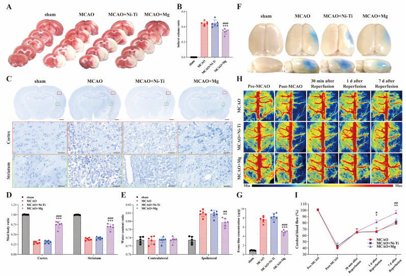
Most acute ischemic stroke patients with large vessel occlusion require stent implantation for complete recanalization. Yet, due to ischemia-reperfusion injury, over half of these patients still experience poor prognoses. Thus, neuroprotective treatment is imperative to alleviate the ischemic brain injury, and a proof-of-concept study was conducted on "biodegradable neuroprotective stent". This concept is premised on the hypothesis that locally released Mg2+/H2 from Mg metal within the bloodstream could offer synergistic neuroprotection against reperfusion injury in distant cerebral ischemic tissues. Initially, the study evaluated pure Mg's neuroactive potential using oxygen-glucose deprivation/reoxygenation (OGD/R) injured neuron cells. Subsequently, a pure Mg wire was implanted into the common carotid artery of the transient middle cerebral artery occlusion (MCAO) rat model to simulate human brain ischemia/reperfusion injury. In vitro analyses revealed that pure Mg extract aided mouse hippocampal neuronal cell (HT-22) in defending against OGD/R injury. Additionally, the protective effects of the Mg wire on behavioral abnormalities, neural injury, blood-brain barrier disruption, and cerebral blood flow reduction in MCAO rats were verified. Conclusively, Mg-based biodegradable neuroprotective implants could serve as an effective local Mg2+/H2 delivery system for treating distant cerebral ischemic diseases.

Keywords: Acute ischemic stroke; Biodegradable implantation; Hydrogen; Magnesium; Neuroprotection.


Conflict of interest statement

Yufeng Zheng is the editor-in-chief for Bioactive Materials and was not involved in the editorial review or the decision to publish this article. All authors declare that they have no known competing financial interests or personal relationships that could have appeared to influence the work reported in this paper.

Figures

Image 1
Graphical abstract
Fig. 1
Fig. 1
The proof-of-concept of the biodegradable neuroprotective stent and the implementation strategies to validate this concept. Biodegradable neuroprotective stent refers to stents that could locally produce neuroprotective substances (like Mg2+/H2) during the degradation of stents within blood flow to the damaged brain tissue, thereby rescuing distal cerebral ischemic tissues. To verify this concept, a middle cerebral artery occlusion rat model was employed, implanting an Mg wire representing Mg stent in the common carotid artery.
Fig. 2
Fig. 2
Computational modeling of Mg wire corrosion within the common carotid artery. (A) The modeling of blood vessels and inserted Mg wires, alongside the corresponding distribution of blood flow velocity within the vessel. (B) The calculated changes in Mg wire diameter over time and the concentration of released Mg2+ and H2 around the wire surface on day 7. (C) The simulated morphological features of the Mg wire over time and the associated contour plots of Mg2+ concentration.
Fig. 3
Fig. 3
In vitro neuroprotection of Mg wires extract on oxygen-glucose deprivation/reoxygenation (OGD/R) injured cells. (A) Raw data for detecting H2 through gas chromatography. (B) Quantification of H2 and Mg2+ in normal Dulbecco's Modified Eagle Medium (DMEM) and magnesium wire immersed DMEM. (C) OGD/R cell experimental flowchart. The neuroprotective effect of Mg wires, with or without ultrasound (US) treatment, was evaluated by (D) cell viability, (E) relative lactic dehydrogenase (LDH) release concentration in the supernatant, (F) intracellular relative fluorescence intensity of reactive oxygen species (ROS) in cells and (G) fluorescent images of intracellular Ca2+ using a fluorescent microscope, scale bars: 20 μm. (H) Quantification of the fluorescence intensity of Ca2+ calculated from (G). Statistical significance is indicated by * p < 0.05, **p < 0.01, ***p < 0.001 vs. OGD/R group; #p < 0.05, ##p < 0.01, ###p < 0.001.
Fig. 4
Fig. 4
In vivo neuroprotection of Mg wires after ischemic injury. (A) Longa score, (B) elevated body swing test (EBST), (C) rotarod test, and (D) adhesive removal Test were administered before the operation and at 1, 3, 5, 7, 10, and 14 days post-operation. (E) The open field test was conducted 7 days after the operation. (F) The walking distance and time in the open field. The sample size for each group was n = 6 for the sham group and n = 12 for the Middle Cerebral Artery Occlusion (MCAO), MCAO + Ni-Ti, and MCAO + Mg groups. Statistical significance is denoted by * p < 0.05, **p < 0.01, ***p < 0.001 for MCAO + Mg vs. MCAO group; ##p < 0.01, ###p < 0.001 for MCAO + Mg vs. MCAO + Ni-Ti group.
Fig. 5
Fig. 5
In vivo brain integrity and cerebral blood flow (CBF) preservation of Mg wires on the 7 days after ischemia/reperfusion injury. (A) TTC stain images of brain slices and (B) semiquantitative analysis of infarction volume. (C) Nissl stain images of brain slices and (D) semiquantitative analysis of Nissl bodies; scale bars: 1 mm and 25 μm. (E) Water content ratio of the contralateral and ipsilateral sides of the ischemic brain. (F) Representative images of Evans blue dye extravasation into the brain and (G) quantification of Evans blue content in brain tissue on the infarcted side. (H) CBF images in rats measured by laser speckle imaging at indicated time points. (I) Temporal CBF profile in rats, derived from (H), with the damaged side CBF expressed as a ratio to the contralateral side. Sample sizes were n = 5 for the sham group and n = 6 for the Middle Cerebral Artery Occlusion (MCAO), MCAO + Ni-Ti, and MCAO + Mg groups. Statistical significance is denoted by * p < 0.05, **p < 0.01, ***p < 0.001 for MCAO + Mg vs. MCAO group; #p < 0.05, ##p < 0.01, ###p < 0.001 for MCAO + Mg vs. MCAO + Ni-Ti group.
Fig. 6
Fig. 6
In vivo corrosion evaluation for Mg wires. (A) 3D reconstructions of the residual Mg wires and corrosion products at 1, 7, 14, and 28 days post-implantation, based on micro-computed tomography analysis. (B) The residual Mg wire volume and the volume of released hydrogen over time. The residual Mg wire volume is derived from simulation results, while the volume of released hydrogen is calculated based on this residual volume. (C) The residual Mg wire volume and the magnesium dissolved/released into the physiological environment over the course of implantation. (D) Scanning electron microscope images and energy-disperse spectrometer (EDS) element mapping of the Mg wires after 1, 7, 14, and 28 days of implantation. The black and white scale bars represent 50 and 10 μm, respectively. (E) The EDS point analysis results of the degradation products on the Mg wires' surface.
Fig. 7
Fig. 7
In vivo Mg2+, H2, reactive oxygen species (ROS) and Ca2+ concentration in blood and brain. The concentration of Mg in the (A) blood and (B) infarction ipsilateral brain (IIB) of rats at 1 and 7 days post-implantation of Mg wires (n = 3). (C) Raw data and (D) quantification of H2 in rat blood tested by gas chromatography. (E) The map and inference of the distribution of Mg2+ and H2 in blood and brain tissue. The relative fluorescence intensity of cerebral (F) ROS and (G) Ca2+ is also depicted. The sample size for each group was n = 5 for the sham group and n = 6 for the Middle Cerebral Artery Occlusion (MCAO), MCAO + Ni-Ti, and MCAO + Mg groups. Statistical significance is indicated by **p < 0.01, ***p < 0.001 for MCAO + Mg vs. MCAO group; ##p < 0.01, ###p < 0.001 for MCAO + Mg vs. MCAO + Ni-Ti group.
Fig. 8
Fig. 8
Illustration of the effect of Mg wire in the ischemic brain. The Mg2+ and hydrogen generated by the degradation of Mg wires travel to the damaged site and cross the BBB into the brain. Mg2+ and hydrogen primarily inhibit the influx of Ca2+ and the production of ROS, respectively, and exhibit a synergistic relationship, ultimately leading to neuron rescue, BBB protection, and enhanced CBF.

References

    1. GBD 2016 Lifetime Risk of Stroke Collaborators Global, regional, and country-specific lifetime risks of stroke, 1990 and 2016. N. Engl. J. Med. 2018;379:2429–2437. - PMC - PubMed
    1. Eren F., Yilmaz S.E. Neuroprotective approach in acute ischemic stroke: a systematic review of clinical and experimental studies. Brain circulation. 2022;8:172–179. - PMC - PubMed
    1. Tong Y., Ding Y., Han Z., Duan H., Geng X. Optimal rehabilitation strategies for early postacute stroke recovery: an ongoing inquiry. Brain circulation. 2023;9:201–204. - PMC - PubMed
    1. Li C., et al. Pleiotropic microenvironment remodeling micelles for cerebral ischemia-reperfusion injury therapy by inhibiting neuronal ferroptosis and glial overactivation. ACS Nano. 2023;17:18164–18177. - PubMed
    1. Ohta T., et al. First-in-human trial of a self-expandable, temporary dilation system for intracranial atherosclerotic disease in patients presenting with acute ischemic stroke. J. Neurointerventional Surg. 2023 jnis-2023-020983. - PMC - PubMed

LinkOut - more resources