Case Reports
. 2024 Fall;24(3):213-218.
doi: 10.31486/toj.24.0014.

Recurrent Intracranial Ewing Sarcoma

Affiliations
Case Reports

Recurrent Intracranial Ewing Sarcoma

Soo Ann Yap et al. Ochsner J. 2024 Fall.

Abstract

Background: Ewing sarcoma is a rare malignant neoplasm that is primarily localized in bone tissues. The prognosis for patients with a newly diagnosed localized Ewing sarcoma has been greatly improved by multimodality treatment. However, treating patients with disseminated or recurrent disease is challenging, with a 5-year overall survival rate of <30%. Case Report: A 17-year-old female with an asymptomatic tumor of the left temple underwent 3 cycles of vincristine, ifosfamide, doxorubicin, and etoposide and achieved partial remission. However, the patient refused further chemotherapy and surgical intervention and was lost to follow-up. After 7 months, the patient presented again with a sizeable tumor on her left temple and worsening symptoms. Chemotherapy with alternating cycles of vincristine, doxorubicin, cyclophosphamide, ifosfamide, and etoposide according to the EURO EWING 2012 trial was initiated. After a positive response, debulking surgery was performed, followed by postsurgical radiation, and partial remission was achieved. Conclusion: Optimal treatment protocols for recurrent Ewing sarcoma are lacking. Treatments are individualized based on the patient's response to treatment and the decisions of tumor boards. Patients with rare tumors such as Ewing sarcoma benefit from multidisciplinary collaboration, resulting in improved quality of care and treatment outcomes.

Keywords: Chemotherapy–adjuvant; neoplasm metastasis; radiotherapy; sarcoma–Ewing; soft tissue neoplasms.


Figures

Figure 1.
Figure 1.
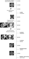
Timeline of disease progression. Dx, diagnosis; Gy, gray; VDC/IE, vincristine, doxorubicin, cyclophosphamide, ifosfamide, and etoposide; VIDE, vincristine, ifosfamide, doxorubicin, and etoposide.
Figure 2.
Figure 2.
(A) Cranial magnetic resonance imaging shows a large tissue mass (54 mm × 26 mm × 59 mm) in the temporal lobe. (B) Cranial computed tomography shows partial remission (6 mm × 10 mm) after 3 cycles of vincristine, ifosfamide, doxorubicin, and etoposide (VIDE).
Figure 3.
Figure 3.
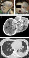
(A) The 20-year-old patient presented with a massive nontender tumor mass on the left temple with visible oral and orbital infiltration. (B) Computed tomography shows a tumor mass of 130 mm × 160 mm × 200 mm in the left hemisphere and (C) pulmonary metastasis.
Figure 4.
Figure 4.
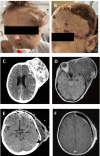
(A) Photograph shows tumor volume reduction and the appearance of necrotic tissue after 4 cycles of chemotherapy. (B) R0 resection was achieved with a successful reconstruction of the calotte. (C) Cranial computed tomography (CT) and (D) cranial magnetic resonance imaging (MRI) show significant shrinkage of the primary tumor (85 mm × 87 mm × 124 mm) after 4 cycles of chemotherapy. (E) Cranial CT shows postsurgical hematoma and fluid retention without evidence of tumor mass. (F) Two months postoperative cranial MRI shows regression of hematoma and fluid retention.

References

    1. Falk S, Alpert M. The clinical and roentgen aspects of Ewing's sarcoma. Am J Med Sci. 1965;250(5):492-508. - PubMed
    1. Fitzer PM, Steffey WR. Brain and bone scans in primary Ewing's sarcoma of the petrous bone. J Neurosurg. 1976;44(5):608-612. doi: 10.3171/jns.1976.44.5.0608 - DOI - PubMed
    1. Turner JL, Sweeney P, Hardy R. Ewing's tumor metastatic to the clivus, simulating meningitis: case report. Neurosurgery. 1980;7(6):619-620. doi: 10.1227/00006123-198012000-00017 - DOI - PubMed
    1. Pankratjevaite L, Eskandarani HA, Lizdenis P, Saladzinskas Z. Challenges in diagnosing an extraosseous Ewing sarcoma: a case report. Int J Surg Case Rep. 2022;91:106708. doi: 10.1016/j.ijscr.2021.106708 - DOI - PMC - PubMed
    1. Sparreboom BD, Trautman J, Yaxley J. Ewing sarcoma: a pictorial review of typical and atypical locations with reference to the updated 2020 WHO classification system. J Med Imaging Radiat Oncol. 2022;66(6):812-818. doi: 10.1111/1754-9485.13456 - DOI - PubMed

Publication types

LinkOut - more resources