. 2024 Aug 28:41:640-656.
doi: 10.1016/j.bioactmat.2024.08.012. eCollection 2024 Nov.

Suspension electrospinning of decellularized extracellular matrix: A new method to preserve bioactivity

Affiliations

Suspension electrospinning of decellularized extracellular matrix: A new method to preserve bioactivity

Sarah Jones et al. Bioact Mater. .

Abstract

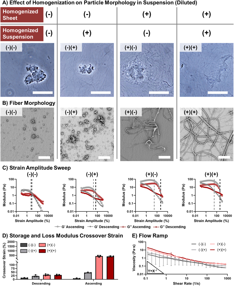
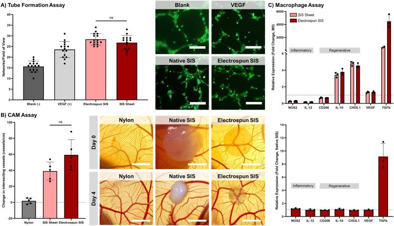
Decellularized extracellular matrices (dECM) have strong regenerative potential as tissue engineering scaffolds; however, current clinical options for dECM scaffolds are limited to freeze-drying its native form into sheets. Electrospinning is a versatile scaffold fabrication technique that allows control of macro- and microarchitecture. It remains challenging to electrospin dECM, which has led researchers to either blend it with synthetic materials or use enzymatic digestion to fully solubilize the dECM. Both strategies reduce the innate bioactivity of dECM and limit its regenerative potential. Herein, we developed a new suspension electrospinning method to fabricate a pure dECM fibrous mesh that retains its innate bioactivity. Systematic investigation of suspension parameters was used to identify critical rheological properties required to instill "spinnability," including homogenization, concentration, and particle size. Homogenization enhanced particle interaction to impart the requisite elastic behavior to withstand electrostatic drawing without breaking. A direct correlation between concentration and viscosity was observed that altered fiber morphology; whereas, particle size had minimal impact on suspension properties and fiber morphology. The versatility of this new method was demonstrated by electrospinning dECM with three common decellularization techniques (Abraham, Badylak, Luo) and tissue sources (intestinal submucosa, heart, skin). Bioactivity retention after electrospinning was confirmed using cell proliferation, angiogenesis, and macrophage polarization assays. Collectively, these findings provide a framework for researchers to electrospin dECM for diverse tissue engineering applications.

Keywords: Biological scaffolds; Electrospinning; Extracellular matrix.


Conflict of interest statement

The authors declare that they have no known competing financial interests or personal relationships that could have appeared to influence the work reported in this paper.

Figures

Image 1
Graphical abstract
Fig. 1
Fig. 1
Schematic of electrospinning method for dECM. A) The preparation of a dECM electrospinning suspension is a five-step process involving decellularization, homogenization, milling, suspension in HFIP, and a second round of homogenization. Macro images for steps 1–5; scale bar = 1 cm. Inset SEM image of dried SIS particle in step 3; scale bar = 300 μm. Inset brightfield image of suspended SIS particle in step 4; scale bar = 60 μm. B) The dECM suspension is electrospun by applying an electric field between the flowing suspension and a collector. Graphic created in BioRender. SEM image of electrospun dECM fibers; scale bar = 30 μm.
Fig. 2
Fig. 2
Effect of homogenization on suspension particle morphology, rheological properties, and fiber collection. A) Brightfield image of dECM particles in an electrospinning suspension. Scale bar = 60 μm. B) SEM images of collection morphology. Scale bar = 20 μm. C) Strain amplitude sweep of dECM suspensions in which solid points were collected with a descending amplitude sweep and hollow points were collected in an ascending manner. The grey dotted line indicates the crossover from the descending amplitude test and the black dotted line indicates the crossover with ascending applied strain. D) The strain amplitude at which the storage and loss moduli cross collected with a descending and ascending strain sweep. E) The rate dependence of the viscosity of the dECM suspensions.
Fig. 3
Fig. 3
Effect of SIS concentration on suspension rheological properties and fiber collection. A) SEM images of fiber morphology. Scale bar = 20 μm. B) Strain amplitude sweep of dECM suspensions in which solid points were collected with a descending strain sweep and hollow points were collected with an ascending strain sweep. The grey dotted line indicates the crossover with descending applied strain and the black dotted line indicates the crossover with ascending applied strain. C) The crossover strain of the storage and loss modulus curves collected with descending and ascending strain sweeps. D) dECM suspension viscosity with respect to shear rate.
Fig. 4
Fig. 4
Effect of dry particle size on suspension particle morphology, rheological properties, and fiber collection. A) Brightfield image of dECM particles in an electrospinning suspension. Scale bar = 60 μm. Inset stereoscope image of dried dECM particles. Scale bar = 3 mm. B) SEM images of fiber morphology. Scale bar = 20 μm. C) Strain amplitude sweep of dECM suspensions in which solid points were collected with a descending strain sweep and hollow points were collected with an ascending strain sweep. The grey dotted line indicates the crossover with descending applied strain and the black dotted line indicates the crossover with ascending applied strain. D) The crossover strain of the storage and loss modulus curves collected with descending and ascending strain sweeps. E) dECM suspension viscosity with respect to shear rate.
Fig. 5
Fig. 5
Effect of SIS decellularization and preparation method (Badylak, Abraham, and Luo) on suspension particle morphology, rheological properties, and fiber collection. A) Brightfield image of dECM particles in an electrospinning suspension. Scale bar = 60 μm. B) SEM images of the electrospun dECM fibers. Scale bar = 20 μm. C) Strain amplitude sweep of dECM suspensions in which solid points were collected with a descending strain sweep and hollow points were collected with an ascending strain sweep. The grey dotted line indicates the crossover with descending applied strain and the black dotted line indicates the crossover with ascending applied strain. D) The crossover strain of the storage and loss modulus curves collected with a descending and ascending strain sweep. E) dECM suspension viscosity with respect to shear rate.
Fig. 6
Fig. 6
Effect of tissue source (SIS, skin, heart) on suspension particle morphology, rheological properties, and fiber collection. A) SEM images of the electrospun dECM fibers. Scale bar = 20 μm. B) Strain amplitude sweep of dECM suspensions in which solid points were collected with a descending strain sweep and hollow points were collected with an ascending strain sweep. The grey dotted line indicates the crossover with descending applied strain and the black dotted line indicates the crossover with ascending applied strain. C) The crossover strain and modulus of the storage and loss modulus curves collected with a descending and ascending strain sweep. D) The viscosity with respect to the shear rate of the dECM suspensions.
Fig. 7
Fig. 7
The effect of electrospinning on SIS composition. A) Differential scanning calorimetry thermogram with the onset temperature, peak temperature, and enthalpy of denaturation. B) Biochemical composition of SIS including total protein, collagen, and sGAG content.
Fig. 8
Fig. 8
Evaluation of bioactivity retention after electrospinning. A) In vitro HUVEC tube formation in SIS-conditioned media. Scale bar = 500 μm. B) In ovo vascularization in a CAM assay after 4 days of sample placement. Vessel density is calculated as the change in the number of vessels intersecting with the sample from Day 0 to Day 4, normalized to the sample perimeter. Scale bar = 3 mm. C) Macrophage gene expression after 72 h of SIS contact with respect to the expression of the M0 control (top) and native SIS (bottom).

References

    1. Badylak S.F. The extracellular matrix as a biologic scaffold material. Biomaterials. 2007/09/01/2007;28(25):3587–3593. - PubMed
    1. Huleihel L., et al. Matrix-bound nanovesicles within ECM bioscaffolds. Sci. Adv. 2016;2(6) - PMC - PubMed
    1. Wilgus T.A. Growth factor-extracellular matrix interactions regulate wound repair. Adv. Wound Care. Dec 2012;1(6):249–254. (in eng) - PMC - PubMed
    1. Badylak S.F., Freytes D.O., Gilbert T.W. Extracellular matrix as a biological scaffold material: structure and function. Acta Biomater. 2009/01/01/2009;5(1):1–13. - PubMed
    1. Li F., Li W., Johnson S., Ingram D., Yoder M., Badylak S. Low-molecular-weight peptides derived from extracellular matrix as chemoattractants for primary endothelial cells. Endothelium. May-Aug 2004;11(3–4):199–206. (in eng) - PubMed

LinkOut - more resources