. 2024 Aug 29:26:663-670.
doi: 10.1016/j.reth.2024.08.013. eCollection 2024 Jun.

Synergistic effect of ex-vivo quality and quantity cultured mononuclear cells and mesenchymal stem cell therapy in ischemic hind limb model mice

Affiliations

Synergistic effect of ex-vivo quality and quantity cultured mononuclear cells and mesenchymal stem cell therapy in ischemic hind limb model mice

Taro Fukuta et al. Regen Ther. .

Abstract

Introduction: Chronic limb-threatening ischemia (CLTI) is a condition characterized by peripheral arterial disease and tissue damage caused by reduced blood flow. New therapies using various cell types, such as mesenchymal stem cells (MSCs) and mononuclear cells (MNCs), have been developed for the patients unresponsive to conventional therapies. MSCs are promising because of their ability to secrete growth factors essential for vascularization, whereas MNCs contain endothelial progenitor cells that are important for blood vessel formation. However, conventional methods for isolating these cells have limitations, especially in patients with diabetes with dysfunctional cells. To overcome this problem, a culture method called quality and quantity cultured peripheral blood MNCs (MNC-QQ) was developed to efficiently produce high-quality cells from small amounts of peripheral blood. Combining MSCs with MNC-QQs has been hypothesized to enhance therapeutic outcomes. This study aimed to examine the angiogenic efficacy of MSCs with MNC-QQs in models with severe lower limb ischemia.

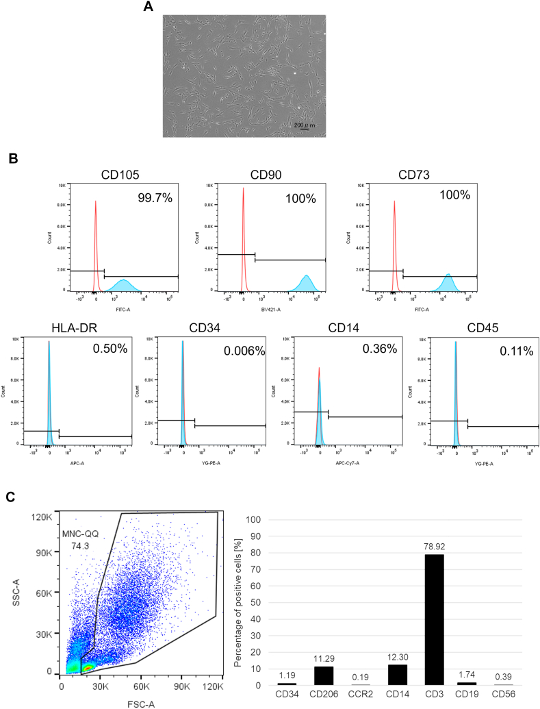
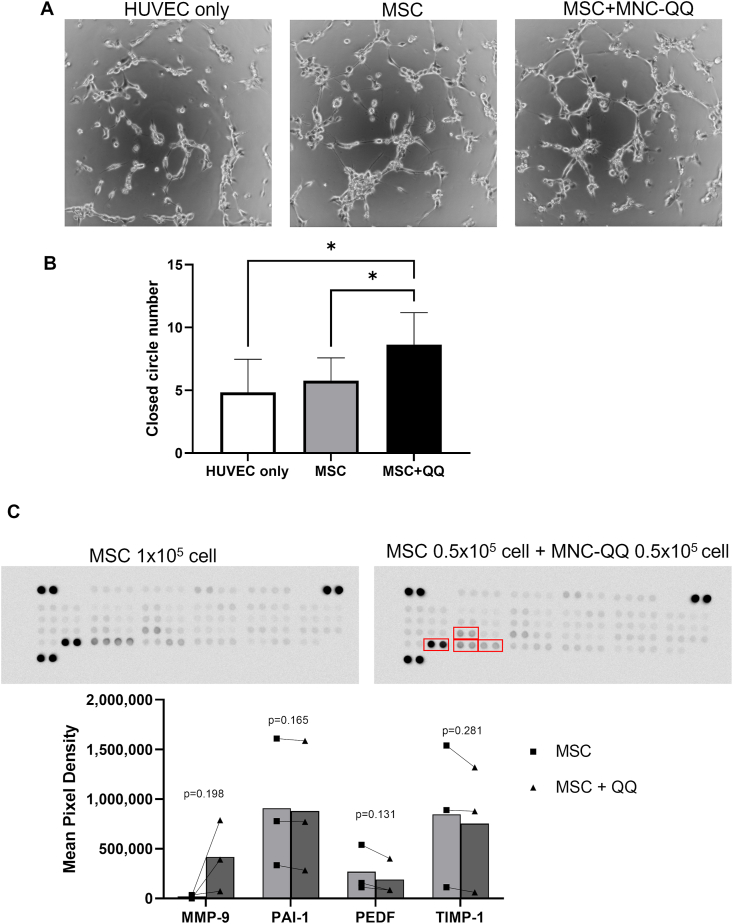
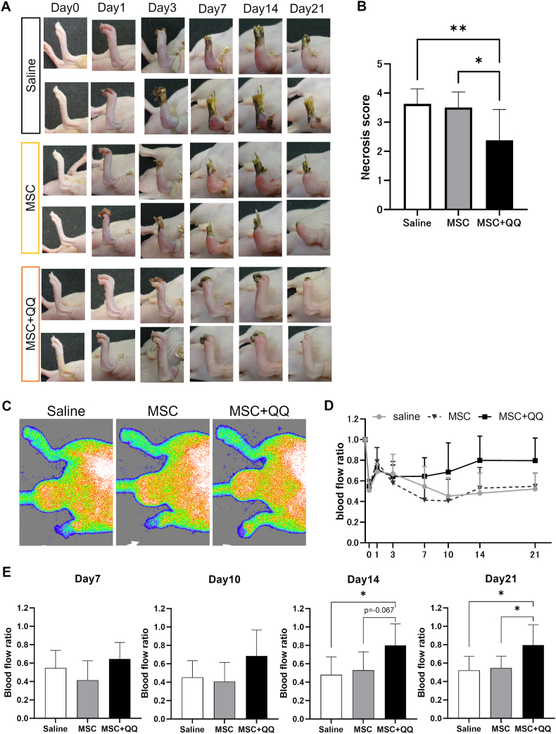
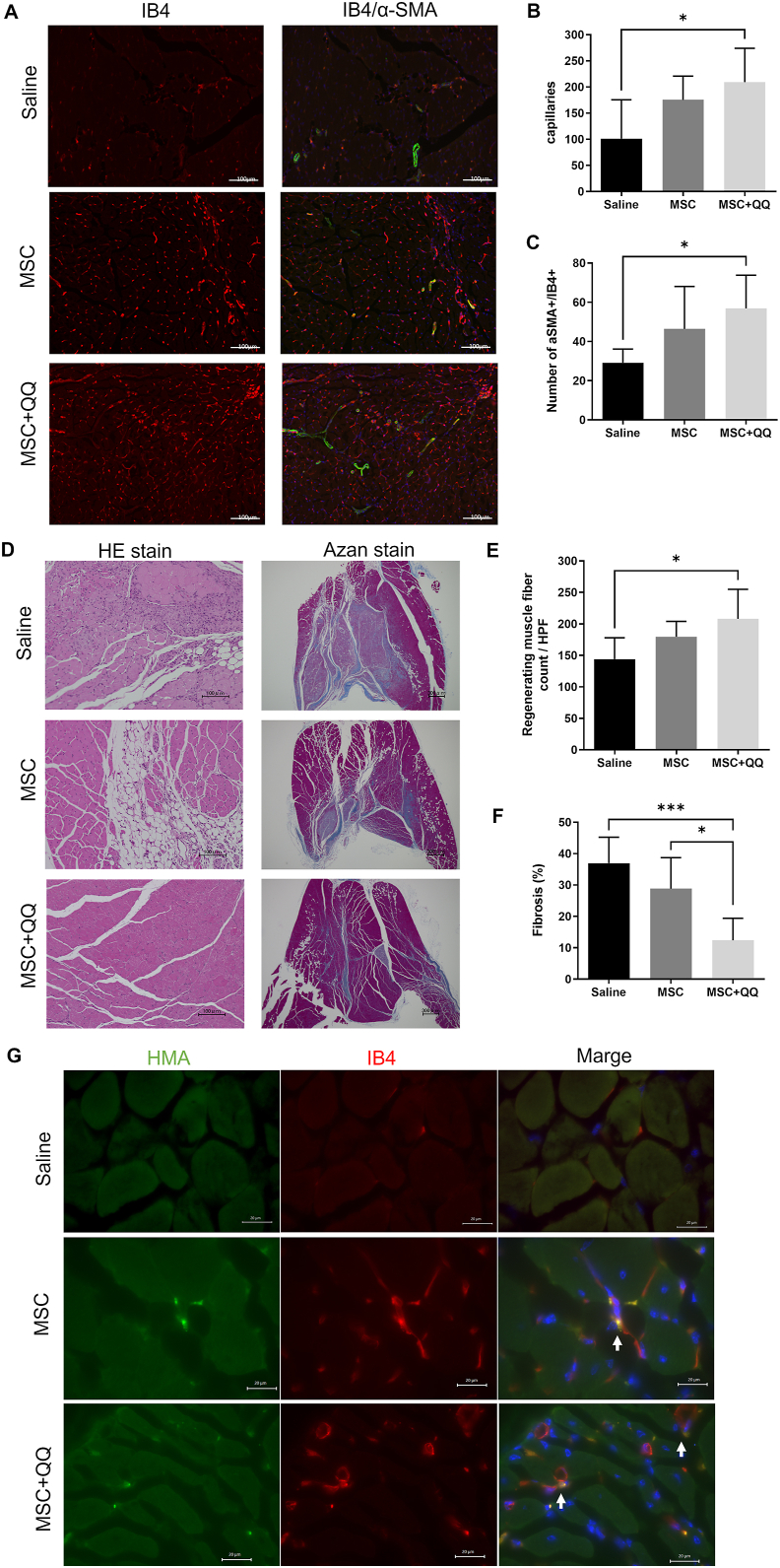
Methods: MNC-QQ was manufactured from the peripheral blood of healthy volunteers, while human bone marrow derived MSCs were purchased. To verify the effects of the MSC and MNC-QQs combination in angiogenesis, we conducted the HUVEC tube formation assay. For in vivo experiments, we created an ischemic limb model using BALB/c nude mice. Saline, MSCs alone, and a combination of MSCs and MNC-QQs were administered intramuscularly into the ischemic limbs. Blood flow was measured over time using laser doppler, and the ischemic limbs were harvested 21 days later for HE staining and immunostaining for histological assessment.

Results: In-vitro studies demonstrated increased angiogenesis when MSCs were combined with MNC-QQs compared with MSCs alone. In vivo experiments using a mouse model of severe lower limb ischemia showed that combination therapy significantly improved blood flow recovery and limb salvage compared with MSCs alone or saline treatment. Histological analysis revealed enhanced vessel density, arteriogenesis, muscle regeneration, and reduced fibrosis in the MSC + MNC-QQ group compared with those in the saline group. Although the specific interactions between MSCs and MNC-QQs have not been fully elucidated, combined therapy leverages the benefits of both cell types, resulting in improved outcomes for vascular regeneration.

Conclusions: This study highlights the potential of the simultaneous transplantation of MSCs and MNC-QQs as a promising therapeutic approach for CLTI, offering sustained long-term benefits for patients.

Keywords: Cell therapy; Mesenchymal stem cells; Peripheral blood mononuclear cell; Vasculogenesis.


Conflict of interest statement

The authors have no conflict of interest to declare.

Figures

Fig. 1
Fig. 1
Identification of MSCs and MNC-QQs. (A) Representative images of MSCs. (B) The expressions of surface marker on MSCs were analyzed by flowcytometry. The expressions of CD105, CD90, and CD73 were positive and HLA-DR, CD34, CD14, and CD45 were negative in MSCs (blue). Red lines indicate isotype control. (C) The expression of CD34, CD206, CCR2, CD14, CD3, CXCR4, CD19, and CD56 in MNC-QQs were analyzed by flowcytometry. MSC, mesenchymal stem cell; MNC-QQs, quality and quantity cultured peripheral blood MNCs.
Fig. 2
Fig. 2
Combination of MSCs and MNC-QQs increased angiogenic potential in vitro. (A) Representative images of HUVEC tube formation assay. (B) The number of closed circle formations was counted. (HUVEC only; n = 6, MSC, MSC + QQ; n = 8) (C) Representative images of human angiogenesis cytokine array and the graphs of their analysis results. The dots in the graph represent individual results (■; MSC, ▲; MSC + QQ) The bars represent the mean value (n = 3). ∗p < 0.05. MSC + QQ, combination of MSC and MNC-QQs; HUVEC, human umbilical vein endothelial cells.
Fig. 3
Fig. 3
Combination of MSC and MNC-QQ transplantations improved limb salvage rate in ischemic hind limb model mice. MSC (2 × 104 cells/body) and the combination of MSC + MNC-QQ (1 × 104 cells/body, each cell) were administered intramuscularly into the ischemic hind limbs immediately after surgery. (A) Representative images of ischemic hind limbs. (B) Necrosis scores of the ischemic hind limbs (0 = no change, I = nail necrosis, II = toe necrosis, III = foot necrosis, IV = knee necrosis, V = total amputation) measured on day 21 (N = 8). (C) The blood flow preservation rate in the affected limb was measured using a laser blood flow imager. Representative images from day 21 are shown. White arrows show the ischemic limbs. (D) (E) The blood flow ratio of the ischemic versus the contralateral hind limb was calculated on days 0, 1, 3, 7, 10, 14, and 21. (N = 8) Graphs show the mean ± SD. ∗p < 0.05, ∗∗p < 0.01. SD, standard deviation.
Fig. 4
Fig. 4
Histological analysis of MSC + QQ transplanted ischemic hind limb reveals significantly increased vessels and regenerating muscles. The gastrocnemius muscles of the ischemic hind limbs were assessed histologically on day 21. (A) The representative images of isolectin GS-IB4 (red) and α-SMA (green). (B) To analyze the number of capillaries, cells positive for endothelial marker IB4 were counted. (C) The number of IB4 and α-SMA double positive vessels were counted. (D) Representative images of hematoxylin and eosin (HE) and azan staining of ischemic hind limbs. (E) Centrally nucleated muscle fibers in HE staining were counted as regenerating muscle fibers. (F) The area containing collagen fibers was measured to assess fibrosis (N = 6). Graphs show the mean ± SD. (G) Representative image of localization of transplanted human cells stained with HMA (green) and isolectin B4 stained vessels (red). White bar in the images represents 20 μm. White allows indicate co-localization of HMA and isolectin B4. ∗p < 0.05, ∗∗p < 0.01. IB4, isolectin GS-IB4; SD, standard deviation; α-SMA, alpha-smooth muscle actin; HMA, human mitochondrial antibody.

Similar articles

References

    1. Conte M.S., Bradbury A.W., Kolh P., White J.V., Dick F., Fitridge R., et al. Global vascular guidelines on the management of chronic limb-threatening ischemia. J Vasc Surg. 2019;69:3S–125S e40. doi: 10.1016/j.jvs.2019.02.016. - DOI - PMC - PubMed
    1. Kawarada O., Fujihara M., Higashimori A., Yokoi Y., Honda Y., Fitzgerald P.J. Predictors of adverse clinical outcomes after successful infrapopliteal intervention. Cathet Cardiovasc Interv. 2012;80:861–871. doi: 10.1002/ccd.24370. - DOI - PubMed
    1. Kim T.I., Vartanian S.S., Schneider P.A. A review and proposed classification system for the No-option patient with chronic limb-threatening ischemia. J Endovasc Ther. 2021;28:183–193. doi: 10.1177/1526602820963911. - DOI - PubMed
    1. Iwase T., Nagaya N., Fujii T., Itoh T., Murakami S., Matsumoto T., et al. Comparison of angiogenic potency between mesenchymal stem cells and mononuclear cells in a rat model of hindlimb ischemia. Cardiovasc Res. 2005;66:543–551. doi: 10.1016/j.cardiores.2005.02.006. - DOI - PubMed
    1. Teraa M., Gremmels H., Wijnand J.G.J., Verhaar M.C. Cell therapy for chronic limb-threatening ischemia: current evidence and future directions. Stem Cells Transl Med. 2018;7(12):842–846. doi: 10.1002/sctm.18-0025. - DOI - PMC - PubMed

LinkOut - more resources