

circ_0114866 promotes the progression and EMT of non-small cell lung cancer via miR-653-5p/MYL6B axis
- PMID: 39281569
- PMCID: PMC11402247
- DOI: 10.1016/j.heliyon.2024.e37062
circ_0114866 promotes the progression and EMT of non-small cell lung cancer via miR-653-5p/MYL6B axis
Abstract
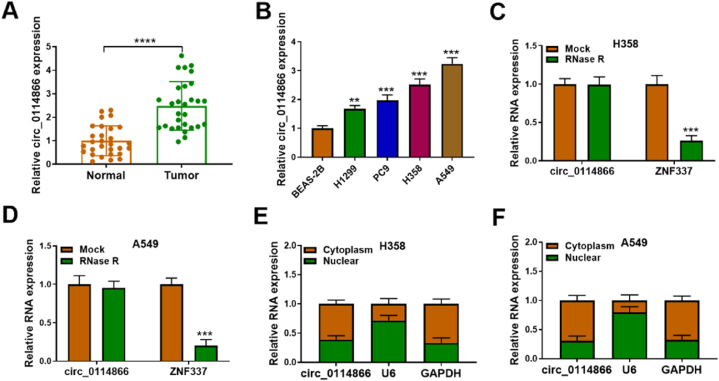
Background: Non-small-cell lung cancer (NSCLC) is the most prevalent form of lung cancer. Circular RNA (circRNA) has emerged as a key player in the development of NSCLC by acting as miRNA sponges. However, the precise role of circ_0114866 in regulating NSCLC process is yet to be elucidated.
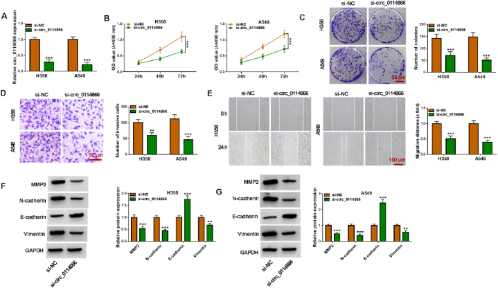
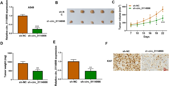
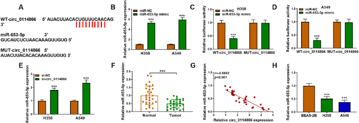
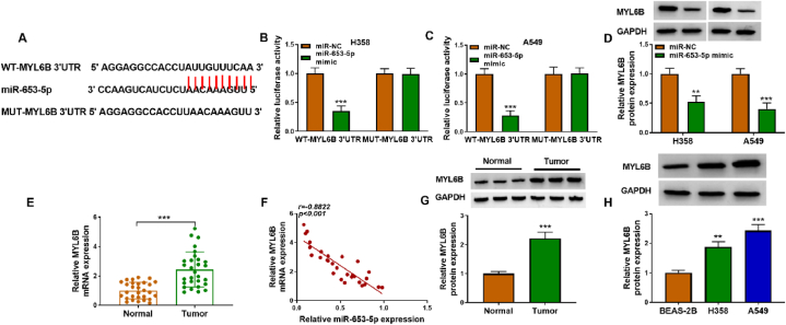
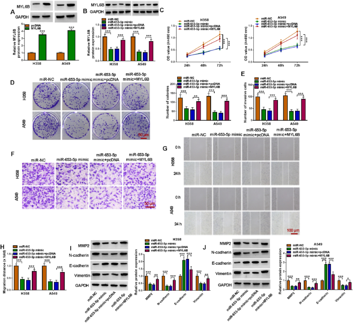
Methods: The expression of circ_0114866, miR-653-5p, and MYL6B were assessed by qPCR. Cell viability, proliferation, invasion, and migration were investigated using CCK-8, colony formation, Transwell, and wound healing assays. The protein levels of MYL6B, MMP-2, N-cadherin, E-cadherin, and vimentin were evaluated through Western blot analysis. Xenograft tumor model were selected to analyze the impact of circ_0114866 on NSCLC tumor growth. Through circBank or Starbase databases, the binding interactions between miR-653-5p and circ_0114866 or MYL6B were predicted. Subsequently, these interactions were verified by dual-luciferase reporter assay.
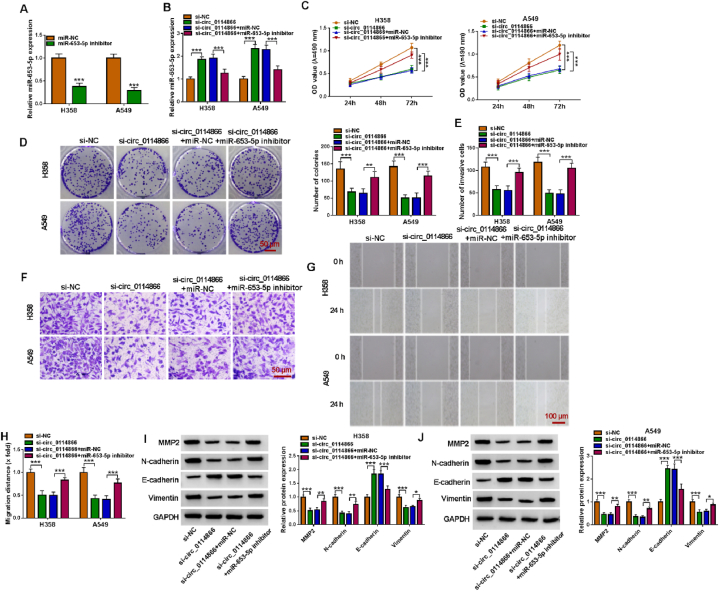
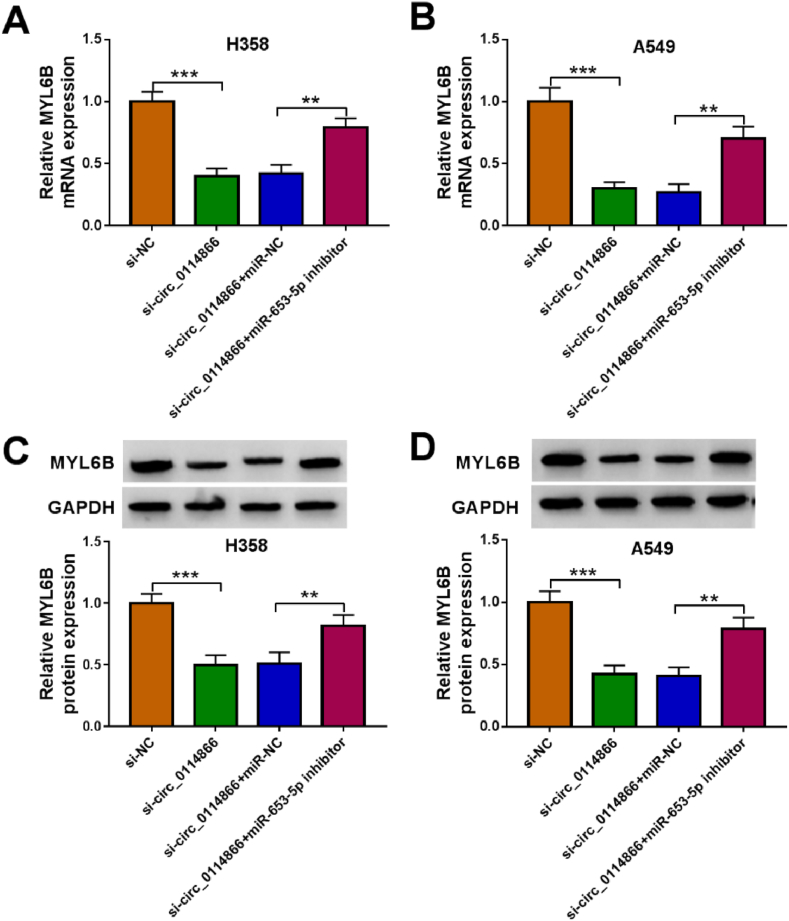
Results: The expression of circ_0114866 and MYL6B were clearly elevated, while miR-653-5p expression was notably reduced in NSCLC tissues and cells. Notably, circ_0114866 knockdown obviously suppressed the proliferation, metastasis, and EMT process in NSCLC cells. Additionally, circ_0114866 functioned as a sponge for miR-653-5p, leading to an increase in MYL6B expression by absorbing miR-653-5p. Furthermore, the inhibitory effects on biological behaviors and EMT process of NSCLC cells induced by circ_0114866 knockdown were reversed by miR-653-5p inhibitor. Moreover, in vivo experiments demonstrated that silencing circ_0114866 resulted in a repression of tumor growth.
Conclusion: Our findings indicate that circ_0114866 knockdown upregulated MYL6B transcription by sponging miR-653-5p, leading to hinder the progression and EMT process of NSCLC.
© 2024 Published by Elsevier Ltd.
Conflict of interest statement
The authors declare that they have no known competing financial interests or personal relationships that could have appeared to influence the work reported in this paper.
Figures
Similar articles
-
Circular RNA circ_0003028 contributes to tumorigenesis by regulating GOT2 via miR-1298-5p in non-small cell lung cancer.Bioengineered. 2021 Dec;12(1):2326-2340. doi: 10.1080/21655979.2021.1935064. Bioengineered. 2021. PMID: 34077306 Free PMC article.
-
Circular circRANGAP1 Contributes to Non-small Cell Lung Cancer Progression by Increasing COL11A1 Expression Through Sponging miR-653-5p.Biochem Genet. 2023 Dec;61(6):2580-2598. doi: 10.1007/s10528-023-10393-x. Epub 2023 May 16. Biochem Genet. 2023. PMID: 37193942
-
Hsa_circRNA_0017620 regulated cell progression of non-small-cell lung cancer via miR-520a-5p/KRT5 axis.J Clin Lab Anal. 2022 Apr;36(4):e24347. doi: 10.1002/jcla.24347. Epub 2022 Mar 18. J Clin Lab Anal. 2022. PMID: 35302673 Free PMC article.
-
Circular RNA hsa_circ_0004396 acts as a sponge of miR-615-5p to promote non-small cell lung cancer progression and radioresistance through the upregulation of P21-Activated Kinase 1.J Clin Lab Anal. 2022 Jun;36(6):e24463. doi: 10.1002/jcla.24463. Epub 2022 May 2. J Clin Lab Anal. 2022. PMID: 35500159 Free PMC article.
-
Circ-CSPP1 knockdown suppresses hepatocellular carcinoma progression through miR-493-5p releasing-mediated HMGB1 downregulation.Cell Signal. 2021 Oct;86:110065. doi: 10.1016/j.cellsig.2021.110065. Epub 2021 Jun 26. Cell Signal. 2021. PMID: 34182091 Review.
Cited by
-
Hsa_circ_0002238 promotes the malignant behavior of colorectal cancer.Front Pharmacol. 2025 Jun 13;16:1541820. doi: 10.3389/fphar.2025.1541820. eCollection 2025. Front Pharmacol. 2025. PMID: 40584619 Free PMC article.
References
LinkOut - more resources
Full Text Sources
Research Materials
Miscellaneous
