. 2025 Jan;12(Suppl 1):S13004.
doi: 10.1117/1.JMI.12.S1.S13004. Epub 2024 Sep 14.

Breathing motion compensation in chest tomosynthesis: evaluation of the effect on image quality and presence of artifacts

Affiliations

Breathing motion compensation in chest tomosynthesis: evaluation of the effect on image quality and presence of artifacts

Maral Mirzai et al. J Med Imaging (Bellingham). 2025 Jan.

Abstract

Purpose: Chest tomosynthesis (CTS) has a relatively longer acquisition time compared with chest X-ray, which may increase the risk of motion artifacts in the reconstructed images. Motion artifacts induced by breathing motion adversely impact the image quality. This study aims to reduce these artifacts by excluding projection images identified with breathing motion prior to the reconstruction of section images and to assess if motion compensation improves overall image quality.

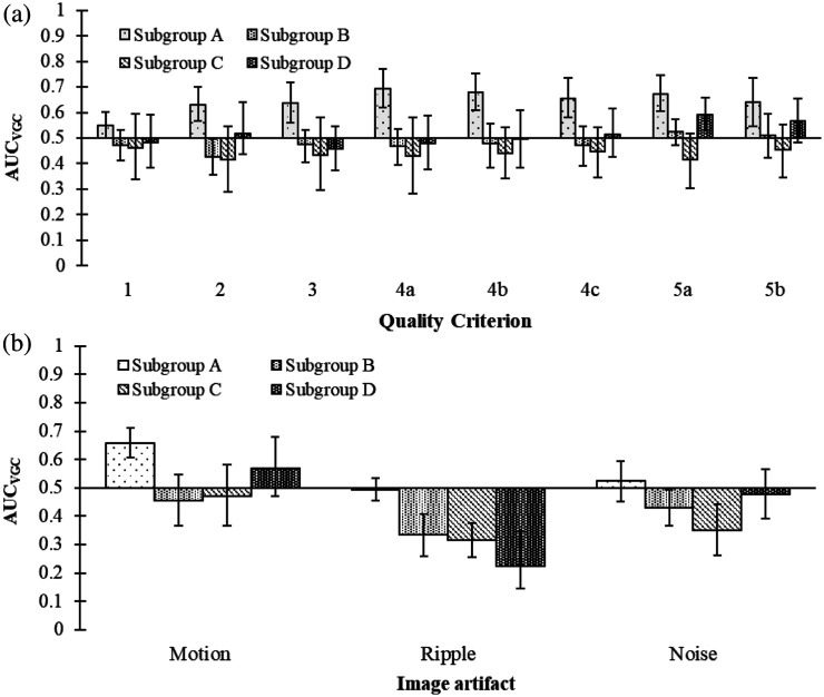
Approach: In this study, 2969 CTS examinations were analyzed to identify examinations where breathing motion has occurred using a method based on localizing the diaphragm border in each of the projection images. A trajectory over diaphragm positions was estimated from a second-order polynomial curve fit, and projection images where the diaphragm border deviated from the trajectory were removed before reconstruction. The image quality between motion-compensated and uncompensated examinations was evaluated using the image quality criteria for anatomical structures and image artifacts in a visual grading characteristic (VGC) study. The resulting rating data were statistically analyzed using the software VGC analyzer.

Results: A total of 58 examinations were included in this study with breathing motion occurring either at the beginning or end ( n = 17 ) or throughout the entire acquisition ( n = 41 ). In general, no significant difference in image quality or presence of motion artifacts was shown between the motion-compensated and uncompensated examinations. However, motion compensation significantly improved the image quality and reduced the motion artifacts in cases where motion occurred at the beginning or end. In examinations where motion occurred throughout the acquisition, motion compensation led to a significant increase in ripple artifacts and noise.

Conclusions: Compensation for respiratory motion in CTS by excluding projection images may improve the image quality if the motion occurs mainly at the beginning or end of the examination. However, the disadvantages of excluding projections may outweigh the benefits of motion compensation.

Keywords: breathing motion; chest tomosynthesis; image quality; motion artifacts; motion compensation.


Figures

Fig. 1
Fig. 1
Examples of two CTS examinations where a second-order polynomial curve has been fitted to the position of the diaphragm border in each projection image (1 to 60). For a patient who stands still and holds their breath during the acquisition, (a) the position of the diaphragm shows almost no deviation from the trajectory, whereas for a patient with respiratory motion, (b) several deviations are seen.
Fig. 2
Fig. 2
Example of the positioning of a fixed ROI over the diaphragm in one of the 60 projection images included in a CTS examination. The border of the diaphragm is detected using gradient analysis in the ROI.
Fig. 3
Fig. 3
Flowchart describing the inclusion of examinations in the study.
Fig. 4
Fig. 4
Examples of the diaphragm border positions with corresponding trajectories for six different examinations included in the study. In the first row [(a)–(c)], motion has been identified in either the beginning or the end of the trajectory. In the second row [(d)–(f)], motion is distributed over the entire trajectory.
Fig. 5
Fig. 5
Resulting AUCVGC with the corresponding 95% confidence interval for all examinations included in the study (n=58). In (a), results for the anatomical quality criteria are presented, and AUCVGC>0.5 indicates higher ratings in the compensated examinations, whereas AUCVGC<0.5 indicates higher ratings in the reference examinations. The quality criteria related to image artifacts are shown in (b), where AUCVGC>0.5 indicates less present and disturbing artifacts in the compensated examinations, whereas AUCVGC<0.5 indicates more present and disturbing artifacts in the reference examinations.
Fig. 6
Fig. 6
Resulting AUCVGC with corresponding 95% confidence interval for subgroups (A to D). In (a), results for the anatomical quality criteria are presented, and AUCVGC>0.5 indicates higher ratings in the compensated examinations, whereas AUCVGC<0.5 indicates higher ratings in the reference examinations. The quality criteria related to image artifacts are shown in (b), where AUCVGC>0.5 indicates less present and disturbing artifacts in the compensated examinations, whereas AUCVGC<0.5 indicates more present and disturbing artifacts in the reference examinations.

Similar articles

Cited by

References

    1. Vikgren J., et al. , “Comparison of chest tomosynthesis and chest radiography for detection of pulmonary nodules: human observer study of clinical cases,” Radiology 249(3), 1034–1041 (2008). 10.1148/radiol.2492080304 - DOI - PubMed
    1. Quaia E., et al. , “Analysis of the impact of digital tomosynthesis on the radiological investigation of patients with suspected pulmonary lesions on chest radiography,” Eur. Radiol. 22(9), 1912–1922 (2012). 10.1007/s00330-012-2440-3 - DOI - PubMed
    1. Johnsson A. A., Vikgren J., Bath M., “Chest tomosynthesis: technical and clinical perspectives,” Semin. Respir. Crit. Care Med. 35(1), 17–26 (2014). 10.1055/s-0033-1363448 - DOI - PubMed
    1. Dobbins J. T., 3rd, et al. , “Digital tomosynthesis of the chest,” J. Thorac. Imaging 23(2), 86–92 (2008). 10.1097/RTI.0b013e318173e162 - DOI - PubMed
    1. Dobbins J. T., 3rd, “Tomosynthesis imaging: at a translational crossroads,” Med. Phys. 36(6), 1956–1967 (2009). 10.1118/1.3120285 - DOI - PMC - PubMed