[Preprint]. 2024 Sep 3:2024.09.02.24312084.
doi: 10.1101/2024.09.02.24312084.

Neuroimaging Biomarkers in Addiction

Affiliations

Neuroimaging Biomarkers in Addiction

Hamed Ekhtiari et al. medRxiv. .

Abstract

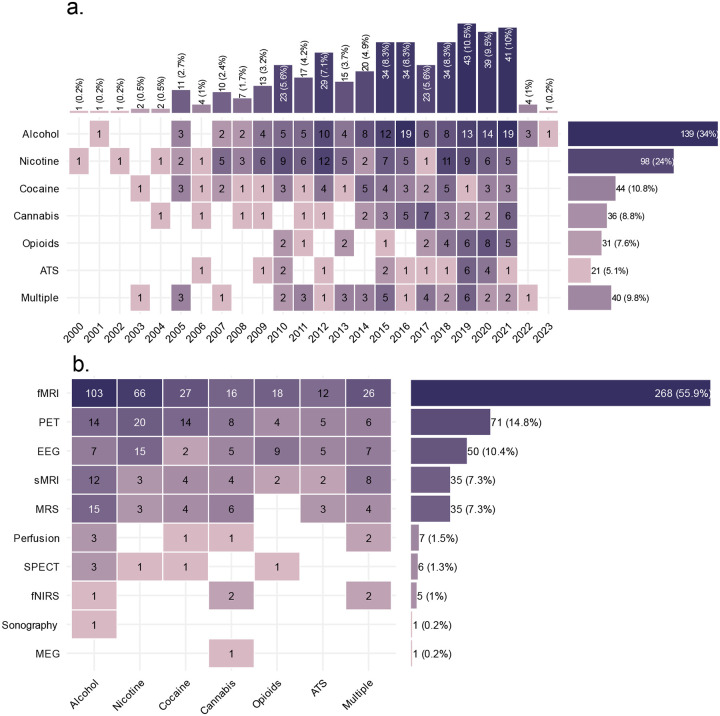
As a neurobiological process, addiction involves pathological patterns of engagement with substances and a range of behaviors with a chronic and relapsing course. Neuroimaging technologies assess brain activity, structure, physiology, and metabolism at scales ranging from neurotransmitter receptors to large-scale brain networks, providing unique windows into the core neural processes implicated in substance use disorders. Identified aberrations in the neural substrates of reward and salience processing, response inhibition, interoception, and executive functions with neuroimaging can inform the development of pharmacological, neuromodulatory, and psychotherapeutic interventions to modulate the disordered neurobiology. Based on our systematic search, 409 protocols registered on ClinicalTrials.gov include the use of one or more neuroimaging paradigms as an outcome measure in addiction, with the majority (N=268) employing functional magnetic resonance imaging (fMRI), followed by positron emission tomography (PET) (N=71), electroencephalography (EEG) (N=50), structural magnetic resonance imaging (MRI) (N=35) and magnetic resonance spectroscopy (MRS) (N=35). Furthermore, in a PubMed systematic review, we identified 61 meta-analyses including 30 fMRI, 22 structural MRI, 8 EEG, 7 PET, and 3 MRS meta-analyses suggesting potential biomarkers in addictions. These studies can facilitate the development of a range of biomarkers that may prove useful in the arsenal of addiction treatments in the coming years. There is evidence that these markers of large-scale brain structure and activity may indicate vulnerability or separate disease subtypes, predict response to treatment, or provide objective measures of treatment response or recovery. Neuroimaging biomarkers can also suggest novel targets for interventions. Closed or open loop interventions can integrate these biomarkers with neuromodulation in real-time or offline to personalize stimulation parameters and deliver the precise intervention. This review provides an overview of neuroimaging modalities in addiction, potential neuroimaging biomarkers, and their physiologic and clinical relevance. Future directions and challenges in bringing these putative biomarkers from the bench to the bedside are also discussed.


Conflict of interest statement

O.C. has received grant funding from Eli Lilly, Inc, and Nestle, Inc. He has provided paid consulting to Novo Nordisk. Dr. Paulus is an advisor to Spring Care, Inc., a behavioral health startup, he has received royalties for an article about methamphetamine in UpToDate. M.P.P. has a consulting agreement with and receives compensation from F. Hoffmann-La Roche Ltd. P.O. is an employee and shareholder of Sage Therapeutics. Other authors report no conflicts of interest. Disclaimer: The views and opinions expressed in this manuscript are those of the authors only and do not necessarily represent the views, official policy or position of the U.S. Department of Health and Human Services or any of its affiliated institutions or agencies.

Figures

Figure 1:
Figure 1:. Distribution of the neuroimaging protocols based on year and substance.
a. Number of protocols starting for each substance each year (n= 409). Years are obtained from the ClinicalTrials.gov database indicating actual or planned start years. b. Number of neuroimaging modalities used in each protocol for each substance. Numbers on this figure sum to 479 for 409 protocols, since 70 protocols used multiple imaging modalities. ATS: Amphetamine-type Stimulants; sMRI: structural MRI, including whole-brain T1 imaging, gray matter volumetry, or diffusion tensor imaging; Perfusion: brain perfusion imaging, including arterial spin labeling, cerebral blood flow imaging, and magnetic resonance angiography; MRS: magnetic resonance-spectroscopy. Data were collected from ClinicalTrials.gov on November 17, 2021.
Figure 2:
Figure 2:. Multi-level characteristics of 688 neuroimaging outcome measures in 409 registered protocols.
These levels include the scales at which neuroimaging modalities have probed the nervous system (structural, biochemical, hemodynamic or electrophysiology), the neuroimaging modality, different paradigms in each modality, and the types of tasks used in task-based functional neuroimaging paradigms. All “structural” paradigms in our database were variants of MRI; “biochemical” paradigms include SPECT, MRS, and PET; “hemodynamic” paradigms include fMRI, fNIRS, less common perfusion imaging modalities, and ultrasound; and EEG and MEG constitute “electrophysiological” imaging paradigms. These modalities have been used for static structural scans of brain gray or white matter and vasculature, resting-state functional scans, or task-related functional scans with various tasks. Note that many protocols have utilized more than one neuroimaging outcome measure and the total number of outcome measures is 688, more than the number of protocols (n=409). Data is collected from ClinicalTrials.gov on November 17, 2021. EEG: electroencephalography; fMRI:functional magnetic resonance imaging; fNIRS: Functional near-infrared spectroscopy; MEG: magnetoencephalography; MRI: magnetic resonance imaging; MRS: magnetic resonance-spectroscopy; PET: positron emission tomography; SPECT: Single-photon emission computed tomography;
Figure 3:
Figure 3:. Multi-level characteristics of 83 neuroimaging outcome measures in 61 meta-analyses.
These levels include the scales at which neuroimaging modalities have probed the nervous system (structural, biochemical, hemodynamic or electrophysiology), the neuroimaging modality, different paradigms in each modality, and the types of tasks used in task-based functional neuroimaging paradigms. All “structural” paradigms in our database were variants of MRI; “biochemical” paradigms include SPECT, MRS, and PET; “hemodynamic” paradigms include fMRI, fNIRS, less common perfusion imaging modalities, and ultrasound; and EEG and MEG constitute “electrophysiological” imaging paradigms. These modalities have been used for assessment of people with different kinds of SUDs. These assessment can be categorized into different biomarker categories. Note that some meta-analyses have utilized more than one neuroimaging outcome measure and the total number of outcome measures is 83, more than the number of total meta-analyses (n=61). Further, 3 of the 83 findings are from mega-analyses rather than meta-analyses, though we use the term meta-analysis to refer to these for simplicity. dMRI: diffusion magnetic resonance imaging; EEG: electroencephalography; fMRI: functional magnetic resonance imaging; fNIRS: Functional near-infrared spectroscopy; MEG: magnetoencephalography; MRI: magnetic resonance imaging; MRS: magnetic resonance-spectroscopy; PET: positron emission tomography; sMRI: structural magnetic resonance imaging; SPECT: Single-photon emission computed tomography; SUD: Substance user disorder
Figure 4:
Figure 4:. Schematic representation of stages in substance use and SUDs and their therapeutic interventions and corresponding biomarker types.
Susceptibility biomarkers can predict transition to substance use or disorder, prognostic biomarkers can predict the future progression of the disorder, diagnostic biomarkers can distinguish clinically-relevant populations, monitoring biomarkers facilitate ongoing information about the course of the disorder with or without intervention, predictive biomarkers can predict treatment response, response biomarkers can reflect the physiological impact of an intervention and potentially be used as surrogate endpoints in lieu of clinical outcomes, and safety biomarkers can help assess the potential hazards of various substances used in clinical or non-clinical settings.
Figure 5:
Figure 5:. Putative neuroimaging biomarkers reported in registered protocols in various substance use disorders (SUDs) and neuroimaging modalities.
Biomarker types are divided between the substance of interest and neuroimaging modalities used in the protocol (510 biomarkers across 409 protocols). The horizontally aligned bars represent the total number of each biomarker type. Note that some of the protocols include more than one biomarker type. Some protocols did not report enough details for neuroimaging modalities in a way that fit any biomarker’s definition. Data is collected from ClinicalTrials.gov on November 17, 2021.
Figure 6:
Figure 6:. Multi-scale brain aberrations as putative neuroimaging biomarkers in trials for substance use disorders (SUDs).
Seven examples of brain aberrations identified in SUDs (yellow boxes) that have been investigated as putative “response” or “predictive” biomarkers or intervention targets in protocols registered in ClinicalTrials.gov (light blue boxes). The relevant literature is referenced in supplementary table 2. FC: Functional Connectivity; FDCR: fMRI Drug Cue Reactivity; PFC: Prefrontal Cortex. tDCS: Transcranial Direct Current Stimulation.
Figure 7:
Figure 7:. Major steps in the development and validation of potential neuroimaging biomarkers for SUDs.
Initially, the context(s) of use for the biomarker is specified and the potential biomarker is precisely defined. Following analytical and clinical validation and cost-benefit analysis, the compiled evidence is presented for regulatory approval. The FDA evaluates the use of biomarkers for drug development through a biomarker qualification process involving submission of a Letter of Intent, a Qualification Plan, and a Full Qualification Package, though a Letter of Support may be issued by the FDA to indicate its support for a biomarker before formal qualification. The use of neuroimgaing biomarkers in clinical contexts also requires initial approval by the FDA, but also the endorsement of a constellation of other institutions (adapted from, reproduced with permission). Surr. Endpoint: Surrogate Endpoint, COU: Context of Use.

References

    1. Degenhardt L, Charlson F, Ferrari A, et al. The global burden of disease attributable to alcohol and drug use in 195 countries and territories, 1990–2016: a systematic analysis for the Global Burden of Disease Study 2016. The Lancet Psychiatry. 2018;5(12):987–1012. doi:10.1016/S2215-0366(18)30337-7 - DOI - PMC - PubMed
    1. Shield KD, Imtiaz S, Probst C, Rehm J. The epidemiology and public health burden of addictive disorders. In: Integrating Psychological and Pharmacological Treatments for Addictive Disorders: An Evidence-Based Guide. 2018:3–31.
    1. National Institutes of Health. National Institute on Drug Abuse. Trends and Statistics. Published online 2020. Accessed May 20, 2020. https://www.drugabuse.gov/related-topics/trends-statistics/
    1. American Psychiatric Association. Diagnostic and Statistical Manual of Mental Disorders. Fifth Edition.; 2013. Accessed September 8, 2020. http://psychiatryonline.org/doi/book/10.1176/appi.books.9780890425596 - DOI
    1. Volkow ND, Boyle M. Neuroscience of Addiction: Relevance to Prevention and Treatment. AJP. 2018;175(8):729–740. doi:10.1176/appi.ajp.2018.17101174 - DOI - PubMed

Publication types