[Preprint]. 2024 Sep 5:rs.3.rs-4636308.
doi: 10.21203/rs.3.rs-4636308/v1.

Inhibition of novel human-HPV hybrid ecDNA enhancers reduces oncogene expression and tumor growth in oropharyngeal cancer

Affiliations

Inhibition of novel human-HPV hybrid ecDNA enhancers reduces oncogene expression and tumor growth in oropharyngeal cancer

Takuya Nakagawa et al. Res Sq. .

Update in

Abstract

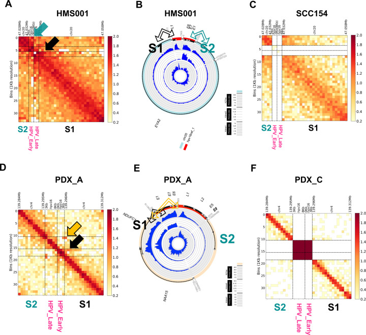
Extrachromosomal circular DNA (ecDNA) have been found in most types of human cancers, and ecDNA incorporating viral genomes has recently been described, specifically in human papillomavirus (HPV)-mediated oropharyngeal cancer (OPC). However, the molecular mechanisms of human-viral hybrid ecDNA (hybrid ecDNA) for carcinogenesis remains elusive. We characterized the epigenetic status of hybrid ecDNA using HPVOPC cell lines and patient-derived tumor xenografts, identifying HPV oncogenes E6/E7 in hybrid ecDNA were flanked by novel somatic DNA enhancers and HPV L1 enhancers, with strong cis-interaction. Targeting of these enhancers by clustered regularly interspaced short palindromic repeats interference or hybrid ecDNA by bromodomain and extra-terminal inhibitor reduced E6/E7 expression, and significantly inhibited in vitro and/or in vivo growth only in ecDNA(+) models. HPV DNA in hybrid ecDNA structures are associated with novel somatic and HPV enhancers in hybrid ecDNA that drive HPV ongogene expression and carcinogenesis, and can be targeted with ecDNA disrupting therapeutics.


Conflict of interest statement

Conflict of interest: P.S.M. is a co-founder, chairs the scientific advisory board (SAB) of and has equity interest in Boundless Bio Inc. (BBI). P.S.M. is also an advisor with equity for Asteroid Therapeutics and is an advisor to Sage Therapeutics. V.B. is a co-founder, consultant, SAB member and has equity interest in Boundless Bio and Abterra. J.T.L is an employee of BBI. J.L. previously consulted for BBI. The other authors have no conflicts of interest to disclose.

Figures

Fig. 1:
Fig. 1:. Identification of hybrid ecDNA in HPVOPC using AA and FastViFI and validation of hybrid ecDNA by multi-FISH.
Examples of circular hybrid ecDNA suggested by AA results in HMS001 (A and C) and PDX tumor PDX_A (B and D). Multi-FISH using each ecDNA specific probe (green) and HPV specific probe (red) for metaphase spread cells showed overlapping of each probe signal in the same place only in hybrid ecDNA(+) samples (yellow allow in E and F). Hybrid ecDNA(+) samples also showed the red HPV signals alone (white arrowhead in E and F), suggesting HPV-only episomes. Hybrid ecDNA(−) cell line SCC154 only showed the red HPV signal (white arrowhead in G). Control cell line NOKSI did not show any signal (H). Scale bar shows 10μm.
Fig. 2:
Fig. 2:. Detecting active enhancer using ChIP-seq, and new identification of HPV integration mechanism in hybrid ecDNA.
ChIP-seq results of Input, K4me3 (promoter), K4me1 (enhancer), and K27ac (activation mark) for NOKSI (normal control), SCC154 (hybrid ecDNA- HPVOPC) and HMS001 (hybrid ecDNA+ HPVOPC) were shown. Promoter is indicated by the light green box, enhancers by the yellow box, and the sequences included in hybrid ecDNA of HMS001 by the light blue box. The active enhancer is marked by the red oval (top) (A). Expanded hybrid ecDNA sequence tracks are shown (bottom), and include anexpanded enhancer map showing HPV integration occurred in the exact center of the active enhancer mark (bottom) (A). The HPV integration site and its ChIP-seq results are also shown. On the other hand, active enhancers already existing originally were not included in hybrid ecDNA (bottom) (A). (B)Hybrid ecDNA in HMS001with ATAC-seq (top) and ChIP-seq(bottom) are shown in the CycleViz plot. A related figure using NOKSI (normal control), PDX_C (hybrid ecDNA- HPVOPC), and PDX_A (hybrid ecDNA+ HPVOPC) is shown (C and D). The newly created active enhancer made a complex with 2 promoters (bottom) (C).
Fig. 3:
Fig. 3:. Human and viral genomes on hybrid ecDNA interacted directly with each other.
Cis-interactions between enhancer and HPV in hybrid ecDNA were analyzed by HiC-seq. Human genome regions on hybrid ecDNA were divided into 2 segments (S1 and S2). The S1 enhancer region closely interacted with the HPV L1 region, and the S2 enhancer region closely interacted with the HPV E6/E7 regions in HMS001 (black arrow in A and B). On the other hand, there was no such interaction in SCC154 that lacked hybrid ecDNA (C). This phenomenon was confirmed in PDX tumors (D-F). In PDX_A, the enhancer existed only in the S1 segment, and the S1 enhancer region closely interacted with the HPV L1 and E6/E7 regions in PDX_A (black and yellow arrows in D and E). On the other hand, there was no such interaction in PDX_C that lacked hybrid ecDNA (F). Although each hybrid ecDNA structure was unique, each of the novel human enhancer regions closely interacted with HPV, confirming the direct interaction of the human and viral genomes on the hybrid ecDNAs.
Fig. 4:
Fig. 4:. CRISPR interference targeting enhancers on hybrid ecDNA blocks HPV oncogene expression.
CRISPR interference, using dCas9-KRAB to target enhancers on the hybrid ecDNA of HMS001, was performed. gRNAs targeting S1: the long part (gRNA#1) and S2: the short part (gRNA#2) of the enhancer on hybrid ecDNA of HMS001 and nontarget controls were used (A). The expression of dCas9 after doxycycline induction was confirmed by qPCR and Western blotting (B and C). MYC and GAPDH were used as controls (C). Western blotting results of E6 and E7 of each CRISPRi condition indicate E6 and E7 expression were reduced by CRISPRi targeting the S2 enhancer (D). Proliferation assay results targeting the S2 enhancer and nonT in HMS001, SCC154, and NOKSI indicate Dox induction inhibited proliferation only when targeting S2 in HMS001 (**P=0.006) (E), but not when targeting SCC154 or NOKSI (F and G).
Fig. 5:
Fig. 5:. Hybrid ecDNA and HPV episomes are associated and reduced after JQ1 treatment.
Multi-probe FISH, using an EYA2 probe and an HPV probe on hybrid ecDNA on super resolution DMSO and JQ1 treatment of HMS001 cells, is shown (A and B). Hybrid ecDNA was observed nearby in the nucleus along with episomal HPV in the “no treatment” condition (A). Each signal (green: EYA2, red: HPV, and blue: DAPI) were also shown separately (bottom). (A). FISH signals of hybrid ecDNA and episomal HPV were reduced after JQ1 treatment (B). Scale bar shows 5μm (0.2μm in expanded picture). Cartoon illustrating how hybrid ecDNA hub disruption may decrease transcription is illustrated (right in A and B).
Fig. 6:
Fig. 6:. JQ1 treatment on hybrid ecDNA significantly blocks HPV oncogene expression and proliferation.
JQ1 treatment on HMS001 and SCC154 were performed. The schema of the experiments were shown (A). qPCR results of MYC and E6/E7 were shown. ACTB was used internal control. MYC and E6/E7 expressions were reduced in a concentration-dependent manner in qPCR (**P = 4 ×10⁻³, ***P = 4 ×10⁻⁴, respectively) at 24h (B). MYC and E6/E7 expressions were reduced in a concentration-dependent manner in western blotting at 6h and 24 h after JQ1 treatment (C). Proliferation assay using JQ1 for HMS001 and SCC154 were shown. JQ1 treatment significantly inhibited tumor growth only in HMS001 in 1uM, but not in SCC154 (*P = 0.03, P = 0.12, respectively) (D).
Fig. 7:
Fig. 7:. JQ1 inhibited proliferation only in hybrid ecDNA(+) HPVOPC PDX tumors.
JQ1 treatment was performed in HPVOPC PDX models. Six PDX_A (hybrid ecDNA+) mice were divided into a vehicle control group and a JQ1 treatment group. Each mouse possessed tumors in both flanks (A). Tumors were harvested after 2 weeks of vehicle control or JQ1 treatment (B). Tumors were harvested after 2 weeks of JQ1 treatment or vehicle control. Tumor volumes of each condition are shown (C-E). In the PDX_A JQ1 treatment group, tumor growth was significantly inhibited compared to the vehicle control group (tumor volume: P = 2×10⁻⁵, tumor weight: P = 1×10⁻⁴ respectively) (C and F).qPCR of E6/E7 is shown between JQ1 treatment and vehicle control. E6/E7 expression was also reduced after JQ1 treatment (P < 1×10⁻⁴) (G). JQ1 treatment for hybrid ecDNA-HPVOPC PDX (PDX_C and PDX004) was also performed (H-M). Neither tumor volume nor tumor weight were inhibited significantly compared to the vehicle control group in PDX_C (H-J) and PDX004 (K-M).

References

    1. Turner K.M. et al. Extrachromosomal oncogene amplification drives tumour evolution and genetic heterogeneity. Nature 543, 122–125 (2017). - PMC - PubMed
    1. Kim H. et al. Extrachromosomal DNA is associated with oncogene amplification and poor outcome across multiple cancers. Nat Genet 52, 891–897 (2020). - PMC - PubMed
    1. Yi E., Chamorro Gonzalez R., Henssen A.G. & Verhaak R.G.W. Extrachromosomal DNA amplifications in cancer. Nat Rev Genet 23, 760–771 (2022). - PMC - PubMed
    1. Wu S. et al. Circular ecDNA promotes accessible chromatin and high oncogene expression. Nature 575, 699–703 (2019). - PMC - PubMed
    1. Zhu Y. et al. Oncogenic extrachromosomal DNA functions as mobile enhancers to globally amplify chromosomal transcription. Cancer Cell 39, 694–707 e7 (2021). - PMC - PubMed

Publication types

LinkOut - more resources