[Preprint]. 2024 Sep 5:2024.09.04.611325.
doi: 10.1101/2024.09.04.611325.

Structure and inhibition mechanisms of Mycobacterium tuberculosis essential transporter efflux protein A

Affiliations

Structure and inhibition mechanisms of Mycobacterium tuberculosis essential transporter efflux protein A

Nitesh Kumar Khandelwal et al. bioRxiv. .

Update in

Abstract

A broad chemical genetics screen in Mycobacterium tuberculosis (Mtb) to identify inhibitors of established or previously untapped targets for therapeutic development yielded compounds (BRD-8000.3 and BRD-9327) that inhibit the essential efflux pump EfpA. To understand the mechanisms of inhibition by these compounds, we determined the structures of EfpA with inhibitors bound at 2.7 - 3.4 Å resolution. Our structures reveal different mechanisms of inhibition for the two inhibitors. BRD-8000.3 binds in a tunnel making contact with the lipid bilayer and extending toward the central cavity to displace the fatty acid chain of a lipid molecule bound in the apo structure, suggesting its blocking of an access route for a natural lipidic substrate, in contrast to its uncompetitive mechanism for the small molecule substrate ethidium bromide which likely enters through an alternative tunnel. Meanwhile, BRD-9327 binds in the outer vestibule without complete blockade of the substrate path to the outside, suggesting its possible inhibition of the dynamical motion necessary for "alternate access" to the two different sides of the membrane, as is characteristic of major facilitator superfamily (MFS) transporters. Both inhibitors may have a role in inhibiting the "alternate access" mechanism that could account for the uncompetitive nature of their efflux of some substrates. Our results explain the basis of the synergy of these inhibitors and their potential for combination in a multi drug strategy for anti-tuberculosis therapy. They also potentially point to a possible function for this essential efflux pump as a lipid transporter. The structures provide a foundation for rational modification of these inhibitors to increase potency.


Conflict of interest statement

Competing interests: N.K.K., M.G., S.B., J.E.G., Z.G., A.Y.E., S.G.B., A.S., D.T.H. and R.M.S. declare no competing interests.

Figures
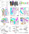
Fig.1|
Fig.1|. Structure of EfpA.
a, BRD-8000 series compounds and BRD-9327 identified by PROSPECT assay. b, The cryoEM density map for the antiparallel dimer of EfpAEM. c, Schematic representation of the topology arrangement of EfpA. Three transmembrane helix (TM) bundles are related to each other by a twofold pseudosymmetry, with two extra linker helix (TM7 and 8). d, EfpA monomer A atomic model along with PG molecules (PG1, PG2 and PG3) with corresponding EM density (left panel). PG2 molecule interaction (middle panel) and rotated 90° forward from the position in the middle panel (right panel). The acyl chain of PG2 that is outside of the protein runs parallel to the TM14 hydrophobic residues (F507 TM14, L506 TM14, M504 TM14 and I500 TM14) and reaches downward to the cytoplasmic surface. The head group present between the dimer interface and its second acyl chain enters EfpA via space present between the G377TM11, G502TM14 residue. This acyl chain is perpendicular to the TM9, TM11, TM12 and TM14 and interact with M320 TM9, A316 TM9, T373 TM11, I374 TM11, L380 TM11,V501 TM14, A498 TM14, I499 TM14, I412 TM12, A415 TM12, V416 TM12 and L419 TM12. e, EfpA monomer (left hand panel D) rotated 90° forward (left panel). PG1 molecule (middle panel) lies between TM9 (I313TM9, A316TM9, M320TM9, L323TM9, I327TM9 and Y330TM9), TM14 (Y487TM14 and L491TM14), TM11 (L380TM11, M384 TM11 and A383 TM11), TM12 (I405 TM12, P404 TM12,I412 TM12 andV416 TM12), TM10 (F346TM10, F349TM10and M353TM10) TM5 (S191TM5) and TM1 (T70TM1). PG3 (right panel) lies between TM2 (I94TM2, V98TM2 and F101TM2), TM7 (C250TM7, A253TM7, V254TM7 and F257TM7), TM9 (F321TM9) and TM13 (M441TM13,S444TM13, L445TM13, P448TM13 and L449TM13). f, Profiles of 361 strains in pooled PROSPECT against BRD-8000 analogue BRD-7158; each strain is shown as a grey line with relevant sensitized strains labeled as in the legend. The x-axis is concentration of BRD-7158 in μM, y-axis is standardized growth rate (sGR) of each strain across 8 doses tested. Right panel shows pathway of PG synthesis highlighting the functions of CoaE and Rv2812c.
Fig. 2|
Fig. 2|. Mode of inhibition of EfpA by BRD-8000.3.
a, BRD-8000.3 binds to EfpAEM in a ligand-observed NMR assay. Intensity of the inhibitor’s aromatic protons is monitored in a titration series of EfpAEM (x-axis), in which peak heights are converted to fractional occupancy (y-axis). A dissociation constant is determined by fitting the data to a standard bimolecular binding model (see methods). Data represents 3 independent titrations. b, Section of cryoEM map of BRD-8000.3 (yellow) bound EfpAEM dimer (left panel). Experimentally determined cryo-EM density of BRD-8000.3 in monomer A (right panel). Map contour level = 0.597 in ChimeraX. c, Tertiary structure of the EfpAEM monomer A with BRD-8000.3 (yellow) with lipid shown in pink, side view left and top view from the extracellular side of monomer A. d, Superposition of BRD-8000.3 (yellow) bound EfpAEM monomer (turquoise) onto the apo structure (orange) with PG2 in apo (pink). BRD8000.3 displaces the PG2 molecule present in the apo structure in the BRD8000.3 bound structure. e, Tertiary structure for BRD-8000.3 binding pocket showing BRD-8000.3 (yellow) and associated side chains viewed from the extracellular face (left) and side view obtained by rotating left panel at 90° on x and y axis. Entry portal glycines G377TM11 and G502TM14 are colored violet and side chain atoms colored by atom, nitrogen (blue), oxygen (red), bromide (brown). Residues where mutation led to resistance to BRD-8000.3 are labelled in pink. f, Dose-response curves showing growth of M. bovis BCG transformed with plasmids overexpressing WT (V319), V319F, and V319A alleles of Mtb EfpA in response to varied concentrations of BRD-8000.3. x-axis is inhibitor concentration and y-axis is culture density after 10 days of growth (OD600). Error bars are standard deviation of 3 replicates.
Fig. 3|
Fig. 3|. Mode of inhibition of EfpA by BRD-9327.
a, Sliced cryo-EM map density of BRD-9327 bound EfpAEM. Both monomers of EfpA shown as yellow-green and gray color respectively. Each monomer has one BRD-9327 molecule (aqua color) and three PG molecules (pink color). b, Tertiary structure of EfpAEM monomer A with BRD-9327 (aqua) bound. Density for lipids PG1, PG2 and PG3 are shown in pink (left panel). Cryo-EM density of BRD-9327 (right panel). Map contour level = 0.679 in ChimeraX. c, Structure of BRD-9327 shows it bound in the external vestibule of EfpAEM between TM1,5,9 and 10. d, Model for BRD-9327 binding site in EfpAEM oriented as in c. e, side view rotated by 70° forward from the position in the d. Residues where mutation led to resistance to BRD-9327 are labelled in pink. Color by atom Nitrogen (blue), oxygen (red) and bromide (brown).
Fig. 4|
Fig. 4|. Model of lipid transport by and inhibition of EfpA.
The inward facing state was predicted by AlphaFold2. One fatty acid chain of a lipid could bind in tunnel 2, transfer to tunnel 3 for release to the outer leaflet via either tunnel 6 or laterally to the outer leaflet via the 4/5 opening as seen for PG3 in the structure. Tunnels were defined by the MOLEonline server in the inward open model and outward open structure, labeled 1,2,3,4,5,and 6. Schematic showing the binding sites of BRD-8000.3 (bottom) and BRD-9327 (top) to the structure. BRD-8000.3 could be competitive with and block access of the substrate into or from the lipids. BRD-9327 binds in the extracellular vestibule and may interfere with dynamics of the alternate access mechanism, or possibly block export of the true substrate through the periplasmic vestibule in Mtb.

Similar articles

References

    1. Organization, W.H. Tuberculosis Global Health Report November 2023. (2023).
    1. Johnson E.O. et al. Large-scale chemical-genetics yields new M. tuberculosis inhibitor classes. Nature 571, 72–78 (2019). - PubMed
    1. Johnson E.O., Office E., Kawate T., Orzechowski M. & Hung D.T. Large-Scale Chemical-Genetic Strategy Enables the Design of Antimicrobial Combination Chemotherapy in Mycobacteria. ACS Infect Dis 6, 56–63 (2020). - PMC - PubMed
    1. Doran J.L. et al. Mycobacterium tuberculosis efpA encodes an efflux protein of the QacA transporter family. Clin Diagn Lab Immunol 4, 23–32 (1997). - PMC - PubMed
    1. Rai D. & Mehra S. The Mycobacterial Efflux Pump EfpA Can Induce High Drug Tolerance to Many Antituberculosis Drugs, Including Moxifloxacin, in Mycobacterium smegmatis. Antimicrob Agents Chemother 65, e0026221 (2021). - PMC - PubMed

Publication types

LinkOut - more resources