[Preprint]. 2024 Sep 8:2024.09.04.611277.
doi: 10.1101/2024.09.04.611277.

Proximity labeling defines the phagosome lumen proteome of murine and primary human macrophages

Affiliations

Proximity labeling defines the phagosome lumen proteome of murine and primary human macrophages

Benjamin L Allsup et al. bioRxiv. .

Abstract

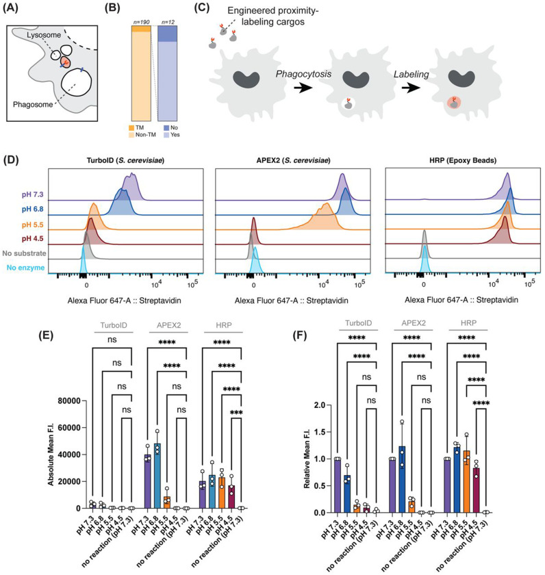
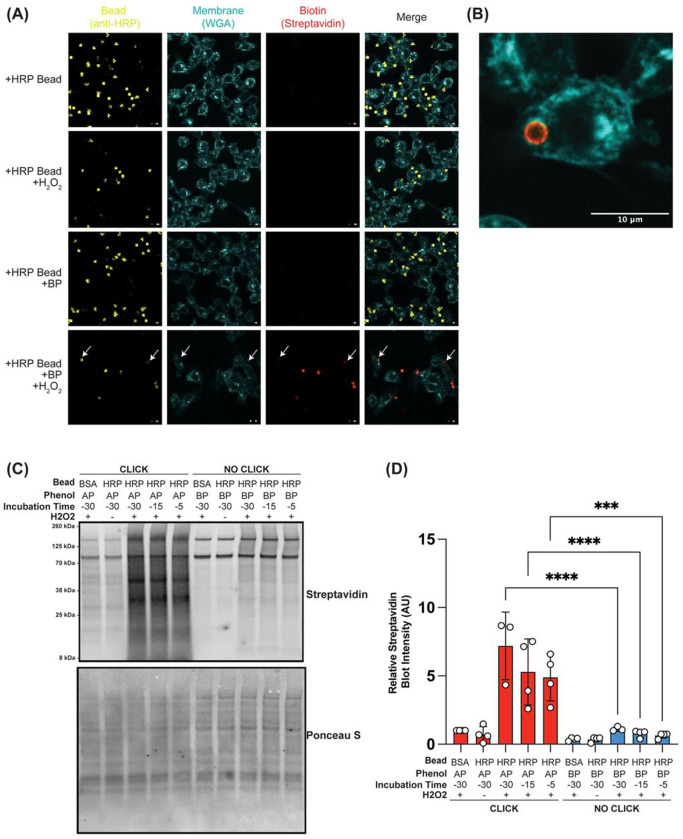
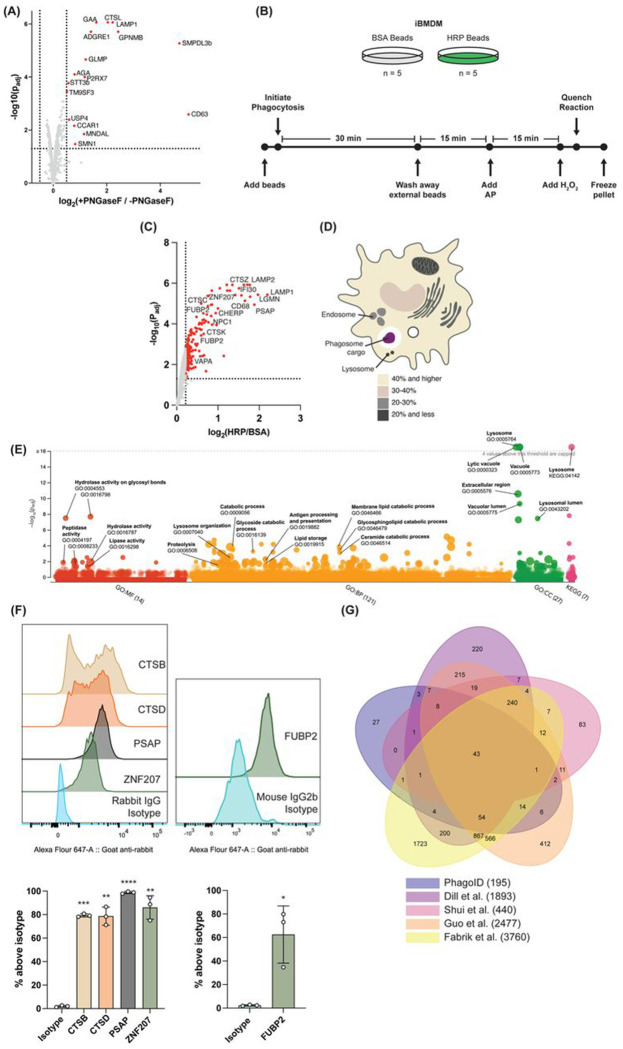
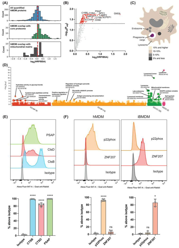
Proteomic analyses of the phagosome has significantly improved our understanding of the proteins which contribute to critical phagosome functions such as apoptotic cell clearance and microbial killing. However, previous methods of isolating phagosomes for proteomic analysis have relied on cell fractionation with some intrinsic limitations. Here, we present an alternative and modular proximity-labeling based strategy for mass spectrometry proteomic analysis of the phagosome lumen, termed PhagoID. We optimize proximity labeling in the phagosome and apply PhagoID to immortalized murine macrophages as well as primary human macrophages. Analysis of proteins detected by PhagoID in murine macrophages demonstrate that PhagoID corroborates previous proteomic studies, but also nominates novel proteins with unexpected residence at the phagosome for further study. A direct comparison between the proteins detected by PhagoID between mouse and human macrophages further reveals that human macrophage phagosomes have an increased abundance of proteins involved in the oxidative burst and antigen presentation. Our study develops and benchmarks a new approach to measure the protein composition of the phagosome and validates a subset of these findings, ultimately using PhagoID to grant further insight into the core constituent proteins and species differences at the phagosome lumen.


Figures

Figure 1:
Figure 1:. HRP-beads enable proximity labeling across the phagosomal pH range.
A Schematic of proximity labeling for membrane bound organelles. A transmembrane protein of a specific cellular compartment is often used as the tether for delivery of a proximity labeling enzyme to a specific compartment. B Analysis of Dill et al. phagosome proteome: of the top 10% most abundant phagosome proteins, 12 proteins were transmembrane (TM) and present at a constant level from early to late phagosome stages. 10 of these 12 proteins were annotated as residing in multiple subcellular locations in the Uniprot database. C Experimental schematic for phagosome lumen proximity labeling mediated by PhagoID. D Representative flow cytometry distributions of TurboID- and APEX2-expressing S. cerevisiae, or HRP-conjugated beads reacted with their appropriate labeling substrate across a range of pH, and self-biotinylation was measured by flow cytometry with fluorescent streptavidin staining. E, F Quantification and statistical analysis of (D) across multiple replicates by (E) absolute fluorescence intensity (F. I.) or (F) relative fluorescence intensity (F. I.). (n=3 independent replicates) Data Information: In (E) and (F), data are presented as mean +/− SD. Reported statistics are from a two-way ANOVA with post-hoc comparison of means and Dunnett’s multiple comparisons test; ns indicates adjusted p-value >= 0.05, *** indicates adjusted p-value <0.001, **** indicates adjusted p-value < 0.0001.
Figure 2:
Figure 2:. Alkyne-phenol yields more abundant phagosomal labeling than biotin-phenol with an optimized protocol.
A iBMDMs phagocytosed HRP-conjugated beads in the presence of 500 μM biotin phenol (BP) for 45 minutes before adding H2O2 to catalyze proximity labeling and stained with fluorescent streptavidin to visualize abundance and localization of biotinylation in the phagosome by confocal microscopy (white arrows indicate fully internalized beads in the 4th row). Scale bars are 10 μm. B Zoomed in field-of-view of an iBMDM with internalized HRP-bead+BP+H2O2. Scale bar is 10 μm. C iBMDMs phagocytosed HRP- or BSA-conjugated beads for 30 minutes at 37° C, followed by washing away extracellular beads and another 30 minutes of incubation at 37° C for phagosome progression. The indicated phenolic labeling substrate (500 μM alkyne phenol (AP) or BP) was added post-wash either 30, 15, or 5 minutes prior to labeling. Labeling was catalyzed with H2O2, click reactions were completed where indicated, and lysates were analyzed by western blot to assess ability of substrate to access the phagosome (representative blot from 3 independent replicates). D Quantification of (C) across independent replicates. Streptavidin band intensities normalized to the Ponceau S intensity indicating total protein abundance (n=3 or 4). Data Information: Reported statistics are from a one-way ANOVA with post-hoc comparison of means and Dunnett’s multiple comparisons test; ns indicates adjusted p-value >= 0.05, *** indicates adjusted p-value <0.001, **** indicates adjusted p-value < 0.0001.
Figure 3:
Figure 3:. PhagoID enables proteomic analysis of iBMDM phagosomes and highlights nuclear-annotated proteins ZNF207 and FUBP2.
A Volcano plot of differentially quantified proteins upon PNGaseF digestion analyzed with the quantitative proteomics workflow (adjusted p < 0.05 and log2FC > 0.5). (n=3 independent lysate preparations) B Experimental schematic for TMT-based PhagoID proteomics experiment: 5 replicates were prepared where iBMDMs phagocytosed BSA beads as a negative control, and 5 replicates were prepared where iBMDMs phagocytosed HRP-beads for PhagoID. C Volcano plot highlighting 195 proteins detected at the iBMDM phagosome lumen following statistical ratiometric analysis and thresholding (adjusted p < 0.05 and log2FC > 0.21). (n=5 independent lysate preparations) D PhagoID-detected proteins were annotated by their subcellular location according to the Uniprot database and labeled with their percent contribution to the phagosome. E PhagoID-detected proteins were subjected to Gene Ontology (GO) and KEGG enrichment analysis via g:Profiler. All tested terms and p-values are reported in Supplemental Table 2. F Single-phagosome resolution of phagosome colocalization for a panel of PhagoID-detected proteins by PhagoFACS. Representative flow cytometry distributions (upper) and quantification of phagosomes stained above isotype control across multiple replicates (lower). (n=3) G Comparison of significant proteins from PhagoID with previous cell-fractionation-based phagosome proteomic datasets (related to Supplementary Table 3). Data Information: In (A) and (C), p-values were calculated using a moderated T-test and adjusted with the Benjamini-Hochberg protocol. In (E), p-values were calculated using a cumulative hypergeometric test and adjusted with the Benjamini-Hochberg protocol. In (F), data are presented as the mean +/− SD. Where more than 1 antibody was compared to an isotype control, a one-way ANOVA with post-hoc comparison of means and Dunnett’s multiple comparisons test was used. When only 1 antibody was compared to an isotype control, a paired T-test was used. ns indicates adjusted p-value >= 0.05, *** indicates adjusted p-value <0.001, **** indicates adjusted p-value < 0.0001.
Figure 4:
Figure 4:. PhagoID enables proteomic analysis of primary human monocyte-derived macrophages and identifies increased abundance of the NADPH oxidase complex.
A Histogram depicting hMDM phagosome lumen quantified log2-foldchange distribution of all proteins (top), proteins among the core phagosome lumen proteome (middle), and proteins among the FP list mitochondrial matrix (bottom). The applied fold-change threshold is indicated with a dashed line. B Volcano plot highlighting 123 proteins detected at the hMDM phagosome following statistical ratiometric analysis and thresholding (adjusted p < 0.05 and log2FC > 0.0538). (n=5 human donors) C PhagoID-detected proteins were annotated by their subcellular location according to the Uniprot database and labeled with their percent contribution to the phagosome. D PhagoID-detected proteins were subjected to Gene Ontology (GO) and KEGG enrichment analysis via g:Profiler. All tested terms and p-values are reported in Supplemental Table 2. E Single-phagosome resolution of phagosome colocalization for a panel of PhagoID-detected proteins by PhagoFACS. Representative flow cytometry distributions (upper) and quantification of phagosomes stained above isotype control across multiple replicates (lower). (n=5 human donors) F PhagoFACS validation of proteins nominated by PhagoID for differential abundance in hMDM and iBMDM. Representative flow cytometry distributions (upper) and quantification of phagosomes stained above isotype control across multiple replicates (lower). (n=5 human donors for hMDM and n=3 for iBMDM). Same data replotted for iBMDM ZNF207 staining as from Fig. 3F for side-by-side comparison with hMDM. Reported statistics from where more than 2 categories are compared are from a one-way ANOVA with Dunnett’s multiple comparisons test. Where 2 categories are compared, a Paired T-test was used. ns indicates adjusted p-value >= 0.05, *** indicates adjusted p-value <0.001, **** indicates adjusted p-value < 0.0001.

Similar articles

References

    1. Flannagan R. S., Jaumouillé V. & Grinstein S. The Cell Biology of Phagocytosis. Annu. Rev. Pathol. Mech. Dis. 7, 61–98 (2012). - PubMed
    1. Jordao L., Bleck C. K. E., Mayorga L., Griffiths G. & Anes E. On the killing of mycobacteria by macrophages. Cell. Microbiol. 10, 529–548 (2008). - PubMed
    1. Pittet M. J., Michielin O. & Migliorini D. Clinical relevance of tumour-associated macrophages. Nat. Rev. Clin. Oncol. 19, 402–421 (2022). - PubMed
    1. Vitale I., Manic G., Coussens L. M., Kroemer G. & Galluzzi L. Macrophages and Metabolism in the Tumor Microenvironment. Cell Metab. 30, 36–50 (2019). - PubMed
    1. Gabandé-Rodríguez E., Keane L. & Capasso M. Microglial phagocytosis in aging and Alzheimer’s disease. J. Neurosci. Res. 98, 284–298 (2020). - PubMed

Publication types

LinkOut - more resources