[Preprint]. 2024 Sep 2:2024.09.02.610853.
doi: 10.1101/2024.09.02.610853.

Methods for high throughput discovery of fluoroprobes that recognize tau fibril polymorphs

Affiliations

Methods for high throughput discovery of fluoroprobes that recognize tau fibril polymorphs

Emma C Carroll et al. bioRxiv. .

Update in

Abstract

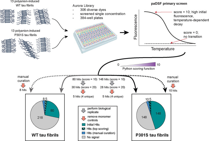
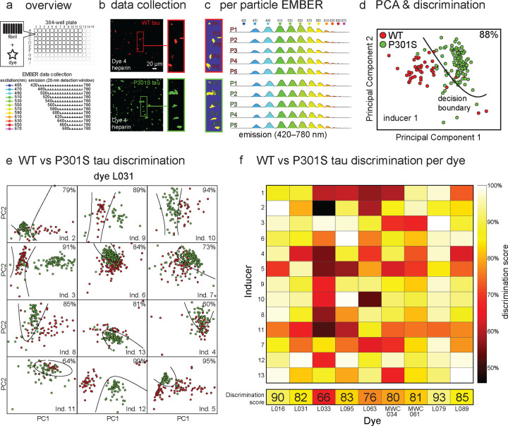
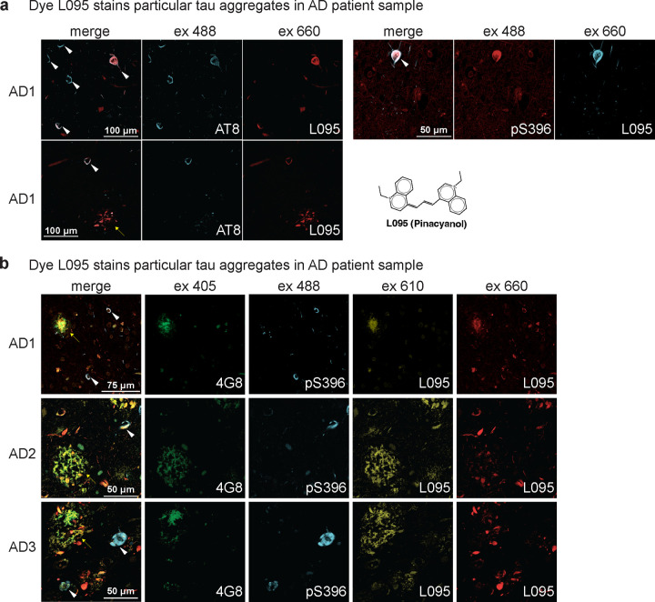
Aggregation of microtubule-associated protein tau (MAPT/tau) into conformationally distinct fibrils underpins neurodegenerative tauopathies. Fluorescent probes (fluoroprobes), such as thioflavin T (ThT), have been essential tools for studying tau aggregation; however, most of them do not discriminate between amyloid fibril conformations (polymorphs). This gap is due, in part, to a lack of high-throughput methods for screening large, diverse chemical collections. Here, we leverage advances in protein adaptive differential scanning fluorimetry (paDSF) to screen the Aurora collection of 300+ fluorescent dyes against multiple synthetic tau fibril polymorphs. This screen, coupled with orthogonal secondary assays, revealed pan-fibril binding chemotypes, as well as fluoroprobes selective for subsets of fibrils. One fluoroprobe recognized tau pathology in ex vivo brain slices from Alzheimer's disease patients. We propose that these scaffolds represent entry points for development of selective fibril ligands and, more broadly, that high throughput, fluorescence-based dye screening is a platform for their discovery.

Keywords: dye; high-throughput screening; molecular recognition; tauopathy; time-resolved fluorescence.


Conflict of interest statement

Competing Interests The authors have no competing interests to disclose.

Figures

Figure 1.
Figure 1.. A high throughput screening platform reveals fluoroprobes that recognize tau fibril polymorphs.
Schematic of the primary screening workflow and summary of the results. Synthetic tau fibrils with diverse conformations were generated using either recombinant WT or P301S tau (0N4R splice isoform), mixed with 13 different inducers (see Fig. S1 and Table S1). These 26 fibril samples were purified and incubated with the Aurora dye library in 384-well plates and then heated to generate temperature vs. fluorescence plots. The resulting data was scored using a Python-based function (see Methods), with the top “hits” (score = 10) being dyes with high initial fluorescence, low background in the control (polyanion inducer; no tau) and a temperature-dependent decay. The highest-scoring hits across two biological replicates (cumulative score = 20) were then compared to the second control (monomeric tau alone; salmon), yielding five hits that reacted with at least one of the WT or P301S fibrils (hashed). This list was supplemented by manual curation of other top performing dyes (blue).
Figure 2.
Figure 2.. High throughput screen results uncovers fluoroprobes that interact with either WT or P301S tau fibril polymorphs.
(a) Heat map of the additive scores for screens performed using WT (top) or P301S (bottom) tau fibrils. Only the highest-scoring fluoroprobes (score = 20) were taken forward for validation. (b) Representative plots of relative fluorescence units (RFU, channel denoted per plot) versus temperature, highlighting the appearance of potentially pan-fibril dyes (L031) and potentially P301S-selective dyes (L017 and L033). Graphs are representative of the two biological replicates. See Extended Data S1 for the full dataset and Table S1 for a list of inducers.
Figure 3.
Figure 3.. EMBER analysis suggests that fluoroprobes bind in distinct chemical environments between WT and P301S tau fibrils.
(a) Overview of the EMBER (excitation multiplexed bright emission recordings) workflow. In the initial screen, the 27 hit dyes from the primary screen (see Fig 2) were screened against 12 synthetic tau fibril samples in 384-well plates. In EMBER, fluorescence data is collected at a range of excitation and emission wavelengths to explore shifts in either wavelength or intensity after dye binding to fibrils. (b) Representative EMBER results, showing individual tau fibril particles composed of either WT tau (red) or P301S tau (green). Insets show the same particles at higher resolution. This example shows WT and P301S tau fibrils with inducer 1 and dye L095. (c) For each particle, Bradley–Roth segmentation is performed across the full wavelength range to provide the EMBER plot (Extended Data 6). The particles are the same as in panel b. (d) Individual EMBER plots are then concatenated for principal component analysis (PCA), followed by quadratic discrimination to quantify polymorph selectivity. In this case, dye L095 was able to discriminate between WT and P301S tau fibrils (in the presence of inducer 1) with 88% accuracy. Boundaries pertaining to the fit discriminants are presented in black lines. (e) Representative data showing that dye L031 is also able to discriminate between WT and P301S tau fibril samples created using other inducers (Ind). PC1 = principal component 1. PC2 = principal component 2. (f) Discrimination heatmap for the full dataset, showing that a subset of the dyes can discriminate between tau fibril samples.
Figure 4.
Figure 4.. Validated fluoroprobe hits are chemically diverse and can detect tau fibril formation in real-time.
(a) Chemical structures of the validated fluoroprobe hits, showing the two clusters (coumarins and polymethines). (b) Histogram of pairwise Tanimoto similarities for all hits compared with one another (left) and compared with Thioflavin T (ThT; right). Tanimoto coefficients calculated using a script created with the RDKit Python package (see Extended Data 3) (c-h) Kinetic aggregation assays. Either ThT or the hit dyes were mixed with WT or P301S tau and aggregation initiated with a polyanion inducer. Raw signal was normalized as a fraction of total signal to fall between 0 and 1 to facilitate comparisons. Data points are the average of replicates (n=2 or n=3) and error bars represent the standard deviation (SD). Note that only one dye was added to each sample, but the results are shown side-by-side. (c) Confirmation that P301S aggregates faster than WT, as shown using ThT and inducer 7. (d) Example of a dye, L031, that has a similar profile to ThT. Shown is P301S tau with inducer 7. (e) Example of a dye, MWC034, that only recognizes P301S tau, and not WT. Reactions contained inducer 7. (f) Example of a dye, L061, that recognizes structures early in the process than ThT. Results from P301S and inducer 7. (g) L033 recognizes relatively early structures, using WT tau and inducer 8. (h) Dye MWC034 recognizes relatively late structures, using P301S tau and inducer 4.
Figure 5.
Figure 5.. Fluoroprobe L095 recognizes tau pathology in brain tissue from human AD patients.
(a) Two samples from an AD patient, labelled with the AT8 or pS396 antibody and with L095. Note that L095 only labels a subset of tau tangles (white arrows), but also labels tau pathology that is consistent with neuropil threads (yellow arrow). (b) Samples from three AD patients, stained with antibodies for either amyloid beta (4G8) or tau pathology (pS396). Note that L095 co-localizes with G48 at 610 nm (yellow arrows), but with AT8 at 660 nm (white arrows), allowing partial spectral discrimination between the two pathologies.
Figure 6.
Figure 6.. Analogs of the coumarin scaffold include both pan-fibril binding and potentially selective fluoroprobes.
(a) Overview of the limited medicinal chemistry campaign. Twenty-four coumarins from the Max A. Weaver Collection (MWC) were screened against 26 fibril samples by paDSF using a pipeline that parallels Figure 1, except that three biological replicates were used. After triage and removal of dyes that bound tau monomer, only 4 coumarins were identified that bind both WT and P301S fibrils. (b) Heat maps of the screening results, showing that most of the analogs failed to recognize either the WT or P301S tau fibril samples (white); whereas a subset produced reproducible signal (green). Here, the top Python score was 30 (10 for each replicate). (b) The chemical structures of the coumarins that bind to both WT and P301S fibrils, suggesting that they are pan-binders. (c) Chemical structures of coumarins with activity against at least one WT or P301S tau fibril conformer. The coumarin scaffold is highlighted in purple.

Similar articles

References

    1. Chung D. C.; Roemer S.; Petrucelli L.; Dickson D. W. (2021) Cellular and pathological heterogeneity of primary tauopathies, Mol Neurodegener 16, 57. - PMC - PubMed
    1. Limorenko G.; Lashuel H. A. (2022) Revisiting the grammar of tau aggregation and pathology formation: How new insights from brain pathology are shaping how we study and target tauopathies, Chemical Society reviews 51, 513–565. - PubMed
    1. Chang C. W.; Shao E.; Mucke L. (2021) Tau: Enabler of diverse brain disorders and target of rapidly evolving therapeutic strategies, Science 371. - PMC - PubMed
    1. Yagishita S.; Itoh Y.; Nan W.; Amano N. (1981) Reappraisal of the fine structure of alzheimer’s neurofibrillary tangles, Acta Neuropathol 54, 239–246. - PubMed
    1. Scheres S. H.; Zhang W.; Falcon B.; Goedert M. (2020) Cryo-em structures of tau filaments, Curr Opin Struct Biol 64, 17–25. - PubMed

Publication types

LinkOut - more resources