[Preprint]. 2024 Sep 3:2024.08.30.610568.
doi: 10.1101/2024.08.30.610568.

Electrospun Fiber Surface Roughness Modulates Human Monocyte-Derived Macrophage Phenotype

Affiliations

Electrospun Fiber Surface Roughness Modulates Human Monocyte-Derived Macrophage Phenotype

Aidan Alemifar et al. bioRxiv. .

Abstract

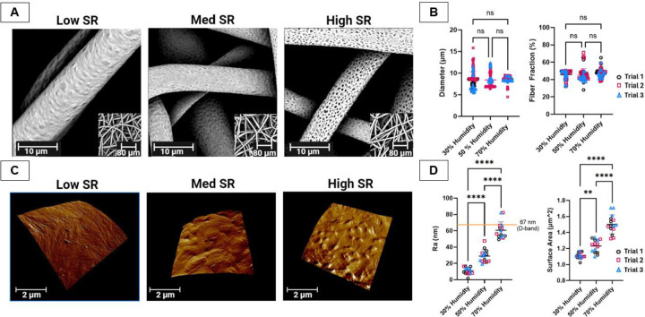
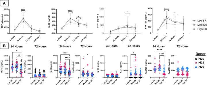
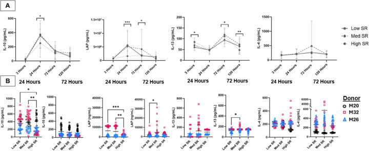
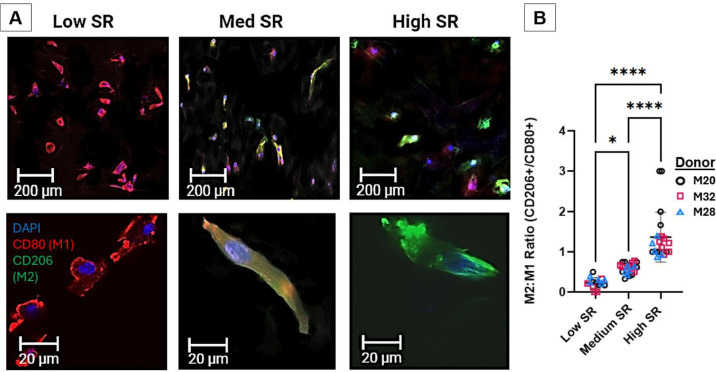
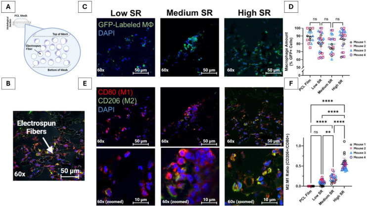
Injuries to fibrous connective tissues have very little capacity for self-renewal and exhibit poor healing after injury. Phenotypic shifts in macrophages play a vital role in mediating the healing response, creating an opportunity to design immunomodulatory biomaterials which control macrophage polarization and promote regeneration. In this study, electrospun poly(-caprolactone) fibers with increasing surface roughness (SR) were produced by increasing relative humidity and inducing vapor-induced phase separation during the electrospinning process. The impact of surface roughness on macrophage phenotype was assessed using human monocyte-derived macrophages in vitro and in vivo using B6.Cg-Tg(Csf1r-EGFP)1Hume/J (MacGreen) mice. In vitro experiments showed that macrophages cultured on mesh with increasing SR exhibited decreased release of both pro- and anti-inflammatory cytokines potentially driven by increased protein adsorption and biophysical impacts on the cells. Further, increasing SR led to an increase in the expression of the pro-regenerative cell surface marker CD206 relative to the pro-inflammatory marker CD80. Mesh with increasing SR were implanted subcutaneously in MacGreen mice, again showing an increase in the ratio of cells expressing CD206 to those expressing CD80 visualized by immunofluorescence. SR on implanted biomaterials is sufficient to drive macrophage polarization, demonstrating a simple feature to include in biomaterial design to control innate immunity.


Figures

Figure 1:
Figure 1:. Modification of Fiber SR with Increasing RH Without Significant Change to Diameter or Porosity.
Electrospun PCL were imaged with SEM (A). Fiber diameter and porosity (B) were then determined and compared. Fiber surface roughness (SR) was visualized and quantified using AFM (C) *p<0.0001
Figure 2:
Figure 2:. Protein Adsorption on Mesh.
Mesh were incubated in 40% FBS (A) or a DAMP solution (B). Adsorbed protein was quantified via a BCA assay. * p ≤ 0.05; **p≤ 0.01: *** p ≤ 0.001; **** p ≤ 0.0001
Figure 3:
Figure 3:. Pro-Inflammatory Cytokine Release.
Cultured media from primary macrophages was tested on an eight-plex Luminex assay at 3, 24, 72, and 120 hours for pro-inflammatory cytokines. Individual figures for 24- and 72-hour timepoints * p ≤ 0.05; **p≤ 0.01: *** p ≤ 0.001; **** p ≤ 0.0001
Figure 4:
Figure 4:. Anti-Inflammatory Cytokine Release.
Cultured media from primary macrophages was tested on an eight-plex Luminex assay at 3, 24, 72, and 120 hours for anti-inflammatory cytokines. Individual figures for 24- and 72-hour timepoints * p ≤ 0.05; **p≤ 0.01: *** p ≤ 0.001; **** p ≤ 0.0001
Figure 5:
Figure 5:. Immunocytochemistry Staining of Primary Macrophages for CD 80 (M1) and CD 206 (M2).
Images of CD 80 and 206 expression in primary macrophages incubated on mesh with increasing SR. * p ≤ 0.05; **** p ≤ 0.0001.
Figure 6:
Figure 6:. Immunohistochemistry Staining of Mesh after Implantation in MacGreen Mice for CD80 (M1) and CD206 (M2).
Schematic (A) and actual (B) visualization of macrophage morphology on fibers. Infiltration (C and D) and polarization (E and F) of macrophages imaged at 60x after subcutaneous implantation in mice. Cropped and zoomed in images of B are shown in the lowest panels. A brightfield image shows interaction between cells and mesh (C). Ratio of cells expressing CD206 and CD80 was measured in ImageJ (D). ** p ≤ 0.01; ****p<0.0001.

Similar articles

References

    1. Yelin E., Weinstein S., and King T., The burden of musculoskeletal diseases in the United States. Semin Arthritis Rheum, 2016. 46(3): p. 259–260. - PubMed
    1. Chiang H. and Jiang C.C., Repair of articular cartilage defects: review and perspectives. J Formos Med Assoc, 2009. 108(2): p. 87–101. - PubMed
    1. Corr D.T. and Hart D.A., Biomechanics of Scar Tissue and Uninjured Skin. Adv Wound Care (New Rochelle), 2013. 2(2): p. 37–43. - PMC - PubMed
    1. Jung H.-J., Fisher M.B., and Woo S.L.Y., Role of biomechanics in the understanding of normal, injured, and healing ligaments and tendons. BMC Sports Science, Medicine and Rehabilitation, 2009. 1(1): p. 9. - PMC - PubMed
    1. Guo S. and Dipietro L.A., Factors Affecting Wound Healing. Journal of Dental Research, 2010. 89(3): p. 219–229. - PMC - PubMed

Publication types