[Preprint]. 2024 Sep 6:2024.09.04.611291.
doi: 10.1101/2024.09.04.611291.

The Pax transcription factor EGL-38 links EGFR signaling to assembly of a cell-type specific apical extracellular matrix in the Caenorhabditis elegans vulva

Affiliations

The Pax transcription factor EGL-38 links EGFR signaling to assembly of a cell-type specific apical extracellular matrix in the Caenorhabditis elegans vulva

Helen F Schmidt et al. bioRxiv. .

Update in

Abstract

The surface of epithelial tissues is covered by an apical extracellular matrix (aECM). The aECMs of different tissues have distinct compositions to serve distinct functions, yet how a particular cell type assembles the proper aECM is not well understood. We used the cell-type specific matrix of the C. elegans vulva to investigate the connection between cell identity and matrix assembly. The vulva is an epithelial tube composed of seven cell types descending from EGFR/Ras-dependent (1°) and Notch-dependent (2°) lineages. Vulva aECM contains multiple Zona Pellucida domain (ZP) proteins, which are a common component of aECMs across life. ZP proteins LET-653 and CUTL-18 assemble on 1° cell surfaces, while NOAH-1 assembles on a subset of 2° surfaces. All three ZP genes are broadly transcribed, indicating that cell-type specific ZP assembly must be determined by features of the destination cell surface. The paired box (Pax) transcription factor EGL-38 promotes assembly of 1° matrix and prevents inappropriate assembly of 2° matrix, suggesting that EGL-38 promotes expression of one or more ZP matrix organizers. Our results connect the known signaling pathways and various downstream effectors to EGL-38/Pax expression and the ZP matrix component of vulva cell fate execution. We propose that dedicated transcriptional networks may contribute to cell-appropriate assembly of aECM in many epithelial organs.


Figures

Figure 1:
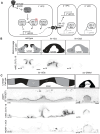
Figure 1:. Zona Pellucida domain (ZP) protein matrix is cell type-specific
A) Position of the vulva and L4.5 stage vulva cells visualized with the membrane marker MIG-2::GFP (muIs28) (Honigberg and Kenyon, 2000). The seven vulva cell types (vulA-F) are highlighted by color. B) Specification of vulva cells. The anchor cell (AC) releases a gradient of LIN-3/EGF to the six equipotent vulva precursor cells (VPCs-ovals). The closest cell receives the strongest EGF signal and adopts the primary (1°) vulva fate, dividing to give rise to vulE/F. The 1° VPC also expresses Delta/Serrate/LAG-2 (DSL) ligands to activate LIN-12/Notch signaling in neighboring VPCs and specify the secondary (2°) vulva fate, which gives rise to vulA/B1/B2/C/D. Transcription factors (TFs) controlling aspects of each cell fate are sometimes broadly expressed in the vulva (e.g. LIN-11/LIM, COG-1/Nkx), or expressed in specific vulva cells (e.g. EGL-38/Pax). C) ZP protein schematics. The LET-653(ZP) fusion consists of the isolated ZP domain and was expressed from a transgene (csIs66) (Cohen et al., 2019); full-length LET-653 has multiple PAN domains at its C-terminus. NOAH-1 and CUTL-18 fusions were each generated by CRISPR-Cas9 genome editing and expressed from the endogenous locus. D) ZP proteins localize to the matrix of specific vulva cells. LET-653(ZP) and CUTL-18 are 1° matrix components on vulE/F. CUTL-18 is also a 2° matrix component on vulB1/B2. NOAH-1 is a 2°-specific matrix component, enriched on vulC. Images are medial confocal slices at the indicated L4 stages. Scale bar 5 μm.
Figure 2:
Figure 2:. ZP protein localization is not explained by their transcription patterns
A. Schematic of transcriptional reporters expressed from the endogenous loci of ZP protein genes. Sequence inserted immediately after stop codon of each ZP protein gene is outlined in red. The gpd-2 3’UTR and trans-spliced leader sequence (SL2) allow the ZP protein and mCh::HIS-44 (H2B) to be expressed as an operon. B. ZP protein gene expression in early- (L4.1) and mid- (L4.4/L4.5) L4 larvae. Top: diagram of approximate position of each vulva nucleus at the indicated stages. Below: Maximum projections of confocal Z stacks through entire vulva of worms expressing the indicated transcriptional reporter. Vulva nuclei are outlined according to the color code below. Lower left: Number of mCherry positive nuclei, (n= number of worms). The cutl-18 transcriptional reporter varied slightly at the mid-L4 stage; it was present in vulA,B1,B2,E, and F (16 total nuclei) in all worms, and also faintly in vulD (18 total nuclei) in 11/14 worms. C. LET-653(ZP) assembles on the surface of 1° cells in absence of its transcription in 1° cells. Mid-L4 vulvas. Top: epifluorescent image of cytoplasmic GFP expressed from a 2233bp let-653 promoter fragment (Hunt-Newbury et al., 2007); GFP is present in 2° cells and faintly in hyp7. Bottom: medial confocal slice of sfGFP::LET-653(ZP) expressed from the same promoter fragment is present inside 2° cells (orange outline) and assembles on 1° cell surfaces. D. The NOAH-1 C-terminus (Cter) is not secreted or cell-type specific. Top: Since NOAH-1::sfGFP(Cter) is predicted to be cleaved N-terminal to the transmembrane domain, the sfGFP remains inside the cells that produced it (Vuong-Brender et al., 2017). Medial confocal slice of L4.5 stage vulva. NOAH-1 C-terminus forms large puncta in all 2° vulva cells and hyp7. Consistent with the transcriptional reporter, NOAH-1::sfGFP(Cter) signal is not distinct between vulC and other 2° cells or hyp7. Below: Schematic of NOAH-1 with sfGFP at its C-terminus, full length (FL), and predicted product after cleavage. Domain symbols as are in Figure 1. All scale bars 5μm.
Figure 3:
Figure 3:. Contributions of EGFR effectors to matrix identity
A) Transcription factors downstream of MPK-1/ERK promote transcription of 1° expressed genes. In the 1° VPC, LET-23/EGFR activates MPK-1/ERK via a Ras signaling cascade. MPK-1 phosphorylates LIN-1/ETS to convert it from a repressor to an activator of 1° identity (Jacobs et al., 1998; Tan et al., 1998). The mediator component SUR-2 acts in parallel to LIN-1 to promote some aspects of 1° identity, such as expression of DSL family ligands that activate LIN-12/Notch to promote the 2° fate in neighboring VPCs (Singh and Han, 1995; Underwood et al., 2017; Zhang and Greenwald, 2011). Unphosphorylated LIN-1 represses 1° expressed genes in the 2° and 3° lineages. B) Matrix identity lin-12/Notch mutants aligns with identity assessed by lineage. Top: schematic of vulva cell types (as assessed by lineage or matrix) in wild type, loss of function (−) lin-12(n137n720) mutants and dominant gain of function (d) lin-12(n137) mutants (Greenwald et al., 1983). Bottom: CUTL-18 is strongly enriched on all cell surfaces lin-12(−) mutants, consistent with a 1° matrix identity, but at faintly on some cell surfaces in lin-12(d) consistent with a 2° matrix identity. Medial confocal slices of mid-L4 stage worms. C) LIN-1/Ets represses, SUR-2/Med promotes 1° matrix identity. Top: schematic of vulva cell fates determined by lineage lin-1(n304) and sur-2(ku9) mutants (Beitel et al., 1995; Howard and Sundaram, 2002; Singh and Han, 1995). Below: LET-653(ZP)::sfGFP (csIs66) and CUTL-18 are enriched on the surface of both 1° lineage (orange arrowhead) and 2° lineage (blue arrow) vulva cells in a lin-1 mutant. Few cells have NOAH-1::mCh on their surface in lin-1 mutants. LET-653(ZP)::sfGFP, CUTL-18 and NOAH-1 are all enriched on a subset of 1° lineage cells in sur-2 mutants. All scale bars 5μm.
Figure 4:
Figure 4:. Vulva matrix identity correlates with EGL-38 expression
A) Schematic of vulva cell types (as assessed by lineage) and summary of matrix assembly as in Figure 3 and (Cohen et al., 2020b) in lin-12/Notch mutants. B) EGL-38 expression in lin-12/Notch mutants aligns with matrix identity. Maximum projections of confocal Z stacks through entire vulva of mid-L4 stage worms. Vulva nuclei expressing EGL-38::GFP are circled - vulD in green, vulE in yellow, vulF in red, and unknown nuclei descended from a 1°-like lineage in orange. + indicates uv1 nuclei. n= number of worms, number of EGL-38 positive vulva nuclei per worm quantified in C. C) Schematic of vulva cell types (as assessed by lineage) and summary of matrix assembly as in Figure 3 in lin-1 and sur-2 mutants. D) LIN-1/Ets represses, SUR-2/Med promotes egl-38 expression consistent with matrix identity. EGL-38::GFP expression in the indicated mutants. Maximum projections of confocal Z stacks through entire vulva of mid-L4 stage worms. Consistent with their matrix identity, the majority of vulva cells expressed EGL-38 in lin-1 mutants, and only some of the vulva cells expressed EGL-38 in sur-2 mutants. n= number of worms, number of EGL-38 positive vulva nuclei per worm quantified in E. All scale bars 5μm. E) Count of the number of EGL-38-positive vulva nuclei in wild type (WT) and lin-12, lin-1, and sur-2 mutants. Presence of EGL-38 was assessed with EGL-38::GFP expressed from the endogenous locus (gu253) or EGL-38::TY1::EGFP::3xFLAG expressed from a transgene (wgIs171). In wild type worms, EGL-38 was always present in 10 vulva nuclei, regardless of method of expression. P values Kruskal–Wallis test. F) Contributions of EGFR effectors to egl-38 expression. LIN-1 represses egl-38 transcription in the absence of EGFR/MAPK signaling. SUR-2 promotes egl-38 transcription in the presence of EGFR/MAPK signaling.
Figure 5:
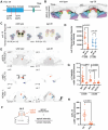
Figure 5:. The Pax transcription factor EGL-38 is required for proper 1° cell ZP matrix assembly
A) Schematic of EGL-38 protein. Red bars indicate the location of hypomorphic missense mutations n578 and sy294 in the conserved Paired box (Pax) domain. B) In egl-38 mutants the vulF cells remain close to each other, blocking the uterine connection. Vulva cells visualized with the membrane marker MIG-2::GFP(muls28) and colored by cell type. C) EGL-38 promotes the transcription of cutl-18 in 1° descendants vulE/F. Maximum projections of confocal Z stacks through entire vulva of mid-L4 of wild type and egl-38(n578) worms expressing the indicated transcriptional reporter. Vulva nuclei are outlined according to the cell colors in A. Lower left: Number of mCherry positive nuclei, (n= number of worms). Wild type Ns represent the same worms as in Figure 2. let-653 and noah-1 transcription were not affected, see Supplemental figure 4. D) Quantification of cutl-18 transcriptional reporter fluorescence in 1°- and 2°- descendant nuclei in wild type and egl-38(n578) mutants. See methods. P values Kruskal–Wallis test. E) EGL-38 is required for proper 1° cell ZP matrix assembly. LET-653(ZP)csls66, mNG::CUTL-18 and NOAH-1::mCherry in wild-type and egl-38(n578) mutants. In egl-38 mutants the 1° matrix (orange arrowheads) does not contain the proper proteins, while 2° cell specific matrices (blue arrows) are maintained. Medial confocal slices at the indicated L4 stages. (n= number of worms). Wild type Ns represent the same worms as in Figure 1. See Supplemental Figure 5 for images of additional alleles and fluorescence quantification of 2° cell surfaces. All scale bars 5 μm. F) Apical enrichment of ZP proteins quantified by fluorescence intensity at the vulE/F surfaces divided by intensity in a box of the same total size in the lumen. See Methods. G and H) Apical enrichment of LET-653(ZP) or CUTL-18 on vulE/F cells. Column labels below indicate wild type worms (WT) or egl-38 alleles (n578 and sy294) and LET-653(ZP) transgenes (csls66 and csls96). All P values Kruskal–Wallis test.
Figure 6:
Figure 6:. Model for 1° matrix fate execution
The 1° lineage is specified through LET-23/EGFR, triggering a signaling cascade to MPK-1/ERK. Signaling promotes egl-38 expression through at least two transcription factors, by relieving LIN-1/Ets repression through direct phosphorylation and promoting SUR-2/Med activity (or that of an unknown partner transcription factor). EGL-38 in turn promotes the expression of unknown 1° matrix organizer(s) that are present on the surface of primary cells and recruit 1° matrix proteins LET-653(ZP) and CUTL-18 and exclude 2° matrix protein NOAH-1.

Similar articles

References

    1. Adams J.R.G., Pooranachithra M., Jyo E.M., Zheng S.L., Goncharov A., Crew J.R., Kramer J.M., Jin Y., Ernst A.M., Chisholm A.D., 2023. Nanoscale patterning of collagens in C. elegans apical extracellular matrix. Nat Commun 14, 7506. 10.1038/s41467-023-43058-9 - DOI - PMC - PubMed
    1. Andrade L.R., Salles F.T., Grati M., Manor U., Kachar B., 2016. Tectorins crosslink type II collagen fibrils and connect the tectorial membrane to the spiral limbus. J Struct Biol 194, 139–146. 10.1016/j.jsb.2016.01.006 - DOI - PMC - PubMed
    1. Bausek N., Waclawek M., Schneider W.J., Wohlrab F., 2000. The Major Chicken Egg Envelope Protein ZP1 Is Different from ZPB and Is Synthesized in the Liver. Journal of Biological Chemistry 275, 28866–28872. 10.1074/jbc.275.37.28866 - DOI - PubMed
    1. Beitel G.J., Tuck S., Greenwald I., Horvitz H.R., 1995. The Caenorhabditis elegans gene lin-1 encodes an ETS-domain protein and defines a branch of the vulval induction pathway. Genes Dev. 9, 3149–3162. 10.1101/gad.9.24.3149 - DOI - PubMed
    1. Birnbaum S.K., Cohen J.D., Belfi A., Murray J.I., Adams J.R.G., Chisholm A.D., Sundaram M.V., 2023. The proprotein convertase BLI-4 promotes collagen secretion prior to assembly of the Caenorhabditis elegans cuticle. PLoS Genet 19, e1010944. 10.1371/journal.pgen.1010944 - DOI - PMC - PubMed

Publication types