[Preprint]. 2024 Sep 5:2024.09.03.611112.
doi: 10.1101/2024.09.03.611112.

A conserved cell-type gradient across the human mediodorsal and paraventricular thalamus

Affiliations

A conserved cell-type gradient across the human mediodorsal and paraventricular thalamus

Anton Schulmann et al. bioRxiv. .

Abstract

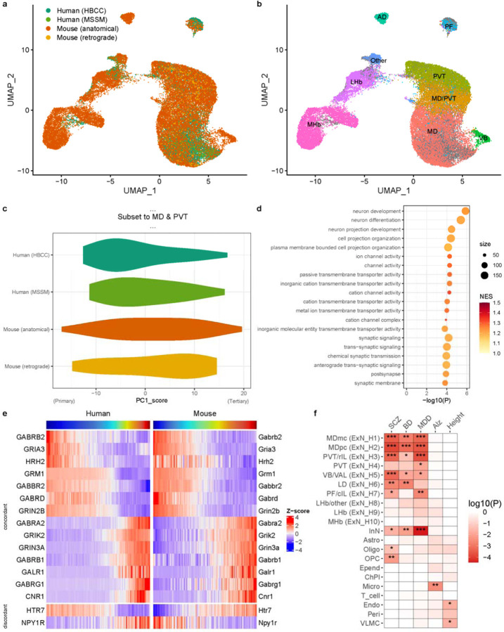
The mediodorsal thalamus (MD) and adjacent midline nuclei are important for cognition and mental illness, but their cellular composition is not well defined. Using single-nucleus and spatial transcriptomics, we identified a conserved excitatory neuron gradient, with distinct spatial mapping of individual clusters. One end of the gradient was expanded in human MD compared to mice, which may be related to the expansion of granular prefrontal cortex in hominids. Moreover, neurons preferentially mapping onto the parvocellular division MD were associated with genetic risk for schizophrenia and bipolar disorder. Midbrain-derived inhibitory interneurons were enriched in human MD and implicated in genetic risk for major depressive disorder.


Conflict of interest statement

Conflict of interest The authors declare no conflicts of interest.

Figures

Fig. 1 –
Fig. 1 –. A cell type atlas of human MD and dorsal midline thalamus.
a. Dissection approach targeting the MD and adjacent midline structures based on gross anatomical landmarks, illustrated by two example photographs of coronal slabs with region of interest highlighted in blue. b. UMAP plot illustrating annotation of major cell classes in human MD and midline thalamus (Excitatory Neurons [ExN]; Inhibitory Neurons [InN]; Astrocytes [Astro]; Oligodendrocytes [Oligo]; Oligodendrocyte Precursor Cells [OPC]; Ependyma [Epend]; Choroid Plexus [ChPl]; Microglia [Micro]; T-cells; Endothelial cells [Endo]; Pericytes [Peri]; Vascular leptomeningeal cells [VLMC]). See supplementary data table 1 for full list of marker genes. c. Dot plot illustrating expression pattern of marker genes associated with major cell classes shown in (b). (b) and (c) are based on HBCC samples; for MSSM see Supplementary fig. 1. d. UMAP plot of excitatory neurons, colored by cluster identities. e. UMAP plot of excitatory neurons colored by first principal component (PC1) score. f. Heatmap of top 15 genes with highest and lowest loadings on PC1. Cells are ordered by their PC1 score. (d), (e), and (f) are based on integration between HBCC and MSSM samples; for details, see Methods and Supplementary fig. 1.
Fig. 2 –
Fig. 2 –. Spatial distribution of neuronal populations in human MD and midline thalamus.
a. Spatial mapping of inferred cell type proportions for four excitatory neuron populations (first four columns) and thalamic inhibitory neurons (last column) across two coronal thalamic sections. Schematic (left) shows position of the 11×11mm wide capture area. Clusters shown preferentially mapped onto MDmc (ExN_H1), MDpc (ExN_H2), intralaminar nuclei, PT and PVT (ExN_H3, ExN_H4). Thalamic inhibitory neuron (InN) were most abundant in MDpc and MDdc. Major findings are summarized in Supplementary table 1. b. RNA-FISH of four marker genes (Primary markers: SHISA6 and GRM1; Secondary/Tertiary markers FOXP2 and SNCA) are shown across panels representative of four (sub)nuclei. MDmc and MDpc preferentially express primary markers, while CM and PVT/PT express tertiary markers with intermediate cells co-expressing markers of both types, especially common in CM.
Fig. 3 –
Fig. 3 –. Comparison of mouse and human thalamic cell-type gradients and functional annotation.
a. UMAP plot illustrating cross-species integration of human and mouse excitatory neurons in medial thalamus, colored by dataset: human data from HBCC and MSSM, and mouse data from this study based on anatomical dissections and a previous study using retrograde labeling from prefrontal cortex. b. UMAP plot of integrated mouse and human data, with mouse cells labeled by their nuclei of origin inferred from integration with public datasets and Allen Brain Atlas ISH data (see also Supplementary fig. 4). Marker genes for individual mouse clusters are shown in Supplementary table 2. c. Violin density plot depicting distribution of PC1 scores for cross-species integrated dataset subset to excitatory neurons from MD or PVT across both human and mouse datasets. The ‘primary’ end is on the negative (left) and the ‘tertiary’ end on the positive (right) side of the x-axis. d. Gene ontology enrichment analysis showing the top 20 terms associated with PC1 from (c) using gene set enrichment analysis. Terms are ordered by theme (neuron projection development; ion channel activity; to synaptic function) and p-value. Size indicates number of genes per term, and color shows the normalized enrichment score (NES). The full test results are shown in supplementary table 3. e. Expression of genes along the cross-species PC1 between MD and PVT, as in (c) and (d), averaged across 100 bins of PC1 scores for each species is shown. Only neurotransmitter receptor-encoding genes with Spearman’s correlation >0.75 (concordant) or <0.5 (discordant) between human and mouse expression across bins are shown. f. MAGMA gene set test results show association of the top 10% most specific gene sets for each cell type with gene-level signal across five large GWAS (SCZ = schizophrenia; BD = bipolar disorder; MDD = major depressive disorder; Alz = Alzheimer’s disease). Asterisks denote significance levels (***: p<.001, **: p<.01, *: p<.05). The full test results are shown in Supplementary table 4.

References

    1. Parnaudeau S., Bolkan S. S. & Kellendonk C. The Mediodorsal Thalamus: An Essential Partner of the Prefrontal Cortex for Cognition. Biol. Psychiatry 83, 648–656 (2018). - PMC - PubMed
    1. Wolff M. & Halassa M. M. The mediodorsal thalamus in executive control. Neuron 112, 893–908 (2024). - PubMed
    1. Penzo M. A. & Gao C. The paraventricular nucleus of the thalamus: an integrative node underlying homeostatic behavior. Trends Neurosci. 44, 538–549 (2021). - PMC - PubMed
    1. Huang A. S. et al. Thalamic Nuclei Volumes in Psychotic Disorders and in Youths With Psychosis Spectrum Symptoms. Am. J. Psychiatry 177, 1159–1167 (2020). - PMC - PubMed
    1. Woodward N. D. & Heckers S. Mapping Thalamocortical Functional Connectivity in Chronic and Early Stages of Psychotic Disorders. Biol. Psychiatry 79, 1016–1025 (2016). - PMC - PubMed

Publication types

LinkOut - more resources