Review
. 2024 Aug 30:12:1421629.
doi: 10.3389/fcell.2024.1421629. eCollection 2024.

Hypoxia-induced epigenetic regulation of breast cancer progression and the tumour microenvironment

Affiliations
Review

Hypoxia-induced epigenetic regulation of breast cancer progression and the tumour microenvironment

Alina L Capatina et al. Front Cell Dev Biol. .

Abstract

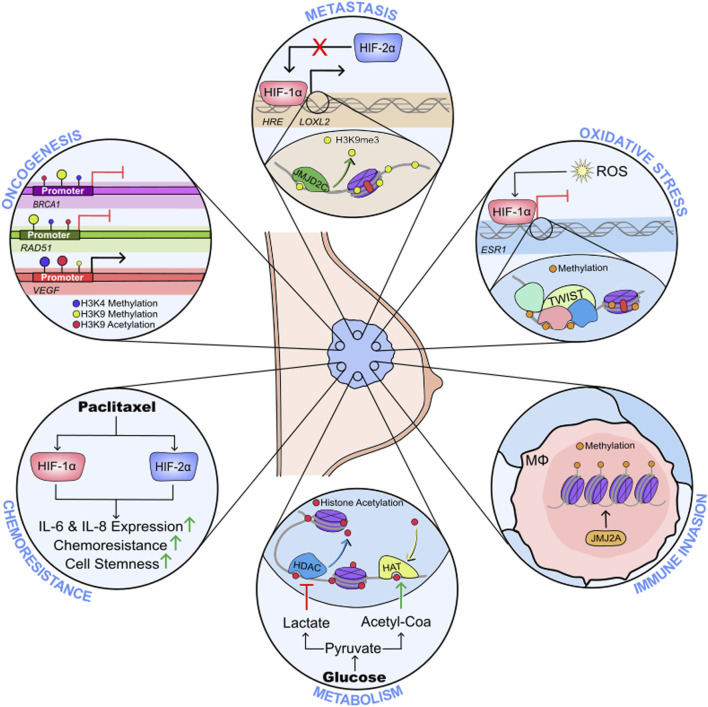
The events that control breast cancer progression and metastasis are complex and intertwined. Hypoxia plays a key role both in oncogenic transformation and in fueling the metastatic potential of breast cancer cells. Here we review the impact of hypoxia on epigenetic regulation of breast cancer, by interfering with multiple aspects of the tumour microenvironment. The co-dependent relationship between oxygen depletion and metabolic shift to aerobic glycolysis impacts on a range of enzymes and metabolites available in the cell, promoting posttranslational modifications of histones and chromatin, and changing the gene expression landscape to facilitate tumour development. Hormone signalling, particularly through ERα, is also tightly regulated by hypoxic exposure, with HIF-1α expression being a prognostic marker for therapeutic resistance in ER+ breast cancers. This highlights the strong need to understand the hypoxia-endocrine signalling axis and exploit it as a therapeutic target. Furthermore, hypoxia has been shown to enhance metastasis in TNBC cells, as well as promoting resistance to taxanes, radiotherapy and even immunotherapy through microRNA regulation and changes in histone packaging. Finally, several other mediators of the hypoxic response are discussed. We highlight a link between ionic dysregulation and hypoxia signalling, indicating a potential connection between HIF-1α and tumoural Na+ accumulation which would be worth further exploration; we present the role of Ca2+ in mediating hypoxic adaptation via chromatin remodelling, transcription factor recruitment and changes in signalling pathways; and we briefly summarise some of the findings regarding vesicle secretion and paracrine induced epigenetic reprogramming upon hypoxic exposure in breast cancer. By summarising these observations, this article highlights the heterogeneity of breast cancers, presenting a series of pathways with potential for therapeutic applications.

Keywords: breast cancer; epigenetics; hypoxia; microenvironment; oestrogen receptor; triple negative breast cancer.


Conflict of interest statement

The authors declare that the research was conducted in the absence of any commercial or financial relationships that could be construed as a potential conflict of interest.

Figures

None
Hypoxia-mediated epigenetic regulation of breast cancer progression.
FIGURE 1
FIGURE 1
Metabolism and epigenetic regulation of breast cancer progression under hypoxia. 1) Glucose availability dictates ERα-dependent metabolic regulation, as such that high glucose promotes an increase in glycolysis, while low glucose induces preferential use of oxidative metabolism (O’Mahony et al., 2012); 2) increased aerobic glycolysis causes intracellular buildup of lactate which inhibits histone deacetylases promoting transcription of oncogenes including c-Myc and SRSF10 (Latham et al., 2012; San-Millán et al., 2019; Martinez-Outschoorn et al., 2011; Pandkar et al., 2023); 3) acetyl CoA promotes histone acetylation and lipogenesis, with the second being a key promoter of malignant transformation (Messier et al., 2016; Menendez and Lupu, 2007); 4) mutations in the TCA enzyme isocitrate dehydrogenase have been associated with depletion of a-ketoglutarate and increased production of 2-hydroxyglutarate (2-HG), which inhibits histone demethylation, while promoting HIF-1α activation (Liu et al., 2023; Lu et al., 2012; Turcan et al., 2012); 5) metabolic receptors not involved in energy metabolism, such as the aryl hydrocarbon receptor (AhR) have been shown to dictate epigenetic reprogramming in breast cancer: AhR has been linked to gene regulation of oncogenes (e.g., BRCA1) through hypermethylation of CpG islands, increased three-methylation and deacetylation of H3K9, increased levels of DNA methyltransferases 1, 3a and 3b, as well as higher levels of methyl binding protein 2 (Thakur et al., 2022; Hockings et al., 2006; Papoutsis et al., 2012).
FIGURE 2
FIGURE 2
Structure of ERs, their cellular locations and activities. Nuclear receptors ERα and ERβ are compartmentalised into distinct functional regions. The N-terminal domain (NTD) houses activation function (AF) domain 1. The DNA binding domain (DBD) interacts with oestrogen response elements (EREs) proximal to target genes. The hinge region (D) contains a nuclear localisation signal and acts as a flexible region between the DBD and ligand binding domain (LBD). The LBD contains a hormone binding pocket where 17β-Estradiol (E2) can associate with the hormone receptors. The LBD is therefore important for the functional activity of the ERs in response to hormone stimulation. The F region is responsible for regulating gene transcription in a ligand-specific manner. ERα and ERβ reside in the cytosol awaiting stimulation by their steroid ligand E2, which induces a conformational change and subsequent translocation of the receptors to the nucleus where they are able to enact their transcriptional activities on target genes, predominantly through receptor binding to EREs (e.g. ERα binds to an ERE upstream of target gene GREB1; ERβ binds to an ERE upstream of target gene TFF1) (Klein-Hitpass et al., 1989; Wärnmark et al., 2003; Schwabe et al., 1993; Kumar et al., 2011). Conversely the GPER is expressed on the plasma membrane and intracellular membranes such as the endoplasmic reticulum, and contains seven transmembrane domains. Like ERα and ERβ, the GPER has high affinity for E2, and its role is to mediate the non-genomic effects of E2 within the cell, including mediating the activation of HIF-1α, and proteins involved in intracellular signalling cascades (Filardo et al., 2000; Filardo et al., 2002; Thomas et al., 2005; De Francesco et al., 2014; Vo et al., 2019).
FIGURE 3
FIGURE 3
Mechanisms of therapy resistance in hypoxic breast cancer cells. In a hypoxic breast cancer cell 1) ESR1 mRNA is significantly reduced in a HIF-1α-dependent manner (Ryu et al., 2011). 2) Hypoxia also causes proteasomal degradation of ERα and ERβ. For ERα, the proteasomal degradation is dependent on a physical interaction between the steroid receptor and HIF-1α. 3) Attenuation of HIF-1α via siRNA inhibits ERα knockdown, and enhances ERβ accumulation, suggesting HIFs are important in regulating the ERα/ERβ ratio (Wolff et al., 2017). 4) Alongside decreased ERα abundance, the transcriptional activity of the steroid receptor is impaired following prolonged culture in hypoxia (Whitman et al., 2019). 5) Reactive oxygen species (ROS) accumulate in hypoxia, which further enhances HIFα stability. ROS impedes ERα activity by preventing dimerization and DNA binding (Whittal et al., 2000; Atsriku et al., 2005; Wang et al., 2007; Kondoh et al., 2013). 6) Furthermore, ROS increases the expression of DNMT1 and MDB4 which may silence ESR1 expression through their methylating activity. This ESR1 silencing can be inhibited by 5-Aza-dC (Mahalingaiah et al., 2015). 7) Further epigenetic modifications silence ESR1; TWIST recruits DNMT3B and HDAC1 to induce hypermethylation of ESR1 promoter and enhance a condensed chromatin structure (Vesuna et al., 2012). 8) Stimulation of ERα36 and GPER by E2 elicits a signalling cascade which ultimately induces NRF2 levels and activity, which further influences redox balance by inducing the glutathione signalling pathway (Ishii and Warabi, 2019). 9) FOXA1 is significantly amplified in endocrine resistant breast cancers, which can drive HIF-2α expression and enhance metastasis (Britschgi et al., 2012; Singh et al., 2013; Fu et al., 2016; Fu et al., 2019). All together, hypoxia and HIFα activity in ERα+ disease cause decreased sensitivity and effectiveness of anti-oestrogens such as Tamoxifen and Fulvestrant, which further drives disease progression, metastasis and correlates to poor disease outcome.

References

    1. Abou Khouzam R., Zaarour R. F., Brodaczewska K., Azakir B., Venkatesh G. H., Thiery J., et al. (2022). The effect of hypoxia and hypoxia-associated pathways in the regulation of antitumor response: friends or foes? Front. Immunol. 13, 828875. 10.3389/fimmu.2022.828875 - DOI - PMC - PubMed
    1. Afzal M. Z., Mercado R. R., Shirai K. (2018). Efficacy of metformin in combination with immune checkpoint inhibitors (anti-PD-1/anti-CTLA-4) in metastatic malignant melanoma. J. Immunother. Cancer 6, 64. 10.1186/s40425-018-0375-1 - DOI - PMC - PubMed
    1. Alam M. W., Persson C. U., Reinbothe S., Kazi J. U., Rönnstrand L., Wigerup C., et al. (2016). HIF2α contributes to antiestrogen resistance via positive bilateral crosstalk with EGFR in breast cancer cells. Oncotarget 7, 11238–11250. 10.18632/oncotarget.7167 - DOI - PMC - PubMed
    1. Angelin A., Gil-de-Gómez L., Dahiya S., Jiao J., Guo L., Levine M. H., et al. (2017). Foxp3 reprograms T cell metabolism to function in low-glucose, high-lactate environments. Cell Metab. 25, 1282–1293. e7. 10.1016/j.cmet.2016.12.018 - DOI - PMC - PubMed
    1. Arita K., Hashimoto H., Shimizu T., Nakashima K., Yamada M., Sato M. (2004). Structural basis for Ca(2+)-induced activation of human PAD4. Nat. Struct. Mol. Biol. 11, 777–783. 10.1038/nsmb799 - DOI - PubMed

LinkOut - more resources