

Comparison of Duplex Ultrasound and Digital Subtraction Angiography for Assessing Tibial Vessel Disease
- PMID: 39282489
- PMCID: PMC11401628
- DOI: 10.7759/cureus.69327
Comparison of Duplex Ultrasound and Digital Subtraction Angiography for Assessing Tibial Vessel Disease
Abstract
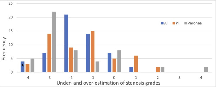
Background Duplex ultrasonography (DUS) is readily available and often used as the first diagnostic test for patients with peripheral artery diseases (PADs). PAD is a disease that affects the general population but more commonly affects diabetics. To date, the role of DUS in the assessment of tibial vessel disease is inconclusive at best. The goal of our study is to assess the validity of DUS in characterizing the presence and severity of tibial diseases via comparison with digital subtraction angiography (DSA) findings. Methods This is a single-center retrospective cohort study analyzing three arterial segments (anterior tibial, posterior tibial, and fibular arteries) in patients who received a duplex study followed by DSA within a 30-day period. All arterial segments were graded from normal (Grade 0) to occluded (Grade 4), based on duplex interpretation and directly compared to direct visualization findings from DSA. Using statistical methods, the sensitivity, specificity, positive predictive value (PPV), negative predictive value (NPV), and accuracy of DUS were determined. Results A total of 171 tibial vessel segments from 57 enrolled subjects with critical limb ischemia symptoms were analyzed in this study. The agreement between both modalities was poor (Kappa=0.19, p < 0.05), with DUS demonstrating a significant underestimation of vessel pathologies. This is also reflected by the overall sub-optimal sensitivity (23%), specificity (84%), PPV (69%), and NPV (41%) in DUS when compared to DSA results as the gold standard. Conclusion Significant disagreements were noted in this study between DUS and DSA findings, primarily significant underestimation of tibial vessel disease by the DUS when compared with the DSA. Caution is advised in the clinical application of DUS in patients with chronic limb-threatening ischemia (CLTI) symptoms and multi-segment tibial vessels due to its demonstrated limitations in this study.
Keywords: digital substraction angiography; duplex ultrasonography; pvd: peripheral vascular disease; tibial vessel disease; vascular surgery.
Copyright © 2024, Neris et al.
Conflict of interest statement
Human subjects: Consent was obtained or waived by all participants in this study. Animal subjects: All authors have confirmed that this study did not involve animal subjects or tissue. Conflicts of interest: In compliance with the ICMJE uniform disclosure form, all authors declare the following: Payment/services info: All authors have declared that no financial support was received from any organization for the submitted work. Financial relationships: All authors have declared that they have no financial relationships at present or within the previous three years with any organizations that might have an interest in the submitted work. Other relationships: All authors have declared that there are no other relationships or activities that could appear to have influenced the submitted work.
Figures
References
-
- Interpretation of arterial duplex testing of lower-extremity arteries and interventions. Hodgkiss-Harlow KD, Bandyk DF. Semin Vasc Surg. 2013;26:95–104. - PubMed
LinkOut - more resources
Full Text Sources