

Myxomatous cause of multiple intracranial aneurysms and cognitive decline: a case report
- PMID: 39283559
- PMCID: PMC11405687
- DOI: 10.1186/s43044-024-00559-2
Myxomatous cause of multiple intracranial aneurysms and cognitive decline: a case report
Abstract
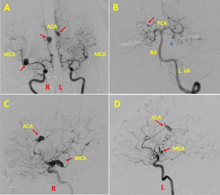
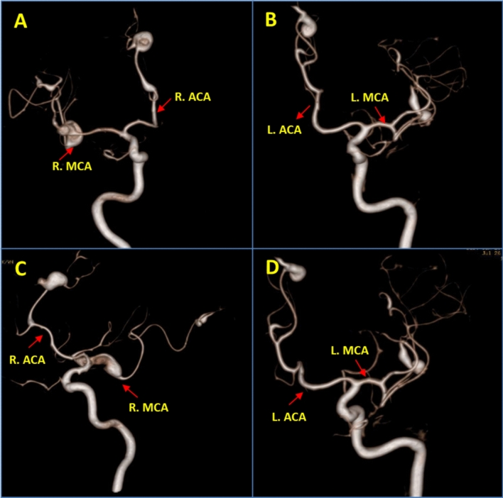
Background: The occurrence of cerebral aneurysm in a case of cardiac myxoma is rare with less than 60 cases reported worldwide. The course of management is still debatable given its rarity. We present a case of multiple intracranial aneurysms secondary to atrial myxoma in a young lady with a brief review of the literature. Case presentation A young lady in her late 30s with a history of right middle cerebral artery territory stroke eight years ago presented with gradually progressive symptoms in the form of holocranial headache, inattention and forgetfulness for the last few years. On neuroimaging, she was found to have multi-territorial lacunar infarcts and multiple intracranial artery aneurysms which was confirmed with a digital subtraction angiogram. A cardiac evaluation revealed a left atrial myxoma. The aetiology of subcortical cognitive decline and intracranial aneurysms was attributed to the myxoma with secondary myxomatous embolism. Other secondary causes were ruled out. She is being followed up medically after resection of the myxoma.
Conclusion: Intracranial aneurysms are rare complications of cardiac myxoma which may present before, concurrent or many years after diagnosis of the myxoma. Nonspecific neurological complaints occasionally are the ominous signs of intracranial aneurysms which mandate a low threshold for neuroimaging in a case of cardiac myxoma. Given the absence of definitive risk factors and unclear natural history, clinical and radiological follow-ups are critical. Learning Points Intracranial aneurysms are rare complications of cardiac myxoma that may present before, concurrent or many years after diagnosis of the myxoma. Special attention must be given to nonspecific neurological complaints with a low threshold for neuroimaging in those with a prior history of cardiac myxoma. Given the absence of definitive risk factors and unclear natural history, clinical and radiological follow-up including conventional angiography and/or magnetic resonance imaging is critical.
Keywords: Atrial myxoma; Intracranial aneurysms; Myxoma; Myxomatous.
© 2024. The Author(s).
Conflict of interest statement
None.
Figures

References
-
- Lenihan D, Yusuf S, Shah A (2019) Tumors affecting the cardiovascular system. In: Braunwald E, editor. Braunwald’s heart disease: a textbook of cardiovascular medicine. Eleventh edition, international edition. Philadelphia, PA: Elsevier; 1662–80.
-
- Iliyas M, Swaminathan N, Ganesan G, Sangareddi V (2020) Left atrial myxoma: a great masquerader with multiple clinical faces—a case series. J Indian Acad Echocardiogr Cardiovasc Imaging 4(1):42 10.4103/jiae.jiae_28_19 - DOI
LinkOut - more resources
Full Text Sources