. 2024 Dec 1;83(12):1060-1075.
doi: 10.1093/jnen/nlae098.

Contribution of major histocompatibility complex class II immunostaining in distinguishing idiopathic inflammatory myopathy subgroups: A histopathological cohort study

Affiliations

Contribution of major histocompatibility complex class II immunostaining in distinguishing idiopathic inflammatory myopathy subgroups: A histopathological cohort study

Lola E R Lessard et al. J Neuropathol Exp Neurol. .

Abstract

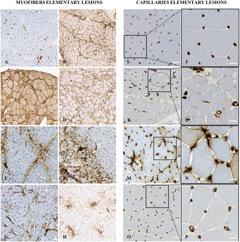
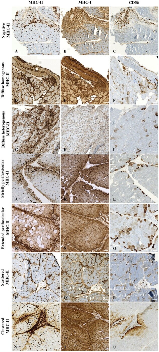
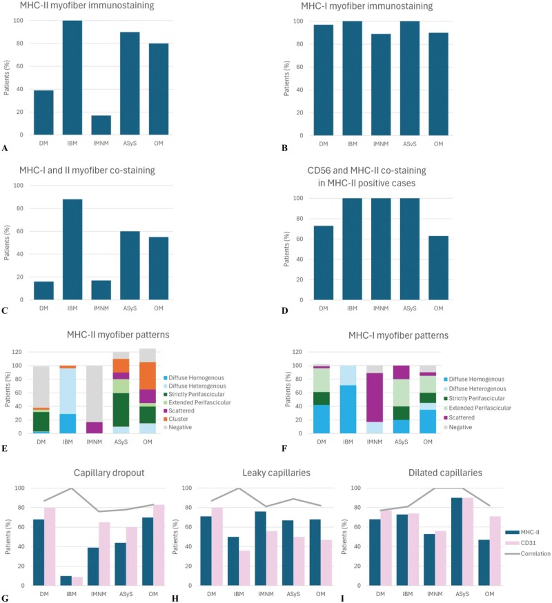
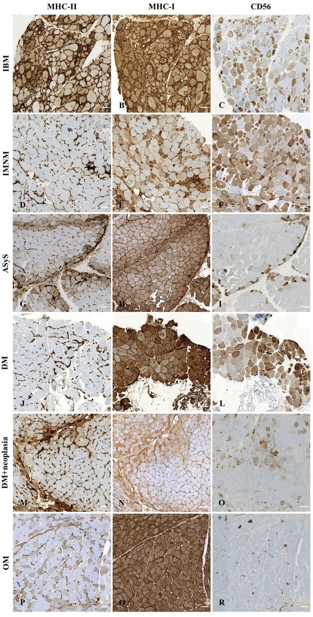
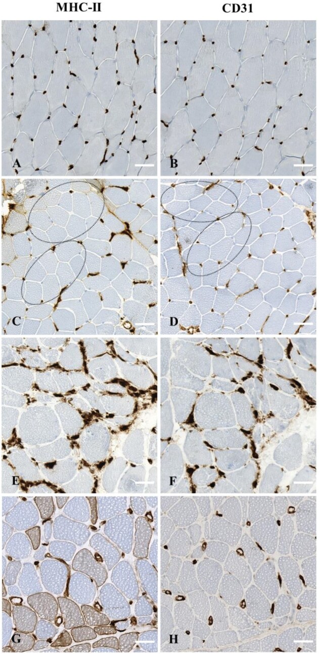
Idiopathic inflammatory myopathies (IIM) are rare, acquired muscle diseases; their diagnosis of is based on clinical, serological, and histological criteria. MHC-I-positive immunostaining, although non-specific, is used as a marker for IIM diagnosis; however, the significance of major histocompatibility complex (MHC)-II immunostaining in IIM remains debated. We investigated patterns of MHC-II immunostaining in myofibers and capillaries in muscle biopsies from 103 patients with dermatomyositis ([DM], n = 31), inclusion body myositis ([IBM], n = 24), anti-synthetase syndrome ([ASyS], n = 10), immune-mediated necrotizing myopathy ([IMNM], n = 18), or overlap myositis ([OM], n = 20). MHC-II immunostaining of myofibers was abnormal in 63/103 of patients (61%) but the patterns differed according to the IIM subgroup. They were diffuse in IBM (96%), negative in IMNM (83%), perifascicular in ASyS (70%), negative (61%) or perifascicular (32%) in DM, and either clustered (40%), perifascicular (30%), or diffuse heterogeneous (15%) in OM. Capillary MHC-II immunostaining also identified quantitative (capillary dropout, n = 47/88, 53%) and qualitative abnormalities, that is, architectural abnormalities, including dilated and leaky capillaries, (n = 79/98, 81%) in all IIM subgroups. Thus, MHC-II myofiber expression patterns allow distinguishing among IIM subgroups. We suggest the addition of MHC-II immunostaining to routine histological panels for IIM diagnosis.

Keywords: capillary; idiopathic inflammatory myopathies; major histocompatibility complex-II; muscle biopsy; myofiber.


Conflict of interest statement

Authors have no disclosures to declare related to the content of this article.

Figures

Figure 1.
Figure 1.
Elementary lesions of myofibers and capillaries observable upon major histocompatibility class II (MHC-II) immunostaining in patients with idiopathic inflammatory myopathies. (A) Normal expression in control muscle biopsy. (B) Diffuse homogenous myofiber positivity in a patient with inclusion body myositis. (C, D) Diffuse heterogeneous myofiber positivity in a patient with inclusion body myositis and overlap myositis, respectively. (E) Strictly perifascicular myofiber positivity in a patient with anti-synthetase syndrome. (F) Extended perifascicular myofiber positivity immunostaining in a patient with dermatomyositis. (G) Scattered myofiber positivity in a patient with immune mediated necrotizing myopathy. (H) Clustered myofiber positivity in a patient with overlap myositis. (I, J) Normal MHC-II expression on capillaries at low magnification (I) and inset of high magnification (J) in a control patient. (K, L) Capillary dropout (dotted ellipse) at low magnification (K) and inset of high magnification (L) in a patient with juvenile dermatomyositis. M-P: Leaky capillaries in a patient with overlap myositis (M, N). Dilated capillaries in a patient with immune mediated necrotizing myopathy (O, P). Low magnifications (M, O) and insets high magnifications (N, P) observable upon MHC-II immunostaining. Scale bars: A-H = 100 μm, I, K, M, O = 50 μm; J, L, N, P = 10 μm.
Figure 2.
Figure 2.
Illustrative images of the different patterns of major histocompatibility complex (MHC) class II, MHC-I, and CD56 myofiber immunostaining on serial sections of muscle biopsies in idiopathic inflammatory myopathies. Left panel shows MHC-II immunostaining, middle panel shows MHCI-I immunostaining, and right panel shows CD56 immunostaining on serial sections of 7 IIM patients. (A-C) Negative MHC-II myofiber immunostaining (A) and corresponding diffuse MHC-I (B) and CD56 (C) immunostaining in a patient with juvenile dermatomyositis. (D-F) Diffuse homogenous MHC-II myofiber positivity (D) and corresponding diffuse MHC-I (E) and CD56 (F) immunostaining in a patient with inclusion body myopathy. (G-I) Diffuse heterogeneous MHC-II myofiber positivity (G) and corresponding diffuse MHC-I (H) and CD56 (I) immunostaining in a patient with inclusion body myopathy. (J-L) Strictly perifascicular MHC-II myofiber positivity (J) and corresponding diffuse MHC-I (K) and CD56 (L) immunostaining in a patient with anti-synthetase syndrome. (M-O) Extended perifascicular MHC-II myofiber positivity (M) and corresponding diffuse MHC-I (N) and CD56 (O) immunostaining in a patient with anti-synthetase syndrome. (P-R) Scattered MHC-II myofiber positivity (P) and corresponding diffuse MHC-I (Q) and CD56 (R) immunostaining in a patient with overlap myositis. (S-U) Clustered MHC-II myofiber positivity (S) and corresponding diffuse MHC-I (T) and CD56 (U) immunostaining in a patient with juvenile dermatomyositis. Scale bars = 100 μm.
Figure 3.
Figure 3.
Graphical representation of myofiber and capillary immunostaining results according to idiopathic inflammatory myopathy subgroup. (A) Major histocompatibility complex (MHC) class II. (B) MHC class I myofiber immunostaining in IIM patients according to subgroup. (C) MHC-I and II patterns of expression of myofibers were compared and classified either as “co-staining” (similar pattern of expression) or “no- co-staining” (different patterns). (D) CD56 and MHC-II patterns of expression of myofibers were compared and classified either as “co-staining” (CD56-positive myofibers were also MHC-II-positive and/or vice versa) or “no- co-staining” (myofibers expressed either CD56 or MHC-II but not the two markers). (E) MHC class II and (F) MHC class I patterns of expression by myofibers in IIM patients according to subgroup. (G) Capillary dropout, (H) leaky, and (I) dilated capillaries assessed by MHC-II and CD31 immunostaining. Correlation was retained when both techniques showed the same pattern of capillary morphological abnormality.
Figure 4.
Figure 4.
Illustrative images of major histocompatibility complex (MHC) class II, MHC-I, and CD56 myofiber immunostaining in each idiopathic inflammatory myopathy subgroup. Left panel shows MHC-II immunostaining, middle panel shows MHCI-I immunostaining, and right panel shows CD56 immunostaining on serial sections of IIM patients. (A-C) IBM: diffuse homogeneous MHC-II (A), diffuse MHC-I (B), and diffuse CD56 positivity (C). (D-F) IMNM: negative MHC-II (D), heterogeneous MHC-I (E), and diffuse CD56 positivity (F). (G-I) ASyS: perifascicular MHC-II (G), extended perifascicular MHC-I (H), and perifascicular CD56 positivity (I). (I-K) DM: negative MHC-II expression (J), diffuse MHC-I (K), and scattered CD56 positivity (L). (M-O) DM (patient with cancer-associated DM): perifascicular MHC-II (M), extended perifascicular MHC-I (N), and scattered CD56 positivity (O). (P-R) OM: heterogenous MHC-II (P), diffuse MHC-I (Q), and scattered CD56 positivity (R). Scale bars = 100 μm.
Figure 5.
Figure 5.
Illustrative images of major histocompatibility complex (MHC) class II and CD31 capillary immunostaining patterns on serial sections of muscle biopsies from controls and patients with idiopathic inflammatory myopathy. Normal MHC-II (A) and corresponding normal CD31 (B) immunostaining in a control patient. Capillary dropout observable upon MHC-II (C) and corresponding CD31 (D) immunostaining (dotted ellipses) in a patient with juvenile dermatomyositis. Leaky capillaries observable upon MHC-II (E) and corresponding CD31 (F) immunostaining in a patient with anti-synthetase syndrome. Dilated capillaries observable upon MHC-II (G) and corresponding CD31 (H) immunostaining in a patient with overlap myositis. Scale bars: 100 μm.

References

    1. Meyer A, Meyer N, Schaeffer M, et al. Incidence and prevalence of inflammatory myopathies: a systematic review. Rheumatology (Oxford). 2015;54:50-63. - PubMed
    1. Lloyd TE, Mammen AL, Amato AA, et al. Evaluation and construction of diagnostic criteria for inclusion body myositis. Neurology. 2014;83:426-433. 29 - PMC - PubMed
    1. Allenbach Y, Mammen AL, Benveniste O, et al. 224th ENMC International Workshop: clinico-sero-pathological classification of immune-mediated necrotizing myopathies Zandvoort, The Netherlands, 14-16 October 2016. Neuromuscul Disord. 2018;28:87-99. - PubMed
    1. Mammen AL, Allenbach Y, Stenzel W, et al. ENMC 239th Workshop Study Group. 239th ENMC International Workshop: classification of dermatomyositis, Amsterdam, the Netherlands, 14-16 December 2018. Neuromuscul Disord. 2020;30:70-92. - PubMed
    1. Zanframundo G, Faghihi-Kashani S, Scirè CA, et al. Defining anti-synthetase syndrome: a systematic literature review. Clin Exp Rheumatol. 2022;40:309-319. - PMC - PubMed

Substances