. 2024 Sep;10(5):e70001.
doi: 10.1002/2056-4538.70001.

The expression of YAP1 and other transcription factors contributes to lineage plasticity in combined small cell lung carcinoma

Affiliations

The expression of YAP1 and other transcription factors contributes to lineage plasticity in combined small cell lung carcinoma

Naoe Jimbo et al. J Pathol Clin Res. 2024 Sep.

Abstract

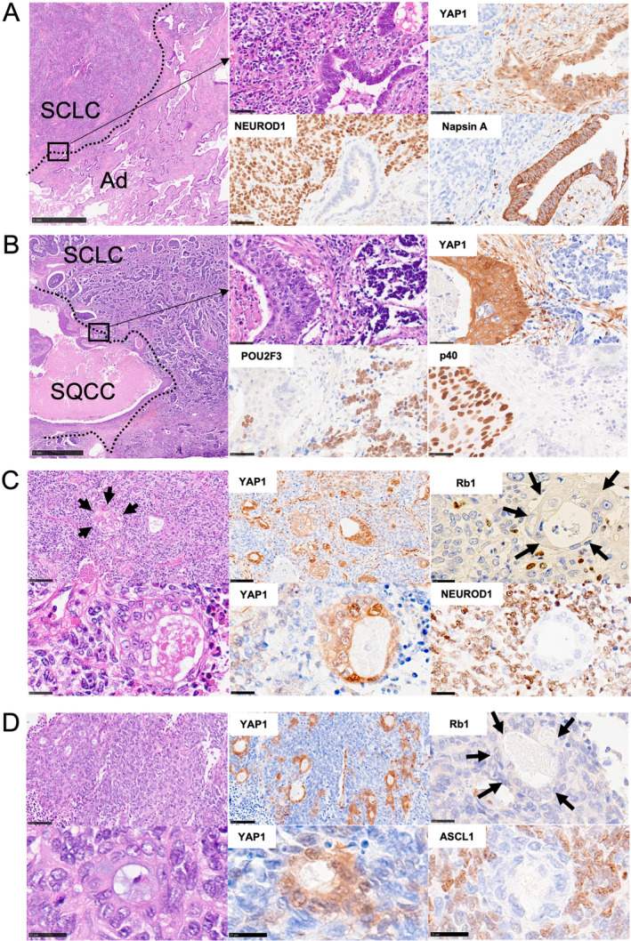
Lineage plasticity in small cell lung carcinoma (SCLC) causes therapeutic difficulties. This study aimed to investigate the pathological findings of plasticity in SCLC, focusing on combined SCLC, and elucidate the involvement of YAP1 and other transcription factors. We analysed 100 surgically resected SCLCs through detailed morphological observations and immunohistochemistry for YAP1 and other transcription factors. Component-by-component next-generation sequencing (n = 15 pairs) and immunohistochemistry (n = 35 pairs) were performed on the combined SCLCs. Compared with pure SCLCs (n = 65), combined SCLCs (n = 35) showed a significantly larger size, higher expression of NEUROD1, and higher frequency of double-positive transcription factors (p = 0.0009, 0.04, and 0.019, respectively). Notably, 34% of the combined SCLCs showed morphological mosaic patterns with unclear boundaries between the SCLC and its partner. Combined SCLCs not only had unique histotypes as partners but also represented different lineage plasticity within the partner. NEUROD1-dominant combined SCLCs had a significantly higher proportion of adenocarcinomas as partners, whereas POU2F3-dominant combined SCLCs had a significantly higher proportion of squamous cell carcinomas as partners (p = 0.006 and p = 0.0006, respectively). YAP1 expression in SCLC components was found in 80% of combined SCLCs and 62% of pure SCLCs, often showing mosaic-like expression. Among the combined SCLCs with component-specific analysis, the identical TP53 mutation was found in 10 pairs, and the identical Rb1 abnormality was found in 2 pairs. On immunohistochemistry, the same abnormal p53 pattern was found in 34 pairs, and Rb1 loss was found in 24 pairs. In conclusion, combined SCLC shows a variety of pathological plasticity. Although combined SCLC is more plastic than pure SCLC, pure SCLC is also a phenotypically plastic tumour. The morphological mosaic pattern and YAP1 mosaic-like expression may represent ongoing lineage plasticity. This study also identified the relationship between transcription factors and partners in combined SCLC. Transcription factors may be involved in differentiating specific cell lineages beyond just 'neuroendocrine'.

Keywords: YAP1; combined small cell lung carcinoma; molecular marker; mosaic; plasticity; small cell carcinoma; small cell lung carcinoma; transcription factors.


Figures

Figure 1
Figure 1
Comparison of clinicopathological findings between combined and pure small cell lung carcinomas. (A) Among 100 cases of small cell lung carcinomas (SCLCs), 35 cases were morphologically classified as combined and 65 as pure SCLCs. (B) Compared with pure SCLCs, combined SCLCs showed a significantly larger size (p = 0.0009). (C) Immunohistochemically, combined SCLCs had higher expression of NEUROD1 compared with pure SCLCs (p = 0.04), with no significant differences in other protein expression or composition of dominant transcription subtypes. (D) Double‐positive transcription factors expression was significantly more common in combined SCLCs, and single marker positivity was more common in pure SCLCs (p = 0.019). (E) Distribution of YAP1 expression (H‐score) of SCLC components in combined SCLCs and pure SCLCs. YAP1 expression (H‐score > 1) was found in 80% of combined SCLCs and 62% of pure SCLCs.
Figure 2
Figure 2
Results of combined small cell lung carcinomas. (A) Summary of results in 35 cases of combined small cell lung carcinoma (SCLC), including dominant transcription subtype, morphological patterns, histopathology details, immunohistochemistry for p53 and Rb1, and genomic analysis. (B) The proportion of SCLC in the total tumour was higher in the order of mosaic, mixture of mosaic and separated, and separated types. A significant difference in the proportion of SCLC was observed between the first two types and the separated type (p = 0.019). (C) The proportion of partner histotype in the total tumour depends on the dominant transcription subtype. As partners, NEUROD1‐dominant combined SCLCs had a significantly higher proportion of adenocarcinoma (p = 0.006), and POU2F3‐dominant combined SCLCs had a significantly higher proportion of squamous cell carcinoma (p = 0.0006). The red lines indicate the median values.
Figure 3
Figure 3
Representative pathological findings in combined small cell lung carcinomas. (A, B) In the separated morphological type, small cell lung carcinoma (SCLC) and each partner were distributed with a clear boundary. At high magnification, they were accompanied by intervening stromal components, which could also be seen in the YAP1 immunostain. (C, D) The boundaries between the SCLC and each partner were indistinct in the mosaic morphological types, and the partners were scattered and mosaic‐like. Foci of squamous cell carcinoma are seen in C (arrows). The distribution of the mosaic‐like pattern was clearly visible in the YAP1 immunostain. High magnification shows direct contact between the SCLC and the partner, with little or no intervening interstitial components, which was also observed with YAP1. In the adenocarcinoma components of combined SCLC showing mosaic morphological types, Rb1 was lost, as in SCLC components (arrows in C and D).
Figure 4
Figure 4
Results of pure small cell lung carcinomas with a focus on YAP1 expression. YAP1 expression pattern in ASCL1‐dominant pure small cell lung carcinomas (SCLCs). (A) YAP1 is completely negative for SCLC and diffusely positive for ASCL1. (B, C) YAP1 is partial and mosaic‐like positive for SCLC, and ASCL1 was almost complementary to YAP1 expression. (D) ASCL1 and YAP1 are diffusely positive.
Figure 5
Figure 5
Unique partners in combined small cell lung carcinomas. (A) In the upper case, apart from conventional squamous cell carcinoma (SQCC), a mixed adenocarcinoma (Ad) and SQCC are observed (*), showing a biphasic pattern with p40‐positive SQCC in the outer layer and adenocarcinoma in the inner layer. This resembles a tumour recognised as a mucoepidermoid carcinoma‐like adenosquamous carcinoma. (B) The second case contains a non‐small cell carcinoma component that could not be morphologically and phenotypically determined as adenocarcinoma or SQCC and showed mixed expression of TTF‐1 (red) and p40 (blue) by double immunohistochemistry. Black‐coloured arrows indicate double‐positive cells. (C) The third case had a rhabdomyosarcoma component that was positive for myogenin. Yellow‐coloured arrows indicate rhabdoid cells.

Similar articles

Cited by

  • Basal cell of origin resolves neuroendocrine-tuft lineage plasticity in cancer.
    Ireland AS, Hawgood SB, Xie DA, Barbier MW, Lucas-Randolph S, Tyson DR, Zuo LY, Witt BL, Govindan R, Dowlati A, Moser JC, Puri S, Rudin CM, Chan JM, Elliott A, Oliver TG. Ireland AS, et al. bioRxiv [Preprint]. 2024 Nov 15:2024.11.13.623500. doi: 10.1101/2024.11.13.623500. bioRxiv. 2024. PMID: 39605338 Free PMC article. Preprint.
  • KLF4 promotes a KRT13+ hillock-like state in squamous lung cancer.
    Izzo LT, Reyes T, Meesala S, Ireland AS, Yang S, Sunil HS, Cheng XC, Tserentsoodol N, Hawgood SB, Patz EF Jr, Witt BL, Tyson DR, O'Donnell KA, Oliver TG. Izzo LT, et al. bioRxiv [Preprint]. 2025 Mar 13:2025.03.10.641898. doi: 10.1101/2025.03.10.641898. bioRxiv. 2025. PMID: 40161723 Free PMC article. Preprint.

References

    1. George J, Lim JS, Jang SJ, et al. Comprehensive genomic profiles of small cell lung cancer. Nature 2015; 524: 47–53. - PMC - PubMed
    1. Rudin CM, Poirier JT, Byers LA, et al. Molecular subtypes of small cell lung cancer: a synthesis of human and mouse model data. Nat Rev Cancer 2019; 19: 289–297. - PMC - PubMed
    1. Lantuejoul S, Fernandez‐Cuesta L, Damiola F, et al. New molecular classification of large cell neuroendocrine carcinoma and small cell lung carcinoma with potential therapeutic impacts. Transl Lung Cancer Res 2020; 9: 2233–2244. - PMC - PubMed
    1. Watanabe K, Kage H, Shinozaki‐Ushiku A, et al. Spontaneous transdifferentiation from small cell lung carcinoma to squamous cell carcinoma. J Thorac Oncol 2019; 14: e31–e34. - PubMed
    1. Quintanal‐Villalonga A, Chan JM, Yu HA, et al. Lineage plasticity in cancer: a shared pathway of therapeutic resistance. Nat Rev Clin Oncol 2020; 17: 360–371. - PMC - PubMed

MeSH terms

Substances