

Brain tumor segmentation by combining MultiEncoder UNet with wavelet fusion
- PMID: 39284311
- PMCID: PMC11540057
- DOI: 10.1002/acm2.14527
Brain tumor segmentation by combining MultiEncoder UNet with wavelet fusion
Abstract
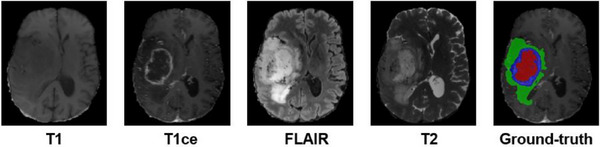
Background and objective: Accurate segmentation of brain tumors from multimodal magnetic resonance imaging (MRI) holds significant importance in clinical diagnosis and surgical intervention, while current deep learning methods cope with situations of multimodal MRI by an early fusion strategy that implicitly assumes that the modal relationships are linear, which tends to ignore the complementary information between modalities, negatively impacting the model's performance. Meanwhile, long-range relationships between voxels cannot be captured due to the localized character of the convolution procedure.
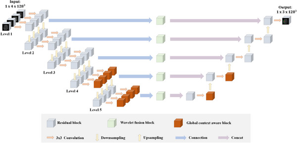
Method: Aiming at this problem, we propose a multimodal segmentation network based on a late fusion strategy that employs multiple encoders and a decoder for the segmentation of brain tumors. Each encoder is specialized for processing distinct modalities. Notably, our framework includes a feature fusion module based on a 3D discrete wavelet transform aimed at extracting complementary features among the encoders. Additionally, a 3D global context-aware module was introduced to capture the long-range dependencies of tumor voxels at a high level of features. The decoder combines fused and global features to enhance the network's segmentation performance.
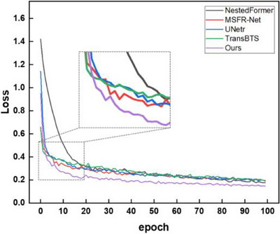
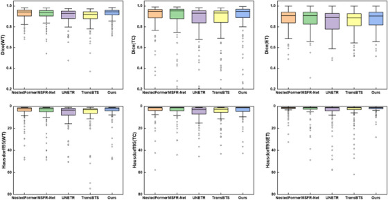
Result: Our proposed model is experimented on the publicly available BraTS2018 and BraTS2021 datasets. The experimental results show competitiveness with state-of-the-art methods.
Conclusion: The results demonstrate that our approach applies a novel concept for multimodal fusion within deep neural networks and delivers more accurate and promising brain tumor segmentation, with the potential to assist physicians in diagnosis.
Keywords: 3D discrete wavelet transformer; brain tumor segmentation; multi‐encoder.
© 2024 The Author(s). Journal of Applied Clinical Medical Physics published by Wiley Periodicals LLC on behalf of American Association of Physicists in Medicine.
Conflict of interest statement
The authors declare no conflicts of interest.
Figures


Similar articles
-
[Fully Automatic Glioma Segmentation Algorithm of Magnetic Resonance Imaging Based on 3D-UNet With More Global Contextual Feature Extraction: An Improvement on Insufficient Extraction of Global Features].Sichuan Da Xue Xue Bao Yi Xue Ban. 2024 Mar 20;55(2):447-454. doi: 10.12182/20240360208. Sichuan Da Xue Xue Bao Yi Xue Ban. 2024. PMID: 38645864 Free PMC article. Chinese.
-
A 3D hierarchical cross-modality interaction network using transformers and convolutions for brain glioma segmentation in MR images.Med Phys. 2024 Nov;51(11):8371-8389. doi: 10.1002/mp.17354. Epub 2024 Aug 13. Med Phys. 2024. PMID: 39137295
-
Joint learning-based feature reconstruction and enhanced network for incomplete multi-modal brain tumor segmentation.Comput Biol Med. 2023 Sep;163:107234. doi: 10.1016/j.compbiomed.2023.107234. Epub 2023 Jul 4. Comput Biol Med. 2023. PMID: 37450967
-
Sparse Dynamic Volume TransUNet with multi-level edge fusion for brain tumor segmentation.Comput Biol Med. 2024 Apr;172:108284. doi: 10.1016/j.compbiomed.2024.108284. Epub 2024 Mar 15. Comput Biol Med. 2024. PMID: 38503086 Review.
-
Efficient brain tumor segmentation using Swin transformer and enhanced local self-attention.Int J Comput Assist Radiol Surg. 2024 Feb;19(2):273-281. doi: 10.1007/s11548-023-03024-8. Epub 2023 Oct 5. Int J Comput Assist Radiol Surg. 2024. PMID: 37796413 Review.
Cited by
-
Multi-level channel-spatial attention and light-weight scale-fusion network (MCSLF-Net): multi-level channel-spatial attention and light-weight scale-fusion transformer for 3D brain tumor segmentation.Quant Imaging Med Surg. 2025 Jul 1;15(7):6301-6325. doi: 10.21037/qims-2025-354. Epub 2025 Jun 30. Quant Imaging Med Surg. 2025. PMID: 40727355 Free PMC article.
References
-
- Pereira S, Pinto A, Alves V, Silva CA. Brain tumor segmentation using convolutional neural networks in MRI images. IEEE Trans Med Imaging. 2016;35(5):1240‐1251. - PubMed
-
- Choi SG, Sohn CB. Detection of HGG and LGG brain tumors using U‐Net. Medico‐legal Update. 2019;19(1):560‐565.
-
- Shukla G, Alexander GS, Bakas S, et al. Advanced magnetic resonance imaging in glioblastoma: a review. Chin Clin Oncol. 2017;6(4):40. - PubMed
-
- Bakas S, Reyes M, Jakab A, et al. Identifying the best machine learning algorithms for brain tumor segmentation, progression assessment, and overall survival prediction in the BRATS challenge. [published online ahead of print April 23, 2019]. Computer Vision and Pattern Recognition. doi:10.48550/arXiv.1811.02629 - DOI
-
- Yue W, Wang Z, Tian B, Pook M, Liu X. A hybrid model‐and memory‐based collaborative filtering algorithm for baseline data prediction of Friedreich's ataxia patients. IEEE Trans Ind Inf. 2020;17(2):1428‐1437.
MeSH terms
Grants and funding
- 62204168/National Natural Science Foundation of China
- 20YDTPJC00160/Tianjin Municipal Science and Technology Program
- 21YDTPJC00780/Tianjin Municipal Science and Technology Program
- 2019KJ101/Science Research Program of Tianjin Education Committee
- 2022KYZ136/Tianjin Research Innovation Project for Postgraduate Students
LinkOut - more resources
Full Text Sources
Medical
