. 2024 Nov 20;34(11):1954-1965.
doi: 10.1101/gr.279396.124.

Measuring X-Chromosome inactivation skew for X-linked diseases with adaptive nanopore sequencing

Affiliations

Measuring X-Chromosome inactivation skew for X-linked diseases with adaptive nanopore sequencing

Sena A Gocuk et al. Genome Res. .

Abstract

X-linked genetic disorders typically affect females less severely than males owing to the presence of a second X Chromosome not carrying the deleterious variant. However, the phenotypic expression in females is highly variable, which may be explained by an allelic skew in X-Chromosome inactivation. Accurate measurement of X inactivation skew is crucial to understand and predict disease phenotype in carrier females, with prediction especially relevant for degenerative conditions. We propose a novel approach using nanopore sequencing to quantify skewed X inactivation accurately. By phasing sequence variants and methylation patterns, this single assay reveals the disease variant, X inactivation skew, and its directionality and is applicable to all patients and X-linked variants. Enrichment of X Chromosome reads through adaptive sampling enhances cost-efficiency. Our study includes a cohort of 16 X-linked variant carrier females affected by two X-linked inherited retinal diseases: choroideremia and RPGR-associated retinitis pigmentosa. As retinal DNA cannot be readily obtained, we instead determine the skew from peripheral samples (blood, saliva, and buccal mucosa) and correlate it to phenotypic outcomes. This reveals a strong correlation between X inactivation skew and disease presentation, confirming the value in performing this assay and its potential as a way to prioritize patients for early intervention, such as gene therapy currently in clinical trials for these conditions. Our method of assessing skewed X inactivation is applicable to all long-read genomic data sets, providing insights into disease risk and severity and aiding in the development of individualized strategies for X-linked variant carrier females.


Figures

Figure 1.
Figure 1.
X inactivation skew sequencing workflow. (A) Adaptive nanopore sequencing of peripheral samples (blood, saliva, and buccal mucosa) to maximize coverage of the X Chromosome. Sequencing of fragments that do not map to the X Chromosome is terminated early. Degraded buccal DNA results in shorter read lengths (B) and reduced X Chromosome enrichment performance (C). The read lengths for (accepted) reads mapping to the X Chromosome are plotted for patient C9 for illustrative purposes. Rejected reads are typically <1 kb. (D) X Chromosome coverage for each patient, split by sample. At least 30× combined X Chromosome coverage was obtained for each patient. (H1) Healthy individual, (C1–C12) female carriers of choroideremia, and (R1–R4) female carriers of RPGR-associated X-linked retinitis pigmentosa.
Figure 2.
Figure 2.
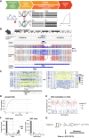
Bioinformatic workflow to measure X inactivation skew with long-read genomics, demonstrated on ground-truth data. (A) Five-step bioinformatic pipeline: basecalling and mapping reads including modified bases, SNV calling, phasing alleles and reads based on SNVs, clustering reads based on their methylation profile, and tabulating the allele/epiallele combinations to calculate the skew. (B) Validation on mouse data with genetically engineered skewed X inactivation: A deletion of the Xist A-repeat (haplotype 2) triggers its constitutive choice as the Xi, as confirmed by the DNA methylation profile of the Xist promoter (red indicates methylated CpG; blue, unmethylated CpG). (C) Example of read clustering by methylation profile for the Phf6 CpG island: reads split into two clusters, one with low methylation (CpG sites with low probability of methylation in yellow; cluster assigned as Xa) and one with high methylation (CpG sites with high probability of being methylated in blue; cluster assigned as Xi). (D) Cumulative sum of CpG islands overlapped by haplotype blocks. Large haplotype blocks overlap many CpG islands, providing additional power to estimate the X inactivation skew in those haplotype blocks. (E) CpG island methylation clustering results for one haplotype block. Each point represents a read. CpG islands with gray reads failed to split into Xi/Xa groups. (F) Nonfolded and folded histograms of the haplotype block-wise skews, binned in 10% bins and scaled by the underlying number of reads. Skew is measured as the proportion of reads supporting haplotype 1 as Xa. Because skewed X inactivation is genetically engineered in this experiment, haplotype blocks are expected to yield skews close to either zero or one. In the case of perfectly balanced X inactivation, haplotype blocks should return skews of 0.5. Folding is done around the x = 0.5 axis, to account for the random assignment of haplotype 1/2 labels to the two alleles in each block. Folding would not be necessary if we could assign haplotype 1 to a single parental haplotype consistently. (G) From the block skews, the sample skew P is calculated by maximizing the joint probability of observing the data. q = 1 − P; for each haplotype block, k is the number of haplotype 1 reads that are Xa and haplotype 2 reads that are Xi (“successes”), and n is the total number of haplotyped and clustered reads (“trials”).
Figure 3.
Figure 3.
X inactivation skew across blood, saliva, buccal swab, and retina in a healthy patient. (A) Collected samples: four quadrants of the retina from one eye (purple discs), blood, saliva, and buccal swab. (B) Cumulative sum of CpG islands overlapped by haplotype blocks. (C) Nonfolded and folded distributions of haplotype blocks’ skews in the seven samples. Blood and saliva show a clear skew, whereas the buccal swab and all four retina samples are not highly skewed. (D) Positive correlation of the nonfolded haplotype blocks’ skews across samples shows that the Xa allelic bias is consistent across tissues (the same X Chromosome in the preferentially active X in all skewed tissues). (E) Skewed X inactivation allows scaffolding haplotype blocks into alleles. Log-likelihood ratio of haplotype 1 being the preferential Xa over Xi for each block. A red line is plotted at y = 1 (haplotype 1 is 10 times more likely than haplotype 2 to be the preferential Xa) and y = −1 (haplotype 2 is 10 times more likely than haplotype 1 to be the preferential Xa). (F) Up to 18% of the X Chromosome for patient H1 can be arranged in a consistent haplotype (higher-order phasing) based on haplotype block skew.
Figure 4.
Figure 4.
X inactivation skew in a cohort of patients with X-linked inherited retinal disorders. (A) Distribution of haplotype blocks’ skews in multiple samples from patients carrying mutations in the CHM gene (C1–C12) or RPGR gene (R1–R4). (B) Correlations of haplotype blocks’ skew between samples indicates consistency in preferential Xa choice across tissues, for each patient for whom two or more samples show skew (P < 0.4). (C) Summary of tissue skews for each patient. (D) Correlation of buccal skew with phenotypic severity. The skew is classified as either deleterious (variant allele is preferentially Xa), protective (variant allele is preferentially Xi), or neutral (no or little skew, P ≥ 0.4). For six CHM variant carriers with skewed buccal swab samples, the skew could not be oriented (nonoriented category). Retinal severity grading presented in this graph is depicted as grades 1–4 to combine retinal phenotypes for choroideremia and RPGR-associated X-linked retinitis pigmentosa (Edwards et al. 2015; Nanda et al. 2018): grade 1 is fine and normal phenotypes, grade 2 is coarse and radial pattern phenotypes, grade 3 is geographic and focal pigmentary retinopathy phenotypes, and grade 4 is male-pattern phenotypes for choroideremia and RPGR-associated X-linked retinitis pigmentosa, respectively.
Figure 5.
Figure 5.
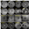
Clinical phenotypes of CHM and RPGR variant carriers and correlation with X inactivation skew. (A) Classification of retinal disease in female carriers of choroideremia based on 55° retinal fundus autofluorescence images, illustrative of the classification by Edwards et al. (2015): fine, coarse, geographic, and male-pattern degeneration. (B) Classification of retinal disease in female carriers of RPGR-associated X-linked retinitis pigmentosa based on 55° retinal fundus autofluorescence images, illustrating the classification by Nanda et al. (2018): normal, radial pattern, focal pigmentary retinopathy, and male-pattern degeneration. (C) Discordant clinical phenotypes of 34-year-old female C10 and her 59-year-old mother, C11, carrying the same pathogenic CHM variant. C10 presents with coarse (grade 2) phenotype, and C11 presents with fine (grade 1) phenotype. C11 had a pronounced buccal swab X inactivation skew (25:75) in favor of expressing the healthy allele, consistent with a milder disease phenotype. The 30° fundus autofluorescence images of right and left eyes are depicted for both carriers. (D) Discordant clinical phenotypes of 73-year-old female C2 and her 64-year-old sister, C3, carrying the same CHM variant. C2 presents with male-pattern degeneration (grade 4; picture also used to illustrate the grade 4 classification in panel A), and C3 presents with a coarse (grade 2) phenotype. C2 and C3 had skews in opposite directions, but the direction relative to the variant could not be determined. The 55° fundus autofluorescence images of right and left eyes are depicted for both carriers.

References

    1. Bittel DC, Theodoro MF, Kibiryeva N, Fischer W, Talebizadeh Z, Butler MG. 2008. Comparison of X-chromosome inactivation patterns in multiple tissues from human females. J Med Genet 45: 309–313. 10.1136/jmg.2007.055244 - DOI - PMC - PubMed
    1. Bonetti G, Cozza W, Bernini A, Kaftalli J, Mareso C, Cristofoli F, Medori MC, Colombo L, Martella S, Staurenghi G, et al. 2023. Towards a long-read sequencing approach for the molecular diagnosis of RPGRORF15 genetic variants. Int J Mol Sci 24: 16881. 10.3390/ijms242316881 - DOI - PMC - PubMed
    1. Britten-Jones AC, Jin R, Gocuk SA, Cichello E, O'Hare F, Hickey DG, Edwards TL, Ayton LN. 2022. The safety and efficacy of gene therapy treatment for monogenic retinal and optic nerve diseases: a systematic review. Genet Med 24: 521–534. 10.1016/j.gim.2021.10.013 - DOI - PubMed
    1. Carss KJ, Arno G, Erwood M, Stephens J, Sanchis-Juan A, Hull S, Megy K, Grozeva D, Dewhurst E, Malka S, et al. 2017. Comprehensive rare variant analysis via whole-genome sequencing to determine the molecular pathology of inherited retinal disease. Am J Hum Genet 100: 75–90. 10.1016/j.ajhg.2016.12.003 - DOI - PMC - PubMed
    1. Caylor RC. 2023. Nonrandom X chromosome inactivation detection. Curr Protoc 3: e748. 10.1002/cpz1.748 - DOI - PubMed

LinkOut - more resources