Review
. 2024 Sep 1;17(9):dmm050814.
doi: 10.1242/dmm.050814. Epub 2024 Sep 16.

Metabolic ripple effects - deciphering how lipid metabolism in cancer interfaces with the tumor microenvironment

Affiliations
Review

Metabolic ripple effects - deciphering how lipid metabolism in cancer interfaces with the tumor microenvironment

Patrick B Jonker et al. Dis Model Mech. .

Abstract

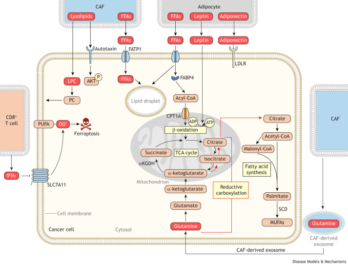
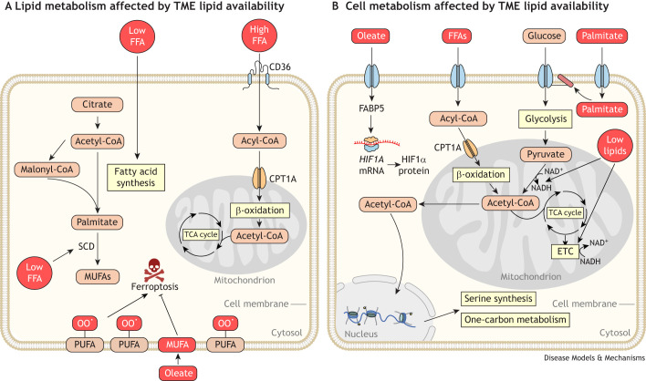
Cancer cells require a constant supply of lipids. Lipids are a diverse class of hydrophobic molecules that are essential for cellular homeostasis, growth and survival, and energy production. How tumors acquire lipids is under intensive investigation, as these mechanisms could provide attractive therapeutic targets for cancer. Cellular lipid metabolism is tightly regulated and responsive to environmental stimuli. Thus, lipid metabolism in cancer is heavily influenced by the tumor microenvironment. In this Review, we outline the mechanisms by which the tumor microenvironment determines the metabolic pathways used by tumors to acquire lipids. We also discuss emerging literature that reveals that lipid availability in the tumor microenvironment influences many metabolic pathways in cancers, including those not traditionally associated with lipid biology. Thus, metabolic changes instigated by the tumor microenvironment have 'ripple' effects throughout the densely interconnected metabolic network of cancer cells. Given the interconnectedness of tumor metabolism, we also discuss new tools and approaches to identify the lipid metabolic requirements of cancer cells in the tumor microenvironment and characterize how these requirements influence other aspects of tumor metabolism.

Keywords: Acidosis; Diet; Hypoxia; Lipid metabolism; Nutrient deprivation; Tumor microenvironment.


Conflict of interest statement

Competing interests The authors declare no competing or financial interests.

Figures

Fig. 1.
Fig. 1.
Cancer cell lipid acquisition and utilization pathways. (A) Cells take up lipids from the environment using several transport mechanisms. Cancer cells can use fatty acid transport proteins (FATPs), fatty acid-binding proteins (FABPs), cluster determinant 36 (CD36) and low-density lipoprotein (LDL) receptor (LDLR) to take up lipids from the environment. Additionally, cancer cells can take up bulk extracellular material via macropinocytosis, which provides a non-receptor-mediated pathway for cancer cells to acquire lipids. (B) Cells can also synthesize fatty acids and sterols from acetyl coenzyme A (CoA) using de novo synthesis metabolic pathways. The expression of enzymes involved in lipid de novo synthesis is transcriptionally regulated by the sterol regulatory element-binding protein (SREBP1 and SREBP2) transcription factors. Synthesizing lipids from acetyl-CoA consumes a considerable amount of cellular nicotinamide adenine dinucleotide phosphate (NADPH) pools, as fatty acid synthase (FASN), 3-hydroxy-3-methyl-glutaryl (HMG)-CoA reductase (HMGCR), stearoyl-CoA desaturase (SCD) and downstream sterol synthesis enzymes all use NADPH as a cofactor. Lipid synthesis also requires nicotinamide adenine dinucleotide (NAD+) to convert mitochondrial pyruvate into acetyl-CoA, which is processed to lipogenic citrate. Thus, via these shared co-factors, lipid synthesis pathways can interact with numerous other metabolic pathways in the cell. (C) Cancer cells can store both fatty acids and sterols as neutral lipids in lipid droplets for later mobilization and use. (D) Cancer cells need sterols and phospholipids, which are the building blocks for the production of cellular membranes. Fatty acids acquired through synthesis and uptake can be charged with CoA by acyl-CoA synthetase long-chain (ACSL) enzymes to allow for further synthesis of complex membrane lipids such as phospholipids. Sterols can also be synthesized de novo or acquired via uptake. Thus, cancer cells can produce membrane precursors by either de novo synthesis or by uptake from the environment. (E) Cancer cells can catabolize fatty acids to generate reducing equivalents and acetyl-CoA, both of which are used by the electron transport chain (ETC) to produce adenosine triphosphate (ATP). Abbreviations: ACC1, acetyl-CoA carboxylase 1, encoded by ACACA; ACLY, gene encoding ATP-citrate synthase; ACSS2, acyl-CoA synthetase short-chain family member 2; ATGL, adipose triglyceride lipase (also known as PNPLA2); CPT1A, carnitine palmitoyltransferase 1A; DGAT, diacylglycerol O-acyltransferase; FFA, free fatty acid; HMGCS, HMG-CoA synthase (encoded by HMGCS1); MUFA, monounsaturated fatty acid; NADH, reduced form of NAD+; TCA, tricarboxylic acid.
Fig. 2.
Fig. 2.
Hypoxia changes lipid metabolism in cancer cells. Hypoxia alters lipid metabolism in cancer cells both via substrate limitation, as oxygen (O2) is required in several lipid metabolic reactions, and via oxygen sensing signaling pathways such as hypoxia-inducible factor 1-α (HIF1α). At the substrate level, limited oxygen directly affects the activity of desaturase enzymes, such as stearoyl coenzyme A (CoA) desaturase (SCD). Thus, hypoxia limits the de novo synthesis of monounsaturated fatty acids (MUFAs). In response, cancer cells uptake lysolipids and release MUFAs from lipid droplets to provide oleate to cell membranes. Low oxygen tension also limits glucose-derived fatty acid synthesis by preventing the regeneration of nicotinamide adenine dinucleotide (NAD+). Hypoxia-driven limitation of NAD+ triggers the reductive use of glutamine as a carbon source for fatty acid synthesis in cancer cells. The red bracket indicates the pathway for reductive carboxylation of glutamine to citrate for de novo fatty acid synthesis. Several oxygen-sensitive signaling pathways also regulate cancer cell lipid metabolism under hypoxic conditions. HIF1α stabilization under hypoxia promotes lipid storage by enhancing lipid uptake through the fatty acid-binding proteins 3, 4 and 7 (FABP3/4/7) and decreasing β-oxidation of stored lipids by inhibiting carnitine palmitoyltransferase 1A (CPT1A). HIF1α facilitates acetate-derived lipid synthesis by promoting the expression of acyl-CoA synthetase short-chain family member 2 (ACSS2), which converts acetate into acetyl-CoA for cytosolic lipid synthesis. HIF1α also contributes to reductive carboxylation of glutamine by enhancing expression of the E3 ubiquitin ligase SIAH2, which ubiquitinates and ultimately degrades α-ketoglutarate dehydrogenase (αKGDH), ensuring that α-ketoglutarate is processed through the reductive side of the tricarboxylic acid (TCA) cycle. Lipid synthesis is also limited under oxygen-limited conditions by the membrane-associated ring-CH-type finger 6 (MARCHF6) oxygen-sensing ubiquitin ligase. MARCHF6 is active under hypoxia and triggers the degradation of SCAP–SREBP complex. Abbreviations: FFA, free fatty acid; SCAP, SREBP cleavage-activating protein; SREBP1, sterol regulatory element-binding protein 1; TCA, tricarboxylic acid; Ub, ubiquitin.
Fig. 3.
Fig. 3.
Amino acid availability constrains lipid metabolism in cancer cells. Amino acid availability alters the metabolic routes that cancer cells use to acquire lipids. Amino acid-replete conditions promote de novo lipid synthesis. For example, glutamine-replete conditions enhance lipid synthesis by stimulating SCAP–SREBP release from the endoplasmic reticulum through ammonia post-translational modifications from glutamine. Similarly, alanine availability promotes lipid synthesis by serving as a source of acetyl coenzyme A (CoA). Starvation of amino acids downregulates lipid synthesis through the general control nonderepressible protein 2 (GCN2) and mammalian target of rapamycin (mTOR) amino acid-sensing pathways. GCN2 activation under amino acid deprivation inactivates SREBP transcription factors, leading to decreased fatty acid synthesis. When mTOR is inactivated under amino acid starvation, fatty acid synthesis decreases and fatty acid storage in lipid droplets increases. Amino acid starvation can also alter how cells acquire biomass for lipid synthesis. For example, under low glutamine conditions, cells increase uptake of citrate for use in lipid synthesis. Abbreviations: S1P/S2P, site-1 protease (encoded by MBTPS1) and site-2 protease (encoded by MBTPS2); SCAP, SREBP cleavage-activating protein; SREBP1, sterol regulatory element-binding protein 1.
Fig. 4.
Fig. 4.
Glucose availability and acidity alter lipid metabolism in cancer cells. Both glucose abundance and acidity alter lipid metabolism in cancer cells. Glucose abundance promotes lipid accumulation by enhancing SREBP-mediated lipid synthesis, which occurs through glycosylation and activation of SCAP, which promotes SREBP maturation. In contrast, glucose depletion decreases lipid synthesis. Glucose starvation leads to AMP-activated protein kinase (AMPK) activation, which inhibits acetyl-CoA carboxylase 1 (ACC1) and de novo lipid synthesis. Low pH also alters lipid metabolism in cancer cells. Acidity increases cluster determinant 36 (CD36)- and low-density lipoprotein (LDL) receptor (LDLR)-mediated lipid uptake and increases fatty acid synthesis by promoting SREBP processing. Thus, acidity promotes lipid acquisition in cancer cells. Acidity also regulates the utilization of lipids. Low pH environments increase β-oxidation of lipids by inhibiting acetyl-CoA carboxylase 2 (ACC2), which restrains β-oxidation. Abbreviations: CoA, coenzyme A; CPT1A, carnitine palmitoyltransferase 1A; FFA, free fatty acid; MUFAs, monounsaturated fatty acids; S1P/S2P, site-1 protease and site-2 protease; SCAP, SREBP cleavage-activating protein; SREBP1/2, sterol regulatory element-binding proteins 1 and 2; TCA, tricarboxylic acid.
Fig. 5.
Fig. 5.
Stromal cells in the tumor microenvironment alter lipid metabolism in cancer cells. Stromal cells alter cancer cell lipid metabolism by providing lipids directly to cancer cells. Cancer-associated fibroblasts (CAFs) and adipocytes can provide fatty acids, sterols and lysophospholipids to cancer cells, which use these lipids for membrane production and for energy production through β-oxidation. Additionally, CAFs promote lipid synthesis by providing cancer cells with glutamine packaged in exosomes for use in reductive lipid synthesis. The red bracket indicates the pathway for reductive carboxylation of glutamine to citrate for de novo fatty acid synthesis. Stromal cells also alter lipid metabolism via cytokine and adipokine signaling. CD8+ T cells release interferon γ (IFNγ) in the tumor microenvironment, which triggers decreased SLC7A11 expression in cancer cells. Decreased SLC7A11 expression in cancer cells leads to increased lipid peroxidation (OO·) and ferroptotic cell death. Adiponectin release from adipocytes alters lipid metabolism in cancer cells by decreasing low-density lipoprotein receptor (LDLR)-mediated lipid uptake, whereas leptin signaling promotes β-oxidation in cancer cells. Abbreviations: αKGDH, α-ketoglutarate dehydrogenase; ADP, adenosine diphosphate; AKT, AKT serine/threonine kinase; ATP, adenosine triphosphate; CoA, coenzyme A; CPT1A, carnitine palmitoyltransferase 1A; FABP4, fatty acid-binding protein 4; FATP1, fatty acid transport protein 1; FFA, free fatty acid; LPC, lysophosphatidylcholine; MUFAs, monounsaturated fatty acids; PC, phosphatidylcholine; PUFA, polyunsaturated fatty acid; SCD, stearoyl-CoA desaturase; SLC7A11, solute carrier family 7 member 11; TCA, tricarboxylic acid.
Fig. 6.
Fig. 6.
Lipid availability alters cancer cell metabolism. (A) Lipid availability in the tumor microenvironment (TME) regulates cancer lipid metabolism. High levels of lipids in the TME promote β-oxidation in cancer cells by providing fatty acids that are taken up by receptors such as cluster determinant 36 (CD36) for subsequent catabolism. Oleate-rich environments, such as the lymph to which metastasizing cancer cells are exposed, lead to increased incorporation of monounsaturated fatty acids (MUFAs) into the membranes of cancer cells. This renders cancer cells resistant to ferroptosis as MUFAs replace oxidation-prone polyunsaturated fatty acids (PUFAs) in cell membranes. In contrast, TMEs that are lipid deplete, such as those in the brain, trigger increased fatty acid synthesis in cancer cells. This change in the lipid acquisition mechanism allows cancer cells to continue to grow even in lipid-poor environments. (B) Lipid availability in the TME also affects metabolism of non-lipid species in cancer cells. Lipid availability serves as a cue that regulates cellular signaling pathways and the expression of metabolic genes to broadly affect cancer cell metabolism. For instance, exposure to oleate alters HIF1α translation in cancer cells by upregulating fatty acid-binding protein 5 (FABP5). Free fatty acids (FFAs) are also a source of acetyl coenzyme A (CoA) that alters histone acetylation, thus altering serine synthesis and one-carbon metabolism in cancer cells. Additionally, lipid availability affects metabolism by impacting nutrient uptake in cancer cells. Palmitate post-translationally modifies nutrient transporters, including glucose transporters. Thus, fatty acid availability increases glucose uptake and glycolysis in cancer cells. Finally, lipid availability impacts other metabolic pathways via the use of shared redox cofactors. For example, lipid deprivation increases lipid synthesis in cancer cells. Increased lipid synthesis consumes and lowers nicotinamide adenine dinucleotide (NAD+) levels in cancer cells. Cancer cells increase oxygen consumption to increase NAD+ regeneration through the electron transport chain (ETC) as a result. Abbreviations: CPT1A, carnitine palmitoyltransferase 1A; NADH, reduced form of NAD+; OO·, peroxidation; SCD, stearoyl-CoA desaturase; TCA, tricarboxylic acid.

Similar articles

Cited by

References

    1. Ackerman, D., Tumanov, S., Qiu, B., Michalopoulou, E., Spata, M., Azzam, A., Xie, H., Simon, M. C. and Kamphorst, J. J. (2018). Triglycerides promote lipid homeostasis during hypoxic stress by balancing fatty acid saturation. Cell Rep 24, 2596-2605.e5. 10.1016/j.celrep.2018.08.015 - DOI - PMC - PubMed
    1. Ackermann, T. and Tardito, S. (2019). Cell culture medium formulation and its implications in cancer metabolism. Trends Cancer Res. 5, 329-332. 10.1016/j.trecan.2019.05.004 - DOI - PMC - PubMed
    1. Agarwala, P. K., Nie, S., Reid, G. E. and Kapoor, S. (2023). Global lipid remodelling by hypoxia aggravates migratory potential in pancreatic cancer while maintaining plasma membrane homeostasis. Biochim. Biophys. Acta Mol. Cell Biol. Lipids 1868, 159398. 10.1016/j.bbalip.2023.159398 - DOI - PMC - PubMed
    1. Alexandrov, T. (2023). Spatial metabolomics: from a niche field towards a driver of innovation. Nat Metab 5, 1443-1445. 10.1038/s42255-023-00881-0 - DOI - PubMed
    1. Alicea, G. M., Rebecca, V. W., Goldman, A. R., Fane, M. E., Douglass, S. M., Behera, R., Webster, M. R., Kugel, C. H., Ecker, B. L., Cecilia Caino, M.et al. (2020). Changes in aged fibroblast lipid metabolism induce age-dependent melanoma cell resistance to targeted therapy via the fatty acid transporter FATP2. Cancer Discov. 10, 1282-1295. 10.1158/2159-8290.CD-20-0329 - DOI - PMC - PubMed