. 2024 Sep 16;15(9):678.
doi: 10.1038/s41419-024-07044-3.

SPHK1 promotes bladder cancer metastasis via PD-L2/c-Src/FAK signaling cascade

Affiliations

SPHK1 promotes bladder cancer metastasis via PD-L2/c-Src/FAK signaling cascade

Wei-Hsiang Kao et al. Cell Death Dis. .

Abstract

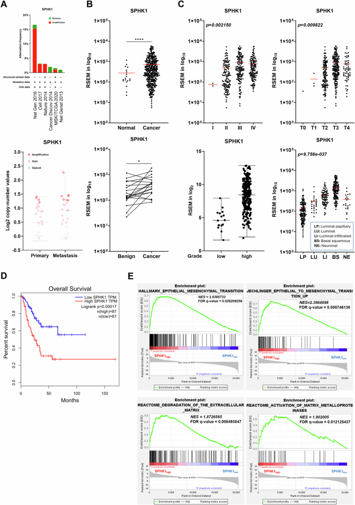
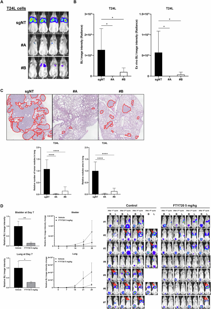
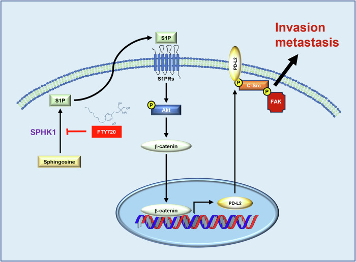
SPHK1 (sphingosine kinase type 1) is characterized as a rate-limiting enzyme in sphingolipid metabolism to phosphorylate sphingosine into sphingosine-1-phosphate (S1P) that can bind to S1P receptors (S1PRs) to initiate several signal transductions leading to cell proliferation and survival of normal cell. Many studies have indicated that SPHK1 is involved in several types of cancer development, however, a little is known in bladder cancer. The TCGA database analysis was utilized for analyzing the clinical relevance of SPHK1 in bladder cancer. Through CRISPR/Cas9 knockout (KO) and constitutive activation (CA) strategies on SPHK1 in the bladder cancer cells, we demonstrated the potential downstream target could be programmed cell death 1 ligand 2 (PD-L2). On the other hand, we demonstrated that FDA-approved SPHK1 inhibitor Gilenya® (FTY720) can successfully suppress bladder cancer metastasis by in vitro and in vivo approaches. This finding indicated that SPHK1 as a potent therapeutic target for metastatic bladder cancer by dissecting the mechanism of action, SPHK1/S1P-elicited Akt/β-catenin activation promoted the induction of PD-L2 that is a downstream effector in facilitating bladder cancer invasion and migration. Notably, PD-L2 interacted with c-Src that further activates FAK. Here, we unveil the clinical relevance of SPHK1 in bladder cancer progression and the driver role in bladder cancer metastasis. Moreover, we demonstrated the inhibitory effect of FDA-approved SPHK1 inhibitor FTY720 on bladder cancer metastasis from both in vitro and in vivo models.


Conflict of interest statement

The authors declare no competing interests.

Figures

Fig. 1
Fig. 1. Clinical correlation between SPHK1 and bladder cancer development.
A The frequency of SPHK1 gene alternation and the status of SPHK1 gene copy number from primary and metastatic samples analyzed from six different bladder cancer TCGA dataset in cBioPortal. B SPHK1 expression in normal bladder vs cancer samples from TCGA database (Benign = 19, Tumor = 408). *p < 0.05, ****p < 0.0001. C The expression of SPHK1 in bladder cancer samples from the TCGA database based on stage, T category in TNM classification, grade, and different subtypes. One-way ANOVA was performed (*p < 0.05, ****p < 0.0001). LP luminal papillary, LU luminal, LI luminal infiltrated, BS basal squamous, NE neuronal. D Significantly different overall survival of bladder cancer patients with high and low SPHK1 expression based on GEPIA website (***p < 0.001). E GSEA analysis of migration/invasion-related pathway in bladder cancer samples from the TCGA database (high SPHK1 group = 117 and low SPHK1 group = 282).
Fig. 2
Fig. 2. The regulation of cell migration and invasion by SPHK1.
A The profile of SPHK1 mRNA and protein expression in 253J-BV bladder cancer cell lines (n = 3). B The characterization of SPHK1 mRNA and protein expression in SPHK1-KO sublines of T24L and 253J-BV cell lines (n = 3). C The effect of SPHK1 on cell migration and invasion of different bladder cancer cell models (n = 3). D The effect of constitutive activate (CA) SPHK1 on cell migration and invasion of 253J and UMUC13 cell lines (n = 3). E The inhibitory effect of SPHK1 inhibitor (FTY720) on cell migration and invasion of T24L and 253J-BV cell lines (n = 3). All results were presented as mean ± SD and statistically calculated (#p < 0.05, **p < 0.01, ***p < 0.001, ####p < 0.0001, ****p < 0.0001).
Fig. 3
Fig. 3. The effect of FTY720 on SPHK1-promoting lung metastases of bladder cancer.
A Determination of lung metastases of T24LsgNT, T24LsgSPHK1#A, and T24LsgSPHK1#B (1 × 106 cells) injected intravenously into female SCID mice via tail vein by whole body BLI (IVIS Spectrum) (n = 4). B The relative imaging intensity of whole body BLI (left panel) and ex vivo BLI (right panel) (n = 4). C The pathological examination and determination of tumor nodules (circled in red) in lung metastases of T24L and SPHK1 KO sublines (n = 4). D The therapeutic effect of FTY720 on primary tumor and lung metastases of orthotopic model of T24L cells. After 2 weeks of cell implantation, IP administration of 5 mg/kg FTY720 (three times per week) was carried out for 2 weeks and followed by and weekly BLI (B: BLI signal from the bladder; L: BLI signal from the lung; red arrow: BLI signal from the lung metastatic nodule) (n = 7). The data were presented as mean ± SD and statistically calculated (*p < 0.05, **p < 0.01, ****p < 0.0001).
Fig. 4
Fig. 4. The role of PD-L2 in SPHK1-elicited cell migration and invasion of bladder cancer.
A GSEA analysis of immune regulation and adhesion-related pathway using bladder cancer samples from TCGA database (high SPHK1 group = 117; low SPHK1 group = 282). B The profile of CD274 (PD-L1) or PDCD1LG2 (PD-L2) expression in different subtypes of bladder cancer from the TCGA database analyzed by one-way ANOVA. C The correlation of PD-L1 or PD-L2 with SPHK1 gene expression in bladder cancer samples from TCGA database. D The profile of PD-L1 and PD-L2 mRNA and protein expression in 253J-BV bladder cancer cell lines (n = 3). E, F The regulation of PD-L2 mRNA and protein expression by SPHK1 activities in bladder cancer cell lines cells (n = 3). G, H The effect of PD-L2 on cell migration and invasion of 253J-BV PD-L2 KO cells and 253J PD-L2 OE (n = 3). All results were presented as mean ± SD and statistically calculated (*p < 0.05, #p < 0.05, **p < 0.01, ##p < 0.01, ***p < 0.001, ****p < 0.0001).
Fig. 5
Fig. 5. The role of PI3K/Akt/β-catenin signaling pathway in SPHK1-induced PD-L2 expression.
A The expression of PD-L2 mRNA in T24L or 253J-BV cell lines treated with various inhibitors for PI3K/Akt, ERK, or NF-κB pathway (n = 3). B The inhibitory effect of LY294002 on PD-L2 mRNA and protein expression in 253J cells treated with S1P (n = 3). C The effect of S1P on Akt activation or PD-L2 protein levels in T24LsgSPHK1 or 253J-BVsgSPHK1 cells (n = 3). D The effect of β-catenin OE on Akt activation or PD-L2 protein levels in T24LsgSPHK1 or 253J-BVsgSPHK1 cells (n = 3). The data were presented as mean ± SD and statistically calculated (*p < 0.05, **p < 0.01, ##p < 0.01, ***p < 0.001, ****p < 0.0001).
Fig. 6
Fig. 6. The central role of PD-L2/c-Src/Y397-FAK-complex in SPHK1-elicited cell migration and invasion of bladder cancer cells.
A The effect of FAK inhibitor on the interaction of activated FAK protein level with PD-L2 in T24L and 253J-BV cell lines. B The effect of FAK inhibitor on cell migration and invasion of 253J-CA or UC13-CA sublines (n = 3). C The effect of FAK inhibitor on cell migration and invasion of 253J PD-L2 OE subline (n = 3). D The effect of FAK inhibitor on cell migration and invasion of T24L and 253J-BV cell lines (n = 3). E The protein–protein interaction between c-Src, activated FAK, and PD-L2 in 253J PD-L2 OE subline. F The effect of c-Src inhibitor (Dasatinib) on the formation of PD-L2/c-Src/FAK protein complex. The data were presented as mean ± SD and statistically calculated (*p < 0.05, **p < 0.01, ***p < 0.001, ****p < 0.0001, ####p < 0.0001).
Fig. 7
Fig. 7. The regulator network of SPHK1 in promoting bladder cancer progression with potential targeted therapeutic strategy.
In bladder cancer cells, SPHK1 phosphorylates sphingosine into S1P and then stimulated its receptors activation for further PD-L2 gene expression through Akt/β-catenin-axis. Furthermore, elevated-PD-L2 facilitates c-Src/FAK complex for promoting bladder cancer migration, invasion, and metastasis. Meanwhile, the clinically FDA-approved SPHK1 inhibitor, FTY720, attenuates the SPHK1-elicited cancer metastasis, suggesting that SPHK1 could be a potential target as novel target therapy for metastatic bladder cancer treatment.

References

    1. Antoni S, Ferlay J, Soerjomataram I, Znaor A, Jemal A, Bray F. Bladder cancer incidence and mortality: a global overview and recent trends. Eur Urol. 2017;71:96–108. 10.1016/j.eururo.2016.06.010 - DOI - PubMed
    1. Lenis AT, Lec PM, Chamie K, Mshs MD. Bladder cancer: a review. JAMA. 2020;324:1980–91. 10.1001/jama.2020.17598 - DOI - PubMed
    1. Al-Zalabani AH, Stewart KF, Wesselius A, Schols AM, Zeegers MP. Modifiable risk factors for the prevention of bladder cancer: a systematic review of meta-analyses. Eur J Epidemiol. 2016;31:811–51. 10.1007/s10654-016-0138-6 - DOI - PMC - PubMed
    1. Zhang X, Zhang Y. Bladder cancer and genetic mutations. Cell Biochem Biophys. 2015;73:65–69. 10.1007/s12013-015-0574-z - DOI - PubMed
    1. Zaghloul MS, Zaghloul TM, Bishr MK, Baumann BC. Urinary schistosomiasis and the associated bladder cancer: update. J Egypt Natl Cancer Inst. 2020;32:44.10.1186/s43046-020-00055-z - DOI - PubMed

MeSH terms