. 2024 Sep 17;7(1):1158.
doi: 10.1038/s42003-024-06806-1.

An intranasal cationic liposomal polysaccharide vaccine elicits humoral immune responses against Streptococcus pneumoniae

Affiliations

An intranasal cationic liposomal polysaccharide vaccine elicits humoral immune responses against Streptococcus pneumoniae

Peng Wei et al. Commun Biol. .

Abstract

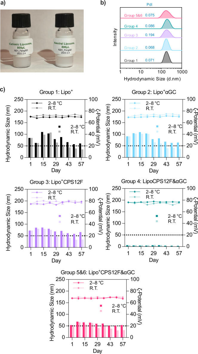
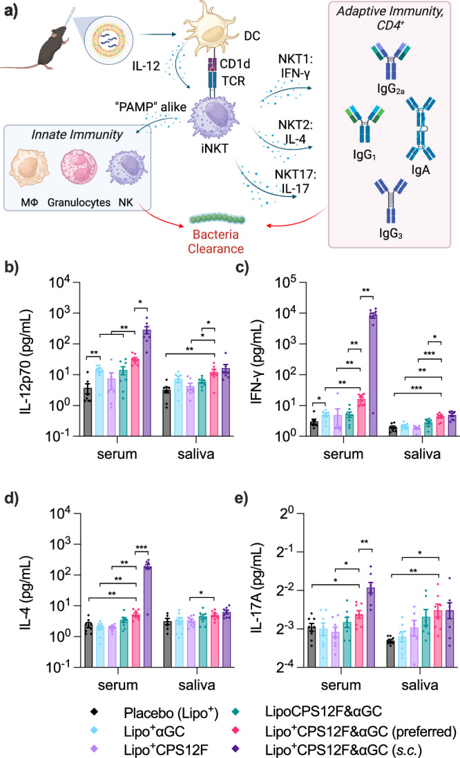
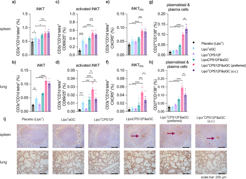
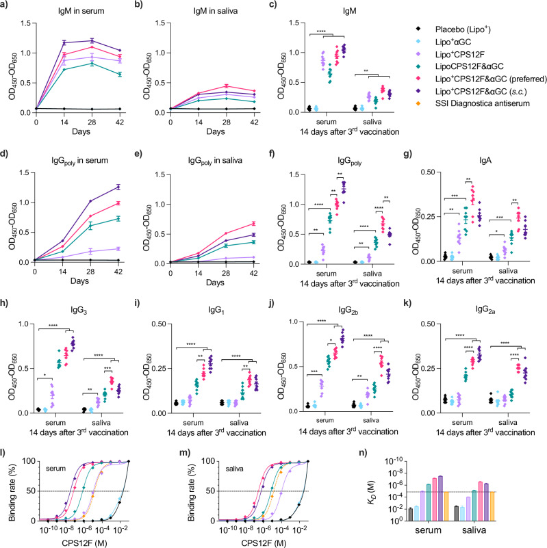
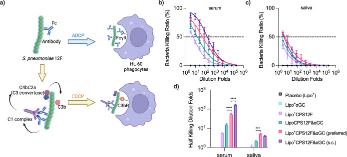
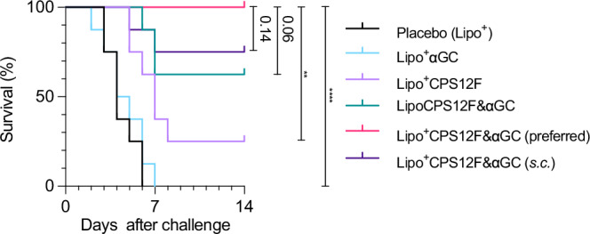
Diseases caused by S. pneumoniae are the leading cause of child mortality. As antibiotic resistance of S. pneumoniae is rising, vaccination remains the most recommended solution. However, the existing pneumococcal polysaccharides vaccine (Pneumovax® 23) proved only to induce T-independent immunity, and strict cold chain dependence of the protein conjugate vaccine impedes its promotion in developing countries, where infections are most problematic. Affordable and efficient vaccines against pneumococcus are therefore in high demand. Here, we present an intranasal vaccine Lipo+CPS12F&αGC, containing the capsular polysaccharides of S. pneumoniae 12F and the iNKT agonist α-galactosylceramide in cationic liposomes. In BALB/cJRj mice, the vaccine effectively activates iNKT cells and promotes B cells maturation, stimulates affinity-matured IgA and IgG production in both the respiratory tract and systemic blood, and displays sufficient protection both in vivo and in vitro. The designed vaccine is a promising, cost-effective solution against pneumococcus, which can be expanded to cover more serotypes and pathogens.


Conflict of interest statement

Technical University of Denmark has filed a patent covering this technology, and the inventors are M.H.C., P.W., C.R., J.R.H., and A.E.H. All other authors declare no competing interests.

Figures

Fig. 1
Fig. 1. Study design.
Molecular weight range a) and chemical structure b) of the pneumococcus serotype 12F capsular polysaccharide. c Chemical structure of the iNKT agonist α -galactosylceramide. d Formulation of the liposomal vaccine Lipo+CPS12F& αGC. e Scheme of the mouse study. The illustration was created with ChemDraw and BioRender.
Fig. 2
Fig. 2. Size distribution, ζ-potential, and stability of liposomal vaccines.
a Photo of cationic and neutral liposomal vaccine solution after static placement. b Hydrodynamic size distribution of liposomal vaccines characterized via DLS with a zetasizer. c Liposomal vaccines were stored at 2–8 C (in a fridge) or 18–24 C (at room temperature). The hydrodynamic size (left axis, curves above) and ζ-potential (right axis, bars below) were measured weekly for 2 months with a zetasizer.
Fig. 3
Fig. 3. Lipo+CPS12F& αGC stimulated elevated pro-inflammatory cytokine secretion compared with other formulations.
a Illustration of cytokines secretion after vaccination, created with BioRender. Concentrations of b IL-12p70, c IFN-γ, d IL-4, and e IL-17A in serum and saliva. BALB/cJRj mice were immunized with Lipo+ (group 1, blank cationic liposomes as placebo), Lipo+αGC (group 2), or 3 nmol antigen (repeat units of the polysaccharides) in Lipo+CPS12F (group 3), LipoCPS12F& αGC (group 4), or Lipo+CPS12F& αGC (group 5 & 6) via intranasal instillation (i.n., group 1–5) or subcutaneous injection (s.c., group 6). Serum and saliva were collected 18 hours after the priming vaccination, and IL-12p70, IFN-γ, IL-4, and IL-17A inside were quantified via cytometric bead array flow cytometry. Data were plotted as mean  ± SEM (n = 8). Welch and Brown-Forsythe ANOVA with multiple comparisons tests were done to assess statistical significance. Group 5 was the preferred group. *, **, ***, **** represent P < 0.05, P < 0.01, P < 0.001, P < 0.0001.
Fig. 4
Fig. 4. Lipo+CPS12F& αGC initiated a higher level of iNKT cell activation and B cell maturation compared with other formulations.
Percentage of a and b iNKT cells (CD3ε+CD1d tetra:αGC+), c and d activated iNKT cells (CD3ε+CD1d tetra:αGC+CD69/25+), e and f follicular helper iNKT cells (CD3ε+CD1d tetra:αGC+CXCR5+), and g and h plasmablast and plasma cells (CD27+CD138+) to total viable cells in the spleen and lung, separately. i Germinal centers (marked by arrows) formed in spleens and morphology of lung tissues after vaccinations. BALB/cJRj mice were immunized with Lipo+ (group 1, blank cationic liposomes as placebo), Lipo+αGC (group 2), or 3 nmol antigen (repeat units of the polysaccharides) in Lipo+CPS12F (group 3), LipoCPS12F& αGC (group 4), or Lipo+CPS12F& αGC (group 5 & 6) via intranasal instillation (i.n., group 1–5) or subcutaneous injection (s.c., group 6) for 3 times at 2 weeks intervals. Spleen and lung were isolated 3 days after the final vaccination and analyzed via multi-color flow cytometry and immunohistochemistry. Data were plotted as mean  ± SEM (n = 8). Welch and Brown–Forsythe ANOVA with multiple comparisons tests were done to assess statistical significance. Group 5 was the preferred group. *, **, ***, **** represent P < 0.05, P < 0.01, P < 0.001, P < 0.0001.
Fig. 5
Fig. 5. Lipo+CPS12F& αGC induced superior high-affinity CPS12F-specific antibody production compared with other formulations.
Change of CPS12F-specific a and b IgM and d and e IgGpoly in serum and saliva after immunization. Levels of CPS12F-specific c IgM, f IgGpoly, g IgA, h IgG3, i IgG1, j IgG2b, k IgG2a in serum and saliva 2 weeks after the final vaccination. Binding rate curve for CPS12F against l serum antibody and m saliva antibody. n Dissociation constants (KD) calculated for antibodies in serum and saliva. BALB/cJRj mice were immunized with Lipo+ (group 1, blank cationic liposomes as placebo), Lipo+αGC (group 2), or 3 nmol antigen (repeat units of the polysaccharides) in Lipo+CPS12F (group 3), LipoCPS12F& αGC (group 4), or Lipo+CPS12F& αGC (group 5 & 6) via intranasal instillation (i.n., group 1–5) or subcutaneous injection (s.c., group 6) for 3 times at 2 weeks intervals. Serum and saliva were collected at the start of the study and 2 weeks after each vaccination. Antibodies inside were quantified via indirect ELISA. Their affinities were evaluated via competitive ELISA, in which the binding rate refers to the ratio of [Ab bound with suspension CPS12F]/[Ab bound with bottom CPS12F], and the KD equals the half-binding concentration of CPS12F. Rabbit antiserum against CPS12F from SSI Diagnostica was used as an external control. Data were plotted as mean  ± SEM (n = 8 for ak, DF = 29 for ln). Curves were fitted using the one-site total binding model. Welch and Brown–Forsythe ANOVA with multiple comparisons tests were done to assess statistical significance. Group 5 was the preferred group. *, **, ***, **** represent P < 0.05, P < 0.01, P < 0.001, P < 0.0001.
Fig. 6
Fig. 6. Lipo+CPS12F& αGC generated antibodies with improved anti-S. pneumoniae 12F activity compared with other formulations.
a Illustration of bacteria-killing mechanisms in the opsonophagocytic killing assay (OPKA), created with BioRender. The ratio of bacteria killed by serum b and saliva c antibodies mediated opsonization and phagocytosis at different dilution folds. d Half-killing dilution folds of the antibodies in serum and saliva samples. BALB/cJRj mice were immunized with Lipo+ (group 1, blank cationic liposomes as placebo), Lipo+αGC (group 2), or 3 nmol antigen (repeat units of the polysaccharides) in Lipo+CPS12F (group 3), LipoCPS12F& αGC (group 4), or Lipo+CPS12F& αGC (group 5 & 6) via intranasal instillation (i.n., group 1–5) or subcutaneous injection (s.c., group 6) for 3 times at 2 weeks intervals. Serum and saliva collected 2 weeks after the final vaccination were pooled-assessed via OPKA (n = 8). Data were fitted in the nonlinear dose-response model. Half-killing dilution folds were interpolated and plotted as mean  ± SEM (DF ≥ 38). Welch and Brown-Forsythe ANOVA with multiple comparisons tests were done to assess statistical significance. Group 5 was the preferred group. *, **, ***, **** represent P < 0.05, P < 0.01, P < 0.001, P < 0.0001.
Fig. 7
Fig. 7. Lipo+CPS12F& αGC vaccination protected mice from S. pneumoniae 12F challenge.
Survival curves of vaccinated mice after bacteria challenge. BALB/cJRj mice were immunized with Lipo+ (group 1, blank cationic liposomes as placebo), Lipo+αGC (group 2), or 3 nmol antigen (repeat units of the polysaccharides) in Lipo+CPS12F (group 3), LipoCPS12F& αGC (group 4), or Lipo+CPS12F& αGC (group 5 & 6) via intranasal instillation (i.n., group 1–5) or subcutaneous injection (s.c., group 6) for 3 times at 2 weeks intervals. Three weeks after the final vaccination, mice (n = 8) were challenged with 1 × 106 CFU S. pneumoniae 12F via i.n. instillation and carefully monitored for the next 14 days. Results were plotted as survival curves. Gehan–Breslow–Wilcoxon nonparametric tests were done to assess statistical significance. Group 5 was the preferred group. *, **, ***, **** represent P < 0.05, P < 0.01, P < 0.001, P < 0.0001.

References

    1. World Health Organization. Pneumococcal disease. https://www.who.int/teams/health-product-policy-and-standards/standards-... (2024).
    1. Global Pneumococcal Sequencing Project. Serotype. https://www.pneumogen.net/gps/#/resources#serotypes (2024).
    1. European Centre for Disease Prevention and Control. Factsheet about pneumococcal disease. https://www.ecdc.europa.eu/en/pneumococcal-disease/facts (2024).
    1. Merck. Effectiveness data for Pneumovax®23 (pneumococcal vaccine polyvalent). https://www.merckvaccines.com/pneumovax23/pneumococcal-vaccine-efficacy (2024).
    1. Emmadi, M. et al. A streptococcus pneumoniae type 2 oligosaccharide glycoconjugate elicits opsonic antibodies and is protective in an animal model of invasive pneumococcal disease. J. Am. Chem. Soc.139, 14783–14791 (2017). - PubMed

Publication types

MeSH terms