. 2024 Dec;38(12):2663-2674.
doi: 10.1038/s41375-024-02401-9. Epub 2024 Sep 16.

Potent combination benefit of the AKT inhibitor capivasertib and the BCL-2 inhibitor venetoclax in diffuse large B cell lymphoma

Affiliations

Potent combination benefit of the AKT inhibitor capivasertib and the BCL-2 inhibitor venetoclax in diffuse large B cell lymphoma

Brandon S Willis et al. Leukemia. 2024 Dec.

Abstract

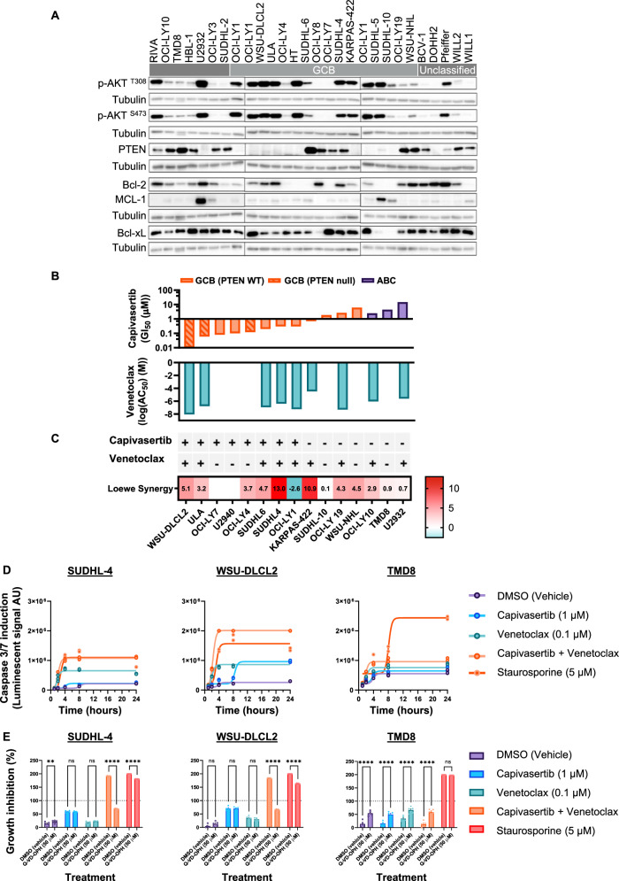
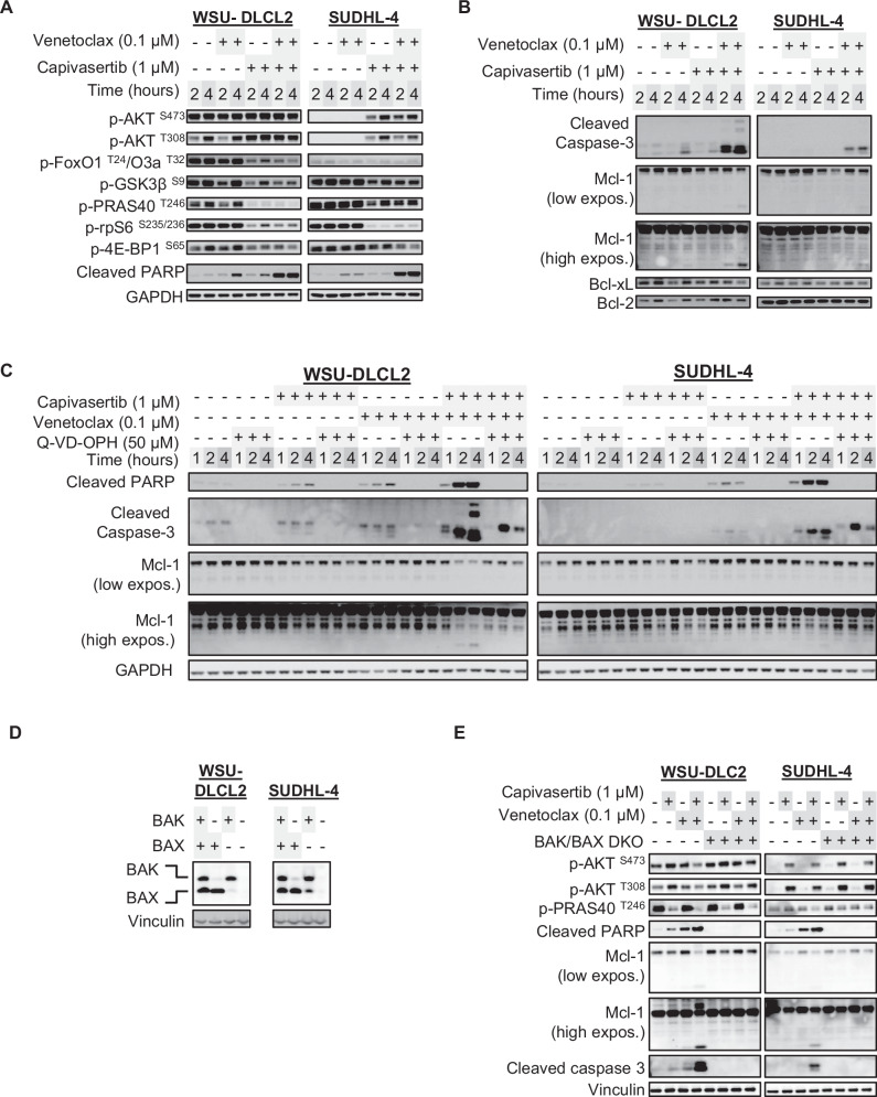
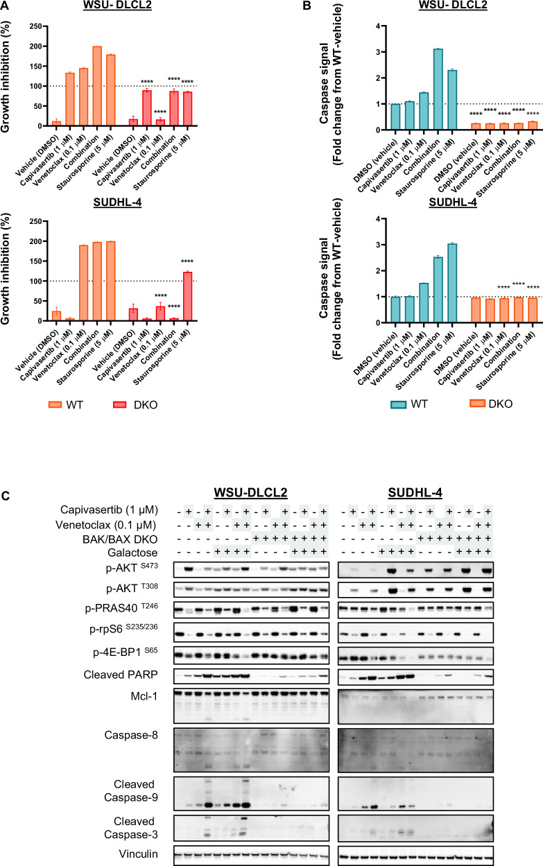
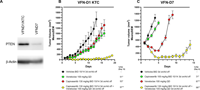
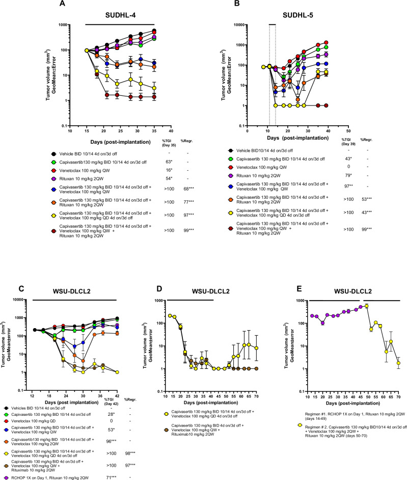
The therapeutic potential of targeting PI3K/AKT/PTEN signalling in B-cell malignancies remains attractive. Whilst PI3K-α/δ inhibitors demonstrate clinical benefit in certain B-cell lymphomas, PI3K signalling inhibitors have been inadequate in relapsed/refractory diffuse large B-cell lymphoma (DLBCL) in part, due to treatment related toxicities. Clinically, AKT inhibitors exhibit a differentiated tolerability profile offering an alternative approach for treating patients with B-cell malignancies. To explore how AKT inhibition complements other potential therapeutics in the treatment of DLBCL patients, an in vitro combination screen was conducted across a panel of DLCBL cell lines. The AKT inhibitor, capivasertib, in combination with the BCL-2 inhibitor, venetoclax, produced notable therapeutic benefit in preclinical models of DLBCL. Capivasertib and venetoclax rapidly induced caspase and PARP cleavage in GCB-DLBCL PTEN wildtype cell lines and those harbouring PTEN mutations or reduced PTEN protein, driving prolonged tumour growth inhibition in DLBCL cell line and patient derived xenograft lymphoma models. The addition of the rituximab further deepened the durability of capivasertib and venetoclax responses in a RCHOP refractory DLBCL in vivo models. These findings provide preclinical evidence for the rational treatment combination of AKT and BCL-2 inhibitors using capivasertib and venetoclax respectively alongside anti-CD20 antibody supplementation for treatment of patients with DLBCL.


Conflict of interest statement

Competing interests: B.W., K.M., H.D., I.N., A.R., M.P., N.B., J.R.R., C.R., J.M., and S.B. are current or former AstraZeneca employees and shareholders.

Figures

Fig. 1
Fig. 1. Capivasertib and venetoclax combination results in rapid caspase-mediated cell death in responsive DLBCL models.
A Base line Western blot biomarker profiling of ABC-, GCB-, and unclassified-DLBCL cell models with the OCI-LY1 cell line serving as reference control. B Capivasertib monotherapy anti-proliferative activity (GI50) (μM) and venetoclax monotherapy apoptotic response (Log AC50) (M) across GCB- and ABC-DLBCL cell lines. Order of cell lines is identical to that shown at the bottom of (C). C Loewe synergy scores for the combination of capivasertib and venetoclax in GCB- and ABC-DLBCL cell lines. + and – indicates each cell line’s monotherapy sensitivity to either capivasertib or venetoclax. Order of cell lines identical to that shown in (B). D Time course of caspase-3/7 activity in a cell model unresponsive to the either monotherapy or combination (TMD8), and two responsive models: WSU-DLCL2 (PTEN-mutant; GCB) and SUDHL-4 (PTEN wild-type; GCB) (n = 3/group). E Relative cell proliferation of SUDHL4, WSU-DLCL2 and TMD8 cell lines pretreated overnight with the pan-caspase inhibitor Q-VD-OPH (50 µM) or vehicle and then dosed with compounds for 24 h (n = 5/group; 2-way ANOVA with Sidak’s multiple comparisons ****p < 0.0001). Staurosporine (5 µM) was a positive control for caspase induction and cell killing.
Fig. 2
Fig. 2. Combination-mediated induction of cell death markers ablated by BAK/BAX codeletion in responsive DLBCL cell models.
A Western blot profiling of the AKT signalling pathway, known AKT substrates, and (B) apoptotic markers in WSU-DLCL2 and SUDHL-4 when treated with capivasertib and venetoclax monotherapies and in combination at 2and 4 h. C Western blot profiling of apoptotic biomarkers time course of WSU-DLCL2 and SUDHL4 cell lines pretreated with vehicle (DMSO) or Q-VD-OPH (50 µM) then dosed with indicated compounds at 1, 2, and 4 h. D CRISPR/Cas9-mediated of single and double knockout (DKO) of BAK and BAX in WSU-DLCL2 and SUDHL4 cell lines. E Comparison of the induction of cell death markers between BAK/BAX wild-type and DKO WSU-DLCL2 and SUDHL4 cell lines when treated with compounds for 4 h.
Fig. 3
Fig. 3. Combination-mediated induction of cell death markers ablated by BAK/BAX codeletion in responsive DLBCL cell models.
A Growth inhibition of BAK/BAX WT and DKO WSU-DLCL2 and SUDHL4 cells treated as indicated in a 72-hr measured by relative Cell Titer Glo signal growth assay (n = 3/group; 2-way ANOVA with Sidak’s multiple comparisons of the means between WT and DKO in the same treatment group ****p < 0.001). B Relative fold change of caspase-8 activity in BAK/BAX WT and DKO WSU-DLCL2 and SUDHL4 cells treated as indicated for 4 h. Staurosporine (5 µM) was a general positive control for caspase induction and cell killing. n = 3/group; 2-way ANOVA with Sidak’s multiple comparisons of the means between WT and DKO in the same treatment group ****p < 0.001. C Western blot profile of BAK/BAX WT and DKO WSU-DLCL2 and SUDHL4 cells treated with compounds for 4 h in normal glucose-rich growth medium or with medium where glucose is substituted with galactose.
Fig. 4
Fig. 4. Combination of capivasertib with venetoclax induces sustained tumour regression in a PTEN-wildtype SUDHL4 xenograft model.
A Tumour growth curves of CB.17 SCID mice bearing the GCB-DLBCL cell line SuDHL4 xenograft tumours treated with capivasertib (130 mg/kg BID 10/14 4-day on/3-day off) monotherapy, venetoclax (100 mg/kg QD) monotherapy and in combination in accordance with schedules captured in Supplementary Table 1. B Assessment of sustained tumour regression (>60 days post dosing). Both capivasertib and venetoclax were administered on a 4-day on/3-day off schedule relative to combinations using reduced frequency of venetoclax treatment. C Assessment of tumour activity of capivasertib dose reduction versus venetoclax dose reduction on anti-tumour response or tumour regression. Data is represented as geometric mean of tumour volumes and standard error of the mean. *p < 0.05, **p < 0.001, ***p < 0.0001.
Fig. 5
Fig. 5. Combination of capivasertib with venetoclax promotes tumour regression in a PTEN-wildtype and PTEN-null DLBCL PDX mouse models.
A Western blot analysis of PTEN expression in the VFN-D7 and VFN-D1_KTC human DLBCL PDX models. B Tumour growth curves of NSG mice bearing tumours with the double-hit GCB DLBCL PDX model, VFN-D7 or (C) the non-GCB DLCBL PTEN deficient PDX model, VFN-D1_KTC treated with capivasertib on a slightly modified schedule (130 mg/kg BID 10/14 3-day on/4-day off) monotherapy, venetoclax (100 mg/kg QD) and in combination with both agents on 3-day on/4-day off schedule. Comparison of combination treatment tumour regression compared to either monotherapy groups. Data is represented as arithmetic mean and standard error of the mean. *p < 0.05, **p < 0.001, ***p < 0.0001.
Fig. 6
Fig. 6. Combination of capivasertib, venetoclax and rituximab promotes tumour regression in GCB-DLBCL cell line xenograft models and in a RHCOP resistant DLBCL xenograft model.
Assessment of tumour regressions following addition of once weekly rituximab to capivasertib and venetoclax relative to the capivasertib and venetoclax combination on 4-day on/3-day off schedule. A, B Tumour growth curves of CB.17 SCID mice bearing with the GCB-DLBCL cell line (A) SUDHL4 and (B) SUDHL5 xenograft models treated with capivasertib (130 mg/kg BID 10/14 4-day on/3-day off) monotherapy, venetoclax (100 mg/kg QD) monotherapy or rituximab (10 mg/kg 2QW) monotherapy and in combination in accordance with schedules shown in Supplementary Table 1. C Comparison of initial tumour response and tumour regression following the triple combination of capivasertib, venetoclax and rituximab in the WSU-DLCL2 xenograft model relative to the doublet combinations and RCHOP (10 mg/kg rituximab 2QW, 25 mg/kg cyclophosphamide once IP, 3 mg/kg doxorubicin hydrochloride once IV, 0.25 mg/kg vincristine sulphate once IV and 0.5 mg/kg prednisone QD5 PO) treated as indicated (see Supp. Table S4). D Assessment of the sustained tumour regressions in the capivasertib venetoclax versus capivasertib venetoclax rituximab groups. E Tumours from the RCHOP treated group shown in (C) that were resistant to treatment (purple dots) were retreated with the capivasertib, venetoclax and rituximab triplet (yellow dots) and tumour regression assessed. All data is represented as geometric mean of tumour volumes and standard error of the mean (*p < 0.05, **p < 0.001, ***p < 0.0001).

References

    1. Swerdlow SH, Campo E, Pileri SA, Harris NL, Stein H, Siebert R, et al. The 2016 revision of the World Health Organization classification of lymphoid neoplasms. Blood. 2016;127:2375–90. - PMC - PubMed
    1. Wright GW, Huang DW, Phelan JD, Coulibaly ZA, Roulland S, Young RM, et al. A Probabilistic Classification Tool for Genetic Subtypes of Diffuse Large B Cell Lymphoma with Therapeutic Implications. Cancer Cell. 2020;37:551–68.e14. - PMC - PubMed
    1. Roschewski M, Phelan JD, Wilson WH. Molecular Classification and Treatment of Diffuse Large B-Cell Lymphoma and Primary Mediastinal B-Cell Lymphoma. Cancer J. 2020;26:195–205. - PMC - PubMed
    1. Tilly H, Morschhauser F, Sehn LH, Friedberg JW, Trneny M, Sharman JP, et al. Polatuzumab Vedotin in Previously Untreated Diffuse Large B-Cell Lymphoma. N. Engl J Med. 2022;386:351–63. - PMC - PubMed
    1. Wang J, Xu-Monette ZY, Jabbar KJ, Shen Q, Manyam GC, Tzankov A, et al. AKT Hyperactivation and the Potential of AKT-Targeted Therapy in Diffuse Large B-Cell Lymphoma. Am J Pathol. 2017;187:1700–16. - PMC - PubMed

MeSH terms