. 2024 Sep 16;81(1):405.
doi: 10.1007/s00018-024-05430-w.

Enhanced complement activation and MAC formation accelerates severe COVID-19

Affiliations

Enhanced complement activation and MAC formation accelerates severe COVID-19

Calder R Ellsworth et al. Cell Mol Life Sci. .

Abstract

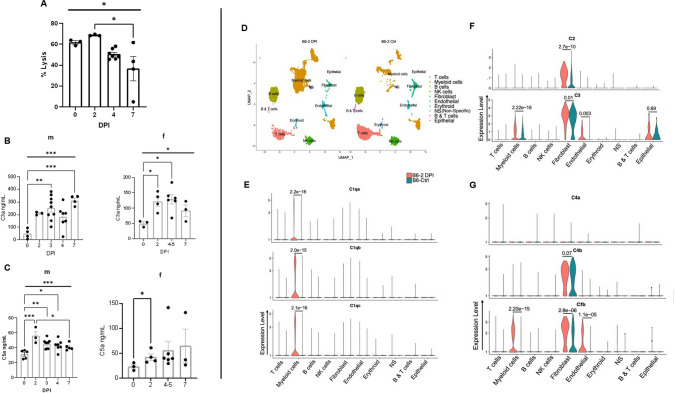
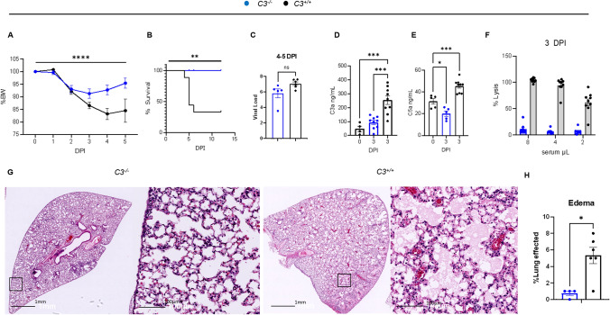
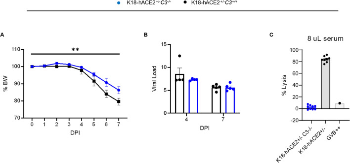
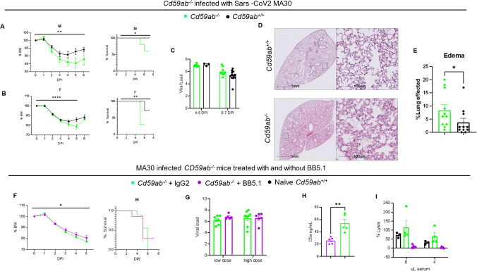
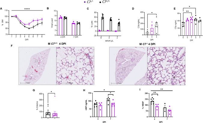
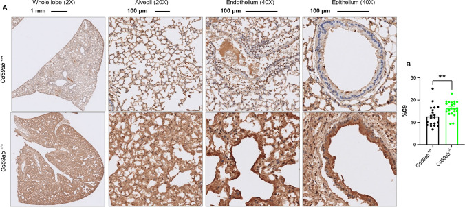
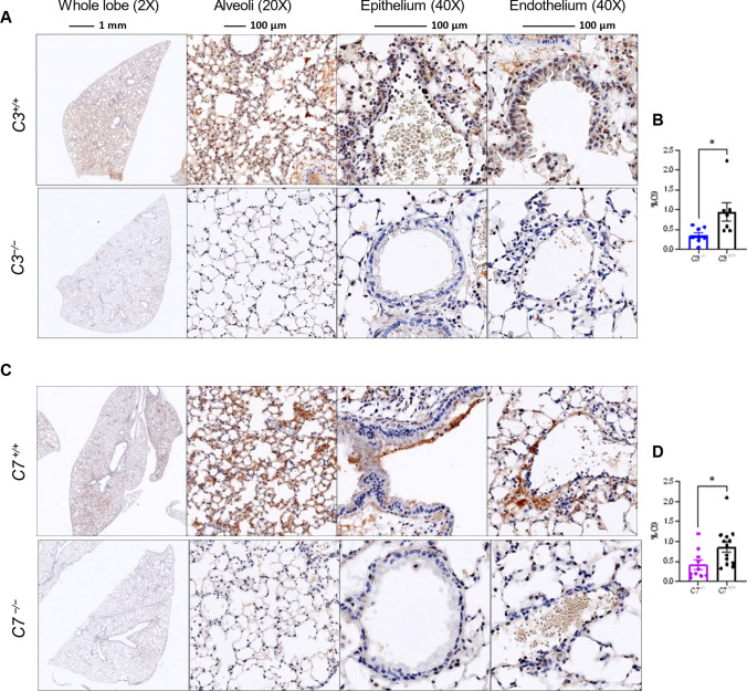
Emerging evidence indicates that activation of complement system leading to the formation of the membrane attack complex (MAC) plays a detrimental role in COVID-19. However, their pathogenic roles have never been experimentally investigated before. We used three knock out mice strains (1. C3-/-; 2. C7-/-; and 3. Cd59ab-/-) to evaluate the role of complement in severe COVID-19 pathogenesis. C3 deficient mice lack a key common component of all three complement activation pathways and are unable to generate C3 and C5 convertases. C7 deficient mice lack a complement protein needed for MAC formation. Cd59ab deficient mice lack an important inhibitor of MAC formation. We also used anti-C5 antibody to block and evaluate the therapeutic potential of inhibiting MAC formation. We demonstrate that inhibition of complement activation (in C3-/-) and MAC formation (in C3-/-. C7-/-, and anti-C5 antibody) attenuates severe COVID-19; whereas enhancement of MAC formation (Cd59ab-/-) accelerates severe COVID-19. The degree of MAC but not C3 deposits in the lungs of C3-/-, C7-/- mice, and Cd59ab-/- mice as compared to their control mice is associated with the attenuation or acceleration of SARS-CoV-2-induced disease. Further, the lack of terminal complement activation for the formation of MAC in C7 deficient mice protects endothelial dysfunction, which is associated with the attenuation of diseases and pathologic changes. Our results demonstrated the causative effect of MAC in severe COVID-19 and indicate a potential avenue for modulating the complement system and MAC formation in the treatment of severe COVID-19.

Keywords: C3; Cd59; Complement; Endothelial dysfunction; MAC; Severe COVID-19.


Conflict of interest statement

The authors have declared that no conflict of interest exists.

Figures

Fig. 1
Fig. 1
SARS CoV2 infection mediates increased systemic complement activation and up-regulates complement transcripts in multiple pulmonary cells in mice. A Complement-mediated hemolytic activity in the infected MA30-infected mice. The 4% of serum was collected from 14-16 weeks old males at different days post infection (DPI) with sublethal dose of SARS-CoV-2 MA30 (2.5×10⁴ TCID50), and were used as source of complement to measure MAC-mediated hemolysis of the antibody-sensitized sheep erythrocytes. BC C3a level (B) and C5a (C) detected by sandwich ELISA from sera of male (m) and female (f) mice collected after infection with sublethal (2.5×10⁴ TCID50) and lethal (5×10⁴ TCID50) doses of SARS-CoV-2 MA30. Overall significances shown in Figure 1A-1C were analyzed by one-way ANOVA and were indicated with horizontal bar, with group comparisons by Tukey test, * p < 0.05, ** p < 0.01, and ***p < 0.001, and **** p < 0.0001. D Major clusters and respective cell types for two 12-week-old female MA30 infected B6 mice (1x10⁴ TCID50) at 2dpi and two 12-week-old non-infected B6 mice by scRNA-seq data. Uniform manifold approximation and projection (UMAP) for dimension reduction plot with major cell types of scRNA-seq. Single-cell suspensions from whole infected lungs at 2 dpi and non-infected lungs were processed and sequenced. We identified 10 major clusters including T cells, myeloid cells, B cells, NK cells, fibroblast, endothelial, erythroid, non-specific, B & T cells, epithelial cells. EG Expression of C1qa, C1qb, C1qc, C2, C4a, C4b, and Cfb in the infected (pink) and non-infected (teal) cell clusters of lungs with p-values indicated
Fig. 2
Fig. 2
Deficiency of C3 protects against severe COVID-19 in MA30-infected B6 mice. AC and GH: C3−/− and C3+/+ mice (n=9/group) aged 14 weeks and of mixed sex (5 males and 4 females per goup) were infected with a lethal dose of MA30 (5×10⁴ TCID50) and sacrificed at 4–5 and 12 DPI. Percent body weight (A) and survival rate (B) after the infection were analyzed by Two-way ANOVA and survival by Log rank (Mantel-Cox) test. C Viral load was quantified by measuring subgenomic N RNA by qPCR in the mice at 4-5 DPI. DF 9 C3−/− and 9 C3+/+ male mice were infected with MA30 5×10⁴ TCID50 and were sacrificed at 3DPI for tissue collection. C3a (D) and C5a (E) serum levels as quantified by sandwich ELISA. Statistics show significance between groups using One-way ANOVA with Tukey’s multiple comparisons. Of note, there is a non-specific C3a signal in C3-/- mice, which may be the result of sample hemolysis and non-specific binding. F Complement activity measured by hemolytic assay with exposure of serial dilution of sera from C3 sufficient and deficient mice at 3 DPI to antibody sensitized sheep erythrocytes. GH Representative images (G) of edema from C3 sufficient and C3 deficient mice after infection at 4-5DPI, and quantification (H) by two-tailed student T-test. * p < 0.05, ** p < 0.01, ***p < 0.001, and **** p < 0.0001 by the respective statistical methods as detailed above
Fig. 3
Fig. 3
Deficiency of C3 protects against severe COVID-19 in K18-hACE2 transgenic mice infected with an original SARS-CoV2 strain. AC C3 deficient and sufficient mice expressing hACE2 under the control of the K18 promoter (C3−/−/K18-hACE2+/− and C3+/+/K18-hACE2+/−) aged 15 weeks with mixed-sex were infected with SARS-CoV-2-WA1 strain (n = 9/group, 5 females and 4 males per group) with 4 mice/group euthanized at 4 DPI and the others at 7 DPI. A Percent body weight changes. p-value = 0.0036 by two-way ANOVA, and B viral load of lung by qPCR showing no significance after infection. C Hemolytic activity of the infected C3−/−/K18-hACE2+/− and C3+/+/K18-hACE2+/− at 3 and 7 DPI using 8% diluted serum as the source of complement
Fig. 4
Fig. 4
Increase and reduction in MAC formation accelerates or partially rescues severe COVID-19, respectively. Administration of an anti-C5 inhibitor BB5.1 partially rescues the deleterious effects of CD59 deficiency. AE Cd59ab−/− (n=10 males, n=10 females) and Cd59ab+/+ mice (n=10 males, n=10 females) were age-matched at 16-20 week old and infected with a sublethal dose of MA30 SARS-CoV2 of 1×10⁴ TCID50. Percent body weight changes and survival of A male mice and B female mice after infection. C Viral load showed no significant difference. D Representative images of lung H/E sections from Cd59ab sufficient and deficient groups with E quantification thereof. FI Cd59ab−/− mice were infected with sublethal doses (1 and 2.5×10⁴ TCID50) of MA30 SARS-CoV-2 and treated with control isotype IgG (n=12) or anti-C5 BB5.1 antibody (n=16). F Percent body weight changes and survival in infected males (n=9/group). G Viral load in the lungs showing no significance with unpaired t-tests. H C5a levels in the sera after infection, significance from unpaired t-test. I Complement activity as measured by hemolytic assay of the infected mice sera at serial dilution
Fig. 5
Fig. 5
Deficiency of C7 protects against severe COVID-19. AI Mixed sex C7+/+ (= 14, 10 males, 4 females) and C7−/− mice (n=11, 7 males, 4 females) aged 15–17 weeks were infected with an infectious dose of MA30 SARS-CoV-2 (2.5×10⁴ TCID50) and euthanized at 4 and 7 DPI. A Percent body weight changes as assessed by Two-way ANOVA. B viral load after infection as measured by qPCR. C Hemolytic assay showing complement activity as measured by serial dilution of the infected sera as a source of the complement to lyse the anti-body sensitized sheep erythrocytes. D-E C3a (D) and C5a (E) measurements of the sera from the infected mice by ELISA with significant differences as assessed by One-way ANOVA with Dunnet’s (D) or Tukey’s (E) multiple comparisons. F-G Representative images of pulmonary edema at 4 DPI (F) and quantification (G) analyzed by Student t-test. H-I vWF (H) and VEGF (I) serum levels (ng/mL) at 4 and 7 DPI. Overall signficiance assessed by One-way ANOVA and indicated by horizontal bar with individual comparisons assessed by two-tailed Student t-test. * p < 0.05, ** p < 0.01, **** p < 0.0001
Fig. 6
Fig. 6
MAC deposition in the lungs of Cd59ab-/- mice as assessed by C9 staining. A-B Age-matched 16-20 week-old male and female Cd59ab−/− and control Cd59ab+/+ mice were sacrificed and tissues collected after death at 4-7 DPI after infection with MA30 (1×10⁴ TCID50). MAC deposition was viewed by staining with polyclonal rabbit anti-rat C9 (kindly obtained from Dr. Paul Morgan) at a 1:400 dilution. A Representative images of the above groups at 5 DPI showing whole lung lobe at magnification 2X, Alveoli at 20X, and epithelium and endothelium at 40X. B Quantification of C9. ** p < 0.01 by two-tailed Student’s t-test
Fig. 7
Fig. 7
MAC deposition in the lungs as assessed by C9 staining. AD Mouse lung sections were stained with an antibody against C9 (USBiological Life Sciences, 362359, Rabbit Anti-C9) used at a 1:1000 dilution. Representative images of the above groups showing whole lung lobe at magnification 2X, Alveoli at 20X, and epithelium and endothelium at 40X. A Representative image at 3DPI of lungs of C3 deficient and sufficient mice which were infected with MA30 at a dose of 5×10⁴ TCID50, and B quantification of C9 staining. C Representative image at 4DPI of lungs of C7 deficient and sufficient mice which were were infected with a dose of MA30 2.5×10⁴ TCID50, and D quantification of C9 staining. *p < 0.05 by two-tailed Student t-test

Similar articles

Cited by

References

    1. Xiao MT, Ellsworth CR, Qin X (2024) Emerging role of complement in COVID-19 and other respiratory virus diseases. Cell Mol Life Sci 81(1):94 10.1007/s00018-024-05157-8 - DOI - PMC - PubMed
    1. Yan B, Freiwald T, Chauss D, Wang L, West E, Mirabelli C et al (2021) SARS-CoV-2 drives JAK1/2-dependent local complement hyperactivation. Sci Immunol. 10.1126/sciimmunol.abg0833 10.1126/sciimmunol.abg0833 - DOI - PMC - PubMed
    1. Hess C, Kemper C (2016) Complement-mediated regulation of metabolism and basic cellular processes. Immunity 45(2):240–254 10.1016/j.immuni.2016.08.003 - DOI - PMC - PubMed
    1. Van Damme KFA, Hoste L, Declercq J, De Leeuw E, Maes B, Martens L et al (2023) A complement atlas identifies interleukin-6-dependent alternative pathway dysregulation as a key druggable feature of COVID-19. Sci Transl Med 15(710):eadi0252 10.1126/scitranslmed.adi0252 - DOI - PubMed
    1. Qin X, Gao B (2006) The complement system in liver diseases. Cell Mol Immunol 3(5):333–340 - PubMed

MeSH terms

LinkOut - more resources