. 2024 Sep 16;15(1):8100.
doi: 10.1038/s41467-024-52372-9.

Agnostic B cell selection approach identifies antibodies against K. pneumoniae that synergistically drive complement activation

Affiliations

Agnostic B cell selection approach identifies antibodies against K. pneumoniae that synergistically drive complement activation

Sjors P A van der Lans et al. Nat Commun. .

Abstract

Antibody-dependent complement activation plays a key role in the natural human immune response to infections. Currently, the understanding of which antibody-antigen combinations drive a potent complement response on bacteria is limited. Here, we develop an antigen-agnostic approach to stain and single-cell sort human IgG memory B cells recognizing intact bacterial cells, keeping surface antigens in their natural context. With this method we successfully identified 29 antibodies against K. pneumoniae, a dominant cause of hospital-acquired infections with increasing antibiotic resistance. Combining genetic tools and functional analyses, we reveal that the capacity of antibodies to activate complement on K. pneumoniae critically depends on their antigenic target. Furthermore, we find that antibody combinations can synergistically activate complement on K. pneumoniae by strengthening each other's binding in an Fc-independent manner. Understanding the molecular basis of effective complement activation by antibody combinations to mimic a polyclonal response could accelerate the development of antibody-based therapies against problematic infections.


Conflict of interest statement

R.J.M., J.S., and F.J.B. are (former) employees at Genmab BV and have ownership interests (including stocks, warrants, patents, etc.). Other authors declare no competing interests.

Figures
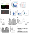
Fig. 1
Fig. 1. Dual staining of human B cells with whole bacterial cells identifies 29 IgGs against K. pneumoniae.
a K. pneumoniae was cultured overnight in the presence of KDO-azide followed by ‘clicking’ cyclooctyne-labeled fluorophores to the azide handles. Fluorescent labeling of KpnO2 with Cy5 or ATTO-488 was assessed via dSTORM microscopy. bd Dual staining of healthy donor B cells with KpnO2-ATTO-488 and KpnO2-Cy5. b, c B cells (CD19+) that bound both KpnO2-ATTO-488 and KpnO2-Cy5 were detected by flow cytometry. The double-bacterium-positive (Q2: ATTO-488+ Cy5+) memory (CD27+) B cells are shown in light blue and the naïve (CD27-) B cells in red. The distribution of memory versus naïve B cells was calculated for the unstained, single stained and dual stained B cells. c Double-bacterium-positive B cells were sorted and analyzed for binding of bacteria by fluorescence microscopy. d Gating strategy to sort K. pneumoniae-specific B cells. Alive B cells (CD19+, death-cell-marker-) that expressed IgG and stained positive for both KpnO2-ATTO-488 and KpnO2-Cy5 were single cell sorted (Q2, right panel). di The heavy and light variable regions (VH and VL, respectively) of single sorted B cells were amplified via RT-PCR and cloned into expression vectors to recombinantly express the antibodies as IgG1 in EXPI293F cells. e Antibody binding of 10-fold diluted expression supernatants to KpnO2. f The unique KpnO2 antibodies were re-expressed and purified to check binding to KpnO2 (at 1 µg/ml). g The VH and VL sequences of unique clones directed against KpnO2 were analyzed for number of base pair alterations compared to germline. h Antibody binding of 10-fold diluted expression supernatants to KpnO1. i The unique KpnO1 antibodies were re-expressed and purified to check binding to KpnO1 (at 2 µg/ml). j The VH and VL sequences of unique clones directed against KpnO1 were analyzed for number of base pair alterations compared to germline. k Summary of number of sorted cells, supernatants containing bacterium specific antibodies and unique purified antibody clones. e, f, h, i Flow cytometry data are expressed as gMFI values of bacterial populations. ae, h Data from a single experiment (f, i) Data represent mean ± SD of three independent experiments.
Fig. 2
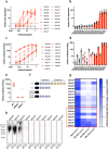
Fig. 2. Antibodies targeting the O2-antigen but not the capsule drive complement activation on KpnO2.
a Antibody-dependent C3b deposition on KpnO2. Bacteria were pre-incubated with a concentration range of KpnO2 IgG1s, followed by addition of 2% KpnO2∆NHS (NHS depleted from KpnO2 specific antibodies) as a complement source. C3b-deposition was detected using anti-hu-C3b-AF647 by flow cytometry. Potent complement activating antibodies in dark blue (more than 2-fold increase of C3b signal at 0.1 µg/ml indicated by the dotted line), moderate activating antibodies in light blue antibodies and non-complement activating antibodies in gray. b Bars represent C3b deposition from (a) in the condition where 0.1 µg/ml IgG1 was added, ranked on C3b deposition level. c Antibody binding of the different KpnO2 specific antibodies to KpnO2 was detected by flow cytometry and depicted with same color coding as in (a). d Bars represent antibody binding data from (c) at 0.1 µg/ml IgG1. e, f KpnO2 transposon mutants that are not recognized by (e) UKpn1 or (f) UKpn2 were single cell sorted. The barcode of the selected mutants was sequenced to determine the location of the transposon insertions (indicated as black bars). e Mutants that were no longer bound by UKpn1 had transposon insertions in genes involved in capsule synthesis. f Mutants that were not recognized by UKpn2 transposon had insertions in the rfb locus. g Antibody binding (at 1 µg/ml) to capsule (∆wbaP) and O-antigen (∆wbbO) deletion mutants was measured by flow cytometry and depicted with the same color coding as used in (a), larger dots indicate UKpn1 (gray) and UKpn2 (dark blue). ad, g Flow cytometry data are represented as relative gMFI of bacterial populations by dividing values by the isotype control. ad Data represent mean ± SD of three independent experiments and (g) average of 2-3 independent experiments.
Fig. 3
Fig. 3. Antibodies against KpnO1 recognize the O1 antigen and differ in their capacity to drive complement activation.
a Antibody-dependent C3b deposition on KpnO1. KpnO1 pre-incubated with anti-KpnO1 IgG1 was incubated with 1% KpnO1∆NHS (NHS depleted from KpnO1 specific antibodies) as a complement source. C3b-deposition was detected using anti-hu-C3b-AF647 by flow cytometry. Potent complement activating antibodies in dark red (>2-fold increase in C3b deposition at 0.1 µg/ml, indicated by dotted line), moderate complement activating antibodies in light red and non-activating antibodies in gray. b Bars represent C3b deposition from (a) at 0.1 µg/ml IgG1, ordered based on C3b deposition intensity. c Antibody binding to KpnO1 was detected by flow cytometry and depicted with the same color coding as in (a). d Bars represent antibody binding from (c) at 0.1 µg/ml IgG1. e Antibody binding (at 1 µg/ml) to KpnO1 wild-type, O-antigen (∆wbbO) and D-galactan II (∆wbbY) was analyzed by flow cytometry. f Schematic representation of the LPS O1- and O2-antigen polysaccharide. The O1-antigen consists of D-galactan-II (orange) on top of D-galactan-I (blue), whereas the O2 antigen contains only D-galactan-I. Deletion of the WbbO and WbbY glycosyltransferase leads to the loss of the complete O-antigen and the loss of the distal D-galactan-II part, respectively. g Antibody binding (at 1 µg/ml) to different O1-antigen expressing strains was analyzed using flow cytometry. The potent complement activating antibodies are highlighted by the red box (h) KpnO1 and an additional clinical strain expressing the O1-antigen (MB-17667) were lysed, and the proteins in the lysate were digested. Western blot was performed using different KpnO1 antibodies followed by goat anti-hu-IgG-HRP. Bands were visualized using ECL. ae, g Flow cytometry data are represented by gMFI values of bacterial populations that were divided by the buffer control. g Data represent log2 transformed data. Data represent (ad) mean ± SD or (e, g) mean of three independent experiments. h A representative experiment of two independent experiments is shown.
Fig. 4
Fig. 4. Hexamerization-enhancing mutation increases complement activation capacity of anti-capsule antibodies.
KpnO2 was incubated with (a) anti-O2 antibodies (UKpn2 and UKpn6) as wildtype and E430G variant or (b) anti-capsule antibodies (UKpn1 and UKpn3) as wildtype and E430G variant or for 30 min at 4 °C. After antibody binding, bacteria were incubated with 3% KpnO2∆NHS as a complement source for 30 min at 37 °C. C3b deposition was detected using anti-hu-C3b-A647 and analyzed by flow cytometry. c KpnO1 was incubated with the antibodies UKpn72, UKpn68 or UKpn82 as wildtype and E430G variant for 30 min at 4 °C. After antibody binding, bacteria were incubated with 1% KpnO1∆NHS as a complement source for 30 min at 37 °C. C3b deposition was detected using anti-hu-C3b-A647 and analyzed by flow cytometry. ac Flow cytometry data are represented by gMFI of bacterial populations. Data represent mean ± SD of three independent experiments.
Fig. 5
Fig. 5. Complement activating monoclonal antibodies promote phagocytosis and killing of K. pneumoniae by neutrophils.
Antibody-dependent phagocytosis of K. pneumoniae KpnO2 (a) and KpnO1 (b). GFP expressing bacteria were incubated in the presence of antibody and 3% ∆NHS (KpnO2) or 0.5% ∆NHS (KpnO1) as a source of complement. Isolated human neutrophils were allowed to phagocytose opsonized bacteria for 15 min at 37 °C, after which the percentage of GFP-positive neutrophils was determined by flow cytometry. Neutrophil killing of K. pneumoniae KpnO2 (c) or KpnO1 (d). Bacteria (2 × 105 bacteria/ml) were incubated with antibodies and 8% KpnO2∆NHS or 1% KpnO1∆NHS. To this mixture 2 × 106 neutrophils/ml were added for 3 h at 37 °C. Samples were serially diluted to determine CFU/ml. The data are depicted as relative survival by dividing the CFU/ml values by the no antibody control. ad Data represent mean ± SD of three independent experiments. c, d Data were analyzed by one-way ANOVA with Dunnett multiple comparison analysis compared to without mAb control. Significant differences are indicated, *p < 0.05, **p < 0.01 and ***p < 0.001.
Fig. 6
Fig. 6. Combining capsule targeting antibodies improves complement activation on KpnO2.
a, b Antibody-dependent C3b deposition. KpnO2 was pre-incubated with antibodies for 30 min at 4 °C. After antibody binding, bacteria were incubated in 3% NHS as a complement source for 30 min at 37 °C. C3b-deposition was detected using anti-hu-C3b-AF647 by flow cytometry. c Phagocytosis of KpnO2-GFP by human neutrophils. KpnO2-GFP was incubated with antibodies in the presence of 3% KpnO2∆NHS. Percentage of neutrophils that phagocytosed was determined by flow cytometry. d Antibody-mediated neutrophil killing was analyzed by incubating neutrophils with KpnO2 at MOI of 0.1 together with 8% KpnO2∆NHS and 0.11 µg/ml antibodies for 2 h at 37 °C. CFU/ml was calculated the next day and expressed as relative survival by dividing with the no antibody control. KpnO2 was pre-incubated with (e) UKpn2 or (f) the combination of UKpn1 and UKpn3 in the presence or absence of 10 µg/ml SpA-B for 30 min at 4 °C. After antibody binding, 3% NHS as complement source with or without SpA-B was added for 30 min at 37 °C. C3b deposition was measured using anti-hu-C3b-A647 by flow cytometry. ad, f In the conditions where antibodies were combined, both antibodies were added in the same concentration. The total antibody concentration in the assay is the same for single or combination of antibodies. a, b, e, f Flow cytometry data are represented as gMFI values of bacterial populations. ac, e, f Data represent mean ± SD of three independent experiments. d Data represents mean ± SD of four independent experiments and were analyzed by one-way ANOVA with Dunnett multiple comparison analysis compared to without mAb control; significant differences are indicated, * p < 0.05.
Fig. 7
Fig. 7. Anti-capsular antibodies strengthen each other’s binding.
K. pneumoniae KpnO2 was incubated with 1 µg/ml directly labeled UKpn3-AF647 (anti-capsule) in the presence of a concentration range of (a) IgG1 antibodies (UKpn1, UKpn2, UKpn3 or UKpn6) or (b) different variants of UKpn1 (IgG1, F(ab’)2 or F(ab)) for 30 min at 4 °C. c K. pneumoniae KpnO2 was incubated with 1 µg/ml UKpn3-AF647 F(ab’)2 in the presence of a concentration range of different UKpn1 variants (IgG1, F(ab’)2 or F(ab)) for 30 min at 4 °C. ac Antibody binding was detected by flow cytometry. The relative antibody binding was calculated by dividing the gMFI of the fluorescent antibody in presence of unlabeled antibody variants with the gMFI of the fluorescent antibody only. d K. pneumoniae KpnO2 was incubated with combinations of UKpn1 and UKpn3 variants for 30 min at 4 °C, followed by washing and addition of 3% NHS as complement source for 30 min at 37 °C. C3b deposition was measured using anti-hu-C3b-A647 by flow cytometry. ad Data represent mean ± SD of three independent experiments.

References

    1. Hajishengallis, G., Reis, E. S., Mastellos, D. C., Ricklin, D. & Lambris, J. D. Novel mechanisms and functions of complement. Nat. Immunol.18, 1288–1298 (2017). 10.1038/ni.3858 - DOI - PMC - PubMed
    1. Diebolder, C. A. et al. Complement is activated by IgG hexamers assembled at the cell surface. Science343, 1260–1263 (2014). 10.1126/science.1248943 - DOI - PMC - PubMed
    1. Abendstein, L. et al. Complement is activated by elevated IgG3 hexameric platforms and deposits C4b onto distinct antibody domains. Nat. Commun.14, 4027 (2023). 10.1038/s41467-023-39788-5 - DOI - PMC - PubMed
    1. Metzemaekers, M., Gouwy, M. & Proost, P. Neutrophil chemoattractant receptors in health and disease: double-edged swords. Cell. Mol. Immunol.17, 433–450 (2020). 10.1038/s41423-020-0412-0 - DOI - PMC - PubMed
    1. Heesterbeek, D. A. et al. Bacterial killing by complement requires membrane attack complex formation via surface‐bound C5 convertases. EMBO J38, e99852 (2019). 10.15252/embj.201899852 - DOI - PMC - PubMed

Publication types

MeSH terms

LinkOut - more resources