. 2024 Sep 16;14(1):21642.
doi: 10.1038/s41598-024-72561-2.

Liver-specific Coxsackievirus and adenovirus receptor deletion develop metabolic dysfunction-associated fatty liver disease

Affiliations

Liver-specific Coxsackievirus and adenovirus receptor deletion develop metabolic dysfunction-associated fatty liver disease

Hong-Gi Kim et al. Sci Rep. .

Abstract

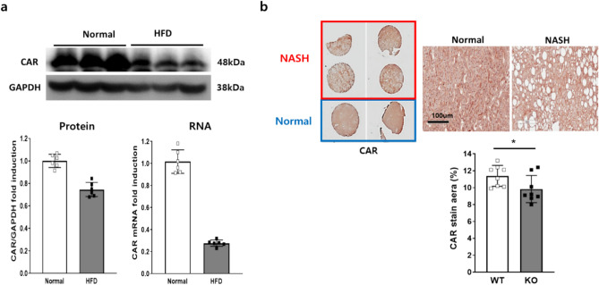
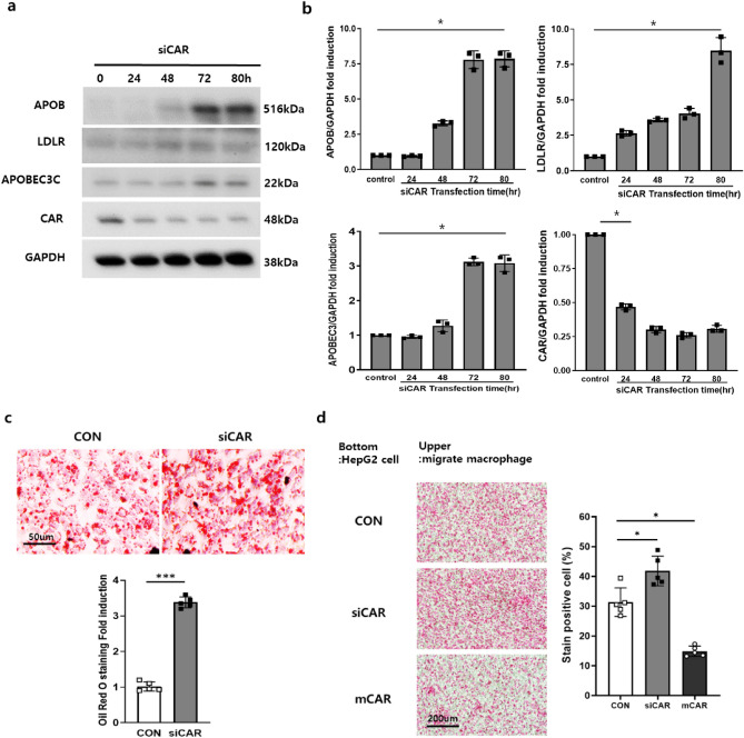
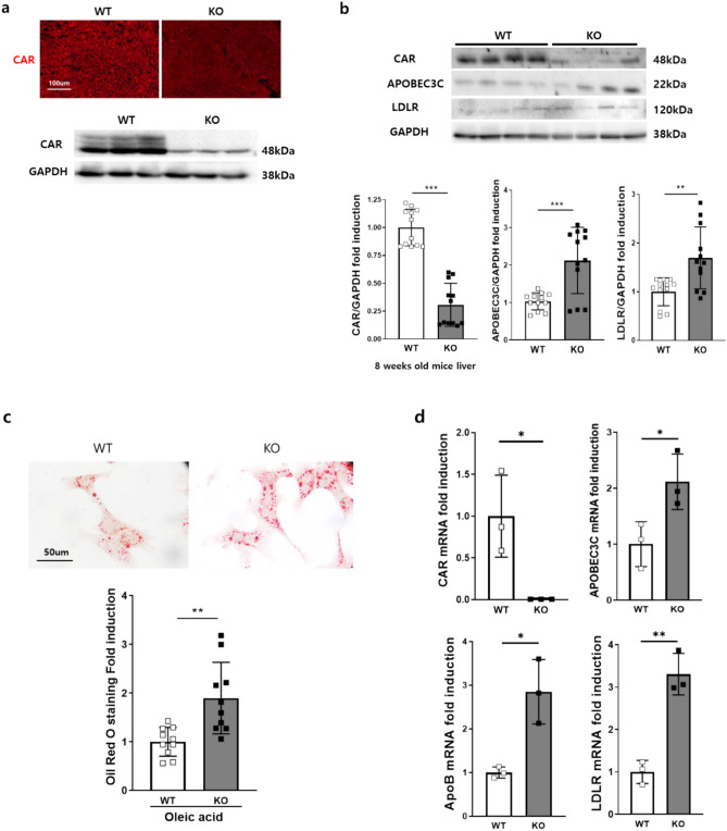
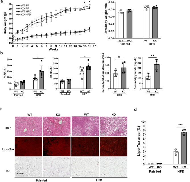
Metabolic dysfunction-associated fatty liver disease (MAFLD) is a common liver disease associated with obesity and is caused by the accumulation of ectopic fat without alcohol consumption. Coxsackievirus and adenovirus receptor (CAR) are vital for cardiac myocyte-intercalated discs and endothelial cell-to-cell tight junctions. CAR has also been reported to be associated with obesity and high blood pressure. However, its function in the liver is still not well understood. The liver of obese mice exhibit elevated CAR mRNA and protein levels. Furthermore, in the liver of patients with non-alcoholic steatohepatitis, CAR is reduced in hepatocyte cell-cell junctions compared to normal levels. We generated liver-specific CAR knockout (KO) mice to investigate the role of CAR in the liver. Body and liver weights were not different between wild-type (WT) and KO mice fed a paired or high-fat diet (HFD). However, HFD induced significant liver damage and lipid accumulation in CAR KO mice compared with WT mice. Additionally, inflammatory cytokines transcription, hepatic permeability, and macrophage recruitment considerably increased in CAR KO mice. We identified a new interaction partner of CAR using a protein pull-down assay and mass spectrometry. Apolipoprotein B mRNA editing enzyme catalytic polypeptide-like 3C (APOBEC3C) demonstrated a complex relationship with CAR, and hepatic CAR expression tightly regulated its level. Moreover, Apolipoprotein B (ApoB) and Low-density lipoprotein receptor (LDLR) levels correlated with APOBEC3C expression in the liver of CAR KO mice, suggesting that CAR may regulate lipid accumulation by controlling APOBEC3C activity. In this study, we showed that hepatic CAR deficiency increased cell-to-cell permeability. In addition, CAR deletion significantly increased hepatic lipid accumulation by inducing ApoB and LDLR expression. Although the underlying mechanism is unclear, CARs may be a target for the development of novel therapies for MAFLD.

Keywords: APOBEC3C; Apolipoprotein B; Coxsackievirus and adenovirus receptor; Metabolic dysfunction–associated fatty liver disease; Paracellular permeability.


Conflict of interest statement

The authors declare no competing interests.

Figures

Fig. 1
Fig. 1
CAR expression decreases in obese mice and liver samples of patients with NASH. Total RNA and protein were extracted from mice livers after a high-fat diet (HFD). (A) CAR mRNA expression in the liver of obese mice was significantly decreased (P < 0.01; n = 3). Total protein was subjected to western blot analysis and then incubated with anti-CAR antibodies. CAR protein expression level was decreased (P < 0.05; n = 3). (B) The liver tissue Histo-dot of patients with NASH was subjected to immunohistochemistry and then incubated with anti-CAR antibodies. Liver cell–cell junction CAR expression levels were decreased in liver specimens of patients with NASH. Data are expressed as mean ± SD, *P < 0.05; **P < 0.01.
Fig. 2
Fig. 2
CAR interacts with a new partner, APOBEC3C. (A) FLAG-CAR was overexpressed and pulled down by anti-FLAG agarose to find an interaction partner. The pull-down protein band was sliced and subjected to mass spectrometry. A new interaction protein, APOBEC3C, was identified. (B) These two protein interactions were confirmed by co-immunoprecipitation after overexpression of FLAG-CAR and GFP-APOBEC3C. Total protein was extracted and subsequently pulled down by anti-FLAG agarose. Agarose binding protein was subjected to western blot analysis using anti-GFP antibodies. (C) The opposite experiment was also performed after APOBEC3C-FLAG overexpression. FLAG agarose pull-down precipitated intracellular proteins were subjected to western blot analysis using anti-CAR antibodies.
Fig. 3
Fig. 3
Transient CAR deletion increases lipid transport regulatory protein and macrophage migration. (A,B) HepG2 cells were transfected with siCAR and scramble RNA as control (Con). Oleic acid treatment and Oil Red O staining were performed to determine lipid uptake levels in CAR deletions. At various time points (24, 48, 72, and 80 h) after siCAR delivery, cellular proteins were extracted and subjected to western blot analysis using anti-ApoB, -LDLR, -APOBEC3C, -CAR, and -GAPDH antibodies. (C) HepG2 cell lipid uptake was investigated by Oil red O staining after siRNA delivery. (D) Transwell chamber macrophage migration assay was performed after siCAR (for deletion) or mouse CAR (mCAR; for overexpression) transfection into bottom HepG2 cells. The upper chamber transwell passed macrophage was stained by Eosin (pink dot). Data are expressed as mean ± SD, *P < 0.05; **P < 0.01; ***P < 0.001.
Fig. 4
Fig. 4
Generation of liver-specific CAR knockout mice and liver protein expression. (A) CAR deletion confirmed liver of 8-weeks-old WT and KO mice by immunofluorescence stain and western blot analysis. (B) Protein was extracted from the livers of WT and KO mice and then subjected to western blot analysis using anti-CAR, -APOBEC3C, and -LDLR antibodies (n = 4 for each group). CAR deletion altered lipid transport regulatory proteins and gene expression in the isolated hepatocytes. (C) The liver hepatocytes of WT and KO mice were isolated and then subjected to a lipid uptake assay. Oil Red O staining showed lipid uptake levels in the hepatocytes. (D) APOBEC3C, ApoB, and LDLR mRNA levels were measured in WT and KO hepatocytes by qPCR. Data are expressed as mean ± SD, *P < 0.05; **P < 0.01; ***P < 0.001.
Fig. 5
Fig. 5
HFD-induced non-alcoholic fatty liver diseases in liver-specific CAR KO mice. (A) The basal phenotype of liver-specific CAR KO mice was confirmed. Body and liver weight were measured under different diet conditions for 16 weeks in pair-fed (■ WT and □ KO) or HFD (◆ WT and ◇ KO) (n = 4 each group) mice. (B) Concentrations of ALT, AST, total cholesterol, and triglyceride (TG) in serum were measured using ELISA (n = 4 in each group). (C,D) After HFD for 16 weeks, the liver was subjected to histological analysis by H&E and neutral lipid Lipo-Tox staining. Peritoneal fat was observed in the paraffin section. Lipid droplets increased in KO mice compared to WT fed an HFD. However, there was no difference between pair-fed mice. Data are expressed as mean ± SD, *P < 0.05 by a two-tailed Student’s t-test.
Fig. 6
Fig. 6
HFDs induced inflammatory cytokine expression and cholesterol accumulation in the liver of KO mice. (A) Liver protein was extracted and then subjected to ELISA. The total cholesterol, triglyceride, and LDL cholesterol levels were measured. (B) Total RNA was extracted from mice liver after being pair-fed or fed an HFD and then subjected to qPCR analysis of ApoB and APOBEC3C. In addition, (C) inflammatory cytokines (IL-1β, IL-6, TNF-α, and MCP-1) and immunocytes (F4/80: macrophage marker; n = 4). (D) Liver subjected to immunofluorescent stain for macrophages. Liver samples were incubated with anti-F4/80 antibodies and Alexa488-labeled secondary antibodies. Infiltrated macrophages presented are by green stain percent area. Data are expressed as mean ± SD, *P < 0.05; **P < 0.01; ***P < 0.001 by a two-tailed Student’s t-test. IL-1β interleukin-1 beta; IL-6, interleukin-6; TNF-α tumor necrosis factor alpha, MCP-1 monocyte chemoattractant protein-1.

References

    1. Machado, M. V. & Cortez-Pinto, H. Non-alcoholic fatty liver disease: What the clinician needs to know. World J. Gastroenterol.20, 12956–12980 (2014). - PMC - PubMed
    1. Behari, J. et al. Liver-specific beta-catenin knockout mice exhibit defective bile acid and cholesterol homeostasis and increased susceptibility to diet-induced steatohepatitis. Am. J. Pathol.176, 744–753 (2010). - PMC - PubMed
    1. Sanyal, A. J. et al. Pioglitazone, vitamin E, or placebo for nonalcoholic steatohepatitis. N. Engl. J. Med.362, 1675–1685 (2010). - PMC - PubMed
    1. Day, C. P. & James, O. F. Steatohepatitis: A tale of two “hits”?. Gastroenterology114, 842–845 (1998). - PubMed
    1. Harrison, S. A., Kadakia, S., Lang, K. A. & Schenker, S. Nonalcoholic steatohepatitis: What we know in the new millennium. Am. J. Gastroenterol.97, 2714–2724 (2002). - PubMed

MeSH terms

Substances

LinkOut - more resources