. 2024 Dec;18(1):94-102.
doi: 10.1080/19336896.2024.2402225. Epub 2024 Sep 16.

Expression of the cellular prion protein by mast cells in white-tailed deer carotid body, cervical lymph nodes and ganglia

Affiliations

Expression of the cellular prion protein by mast cells in white-tailed deer carotid body, cervical lymph nodes and ganglia

Anthony E Kincaid et al. Prion. 2024 Dec.

Abstract

Chronic wasting disease (CWD) is a transmissible and fatal prion disease that affects cervids. While both oral and nasal routes of exposure to prions cause disease, the spatial and temporal details of how prions enter the central nervous system (CNS) are unknown. Carotid bodies (CBs) are structures that are exposed to blood-borne prions and are densely innervated by nerves that are directly connected to brainstem nuclei, known to be early sites of prion neuroinvasion. All CBs examined contained mast cells expressing the prion protein which is consistent with these cells playing a role in neuroinvasion following prionemia.

Keywords: Carotid body; chronic wasting disease; lymph node; mast cell; nodose ganglion; prions; superior cervical ganglion; white-tailed deer.


Conflict of interest statement

No potential conflict of interest was reported by the author(s).

Figures

Carotid bodies collected from white-tailed deer are easily identified using toluidine blue and hematoxylin stains in light micrographs due to their characteristic clusters of cells surrounded by a connective tissue capsule and many capillaries.
Figure 1.
Carotid bodies (CB) from white-tailed deer (WTD; animal #1450) can be identified by staining tissue sections with either toluidine blue (TB; panels a, b) or haematoxylin and eosin (H&E; panels c, d). Low power views (panels a, c) show that CBs are irregular-shaped lobulated structures surrounded by connective tissue capsules (indicated by arrows; more obvious in H&E sections). Bands of connective tissue extend towards the middle of the CBs separating the structure into clusters of cells. Medium power views (panels b, d) show the connective tissue bands separating the clusters of cells (indicated by asterisks). Note the presence of numerous blood vessels, some of which contain red blood cells (indicated by arrows). Scale bars: panels a, c = 200 µm; panels b, d = 50 µm.
The 8H4 antibody shows the presence of the misfolded prion protein in the obex of an infected white-tailed deer and the absence of the misfolded prion protein in the obex of an uninfected white-tailed deer.
Figure 2.
The 8H4 antibody was used to demonstrate the presence of the prion protein in WTD tissue. Tissue sections collected from the obex of an infected WTD (panel a) shows the presence of the pathogenic isoform of the prion protein (PrPCWD) in a clinically-ill deer (animal #817). The area inside the box is enlarged in the inset. The plaque-like immunoreactivity was only noted in the obex of infected animals. Tissue sections taken from the obex of an uninfected deer (animal #955) and processed concurrently (panel b) show a lack of PrPCWD immunoreactivity. Scale bars: panels a, b = 200 µm.
Cells identified with an antibody specific to mast cells are similar in size, shape, and distribution in white-tailed deer carotid bodies as cells that are identified using an antibody against the prion protein. When stains are combined on the same tissue sections the cells are identified as a single population; thus, mast cells express the prion protein in carotid bodies of white-tailed deer.
Figure 3.
Mast cells in the WTD CB express the prion protein. CBs from an animal (animal # 1436) were identified using the prion antibody, 8H4, (panel a) or a mast cell specific antibody (panel b). Note that the distribution, size and morphology of the cells is the same in the two panels. Arrows indicate a single cell enlarged in the insets of panels a and b. Note the presence of large granules in the cells, a characteristic feature of mast cells, which were immunoreactive using either antibody. Replacement of the primary antibodies with the appropriate isotype control resulted in the complete absence of any PrPC immunoreactivity (Figure 3c) or mast cell immunoreactivity (Figure 3d). Mast cells can also be identified by staining with TB/AS (panel e). To confirm that mast cells express PrPC tissue sections were IHC processed using the 8H4 antibody followed by counterstaining with TB/AS. Only one population of cells was stained and was immunoreactive for 8H4 and contained the granules characteristic of mast cells (panel f; animal # 1438). Arrows indicate stacks of red blood cells in capillaries adjacent to the mast cells, demonstrating the proximity of mast cells to blood in the CBs (panels e, f). Scale bars: panels a-b = 200 µm. Panels c-d = 100 µm. Panels e, f = 10 µm.
Mast cells express the prion protein in cervical lymph nodes, the superior cervical ganglion, nodose ganglion and nerves located near the carotid bodies.
Figure 4.
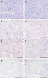
Mast cells in WTD structures located near the CBs also express the prion protein. Tissue sections containing cervical lymph nodes (panels a, b; animal #1442), superior cervical ganglia (panels c, d; animal #1306), nodose ganglia (panels e, f; animal #1438) and nerve (panels g, h; animal #1438) were stained using either 8H4 antibody (panels a, c, e, g) or mast cell antibody (panels b, d, f, h). Note the number, distribution, size and morphology of the cells is the same in tissue sections processed using either antibody and that there was no difference between tissues collected from infected or uninfected tissues. Scale bars: panels a-h = 100 µm.
A number of structures associated with carotid bodies serve as potential routes for neuroinvasion of prions circulating in the blood, these include the superior cervical ganglion, nodose ganglion and cervical lymph nodes.
Figure 5.
Schematic representation of potential routes of neuroinvasion following CB exposure to prions in blood. Black circles represent neuronal cell bodies, lines represent axons, and the ‘v’ line split represents axon terminals. Structures shown within grey shaded area of the figure were located within tissue blocks taken from WTD exposed to prion-infected tissues as listed in Table 1 (not all structures were in all tissue blocks). Two potential routes of prion neuroinvasion from CBs are synaptically linked to early sites of prion neuroinvasion: the nucleus of the solitary tract (NTS) and the intermediolateral cell column of the thoracic spinal cord (IML). The first pathway is via branches of the carotid sinus nerve that terminates in the NTS in the brainstem. The sensory cell bodies of these axons are located in the petrosal ganglion. The NTS is synaptically linked to the dorsal motor nucleus of the vagus (DMNV). The second pathway is via the sympathetic postganglionic fibres that innervate the CBs; the cell bodies of these fibres are located in the superior cervical ganglion (SCG). These neurons are synaptically linked to sympathetic preganglionic fibres that originate from sympathetic preganglionic neurons in the IML of the thoracic spinal cord. The arrows indicate the direction of transport of prions into the CNS. Tissue blocks also included the nodose ganglion (NG) which is the location of vagal sensory neurons that innervate thoracic and abdominal viscera, including the Peyer’s patches of the ileum, known to contain PrPCWD following oral exposure [13]; the fibres of these cells terminate in the NTS. Some of the sections included cervical lymph nodes (CLNs), known sites for prion replication [40]. The CBs, SCG, the NG, the nerves and CLNs all contained mast cells that expressed PrPC. This figure was created using BioRender.com.

References

    1. Williams ES, Young S.. Chronic wasting disease of captive mule deer: a spongiform encephalopathy. J Wildl Dis. 1980;16(1):1689–1698. doi: 10.7589/0090-3558-16.1.89 - DOI - PubMed
    1. Chronic wasting disease: occurrence. Centers for Disease Control and Prevention. [cited 2023 Dec 20]. Available from: https://www.cdc.gov/prions/cwd/occurrence.html
    1. Miller MW, Williams ES. Horizontal prion transmission in mule deer. Nature. 2002;425(6953):35–36. doi: 10.1038/425035a - DOI - PubMed
    1. Mathiason CK, Hays SA, Powers J, et al. Infectious prions in pre-clinical deer and transmission of chronic wasting disease solely by environmental exposure. PLOS ONE. 2009;4(6):e5916. doi: 10.1371/journal.pone.0005916 - DOI - PMC - PubMed
    1. Selariu A, Powers JG, Nalls A, et al. In utero transmission and tissue distribution of chronic wasting disease-associated prions in free-ranging Rocky Mountain Elk. J Gen Virol. 2015;96(11):3444–3455. doi: 10.1099/jgv.0.000281 - DOI - PMC - PubMed

Publication types

LinkOut - more resources