

Monascus pigment-protected bone marrow-derived stem cells for heart failure treatment
- PMID: 39285916
- PMCID: PMC11403898
- DOI: 10.1016/j.bioactmat.2024.08.038
Monascus pigment-protected bone marrow-derived stem cells for heart failure treatment
Abstract
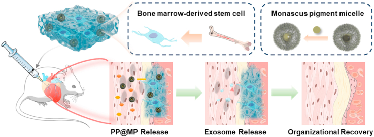
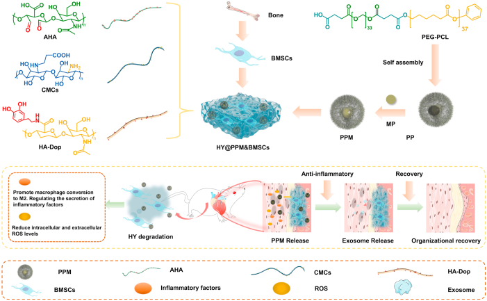
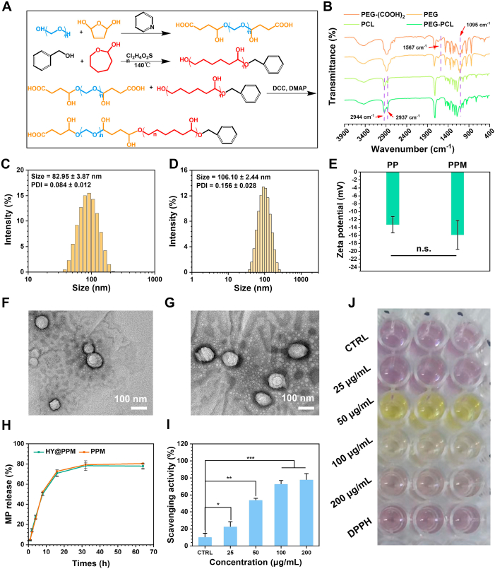
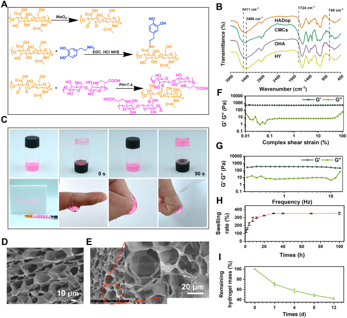
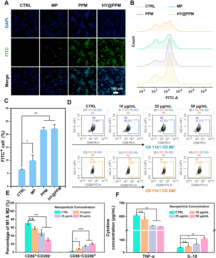
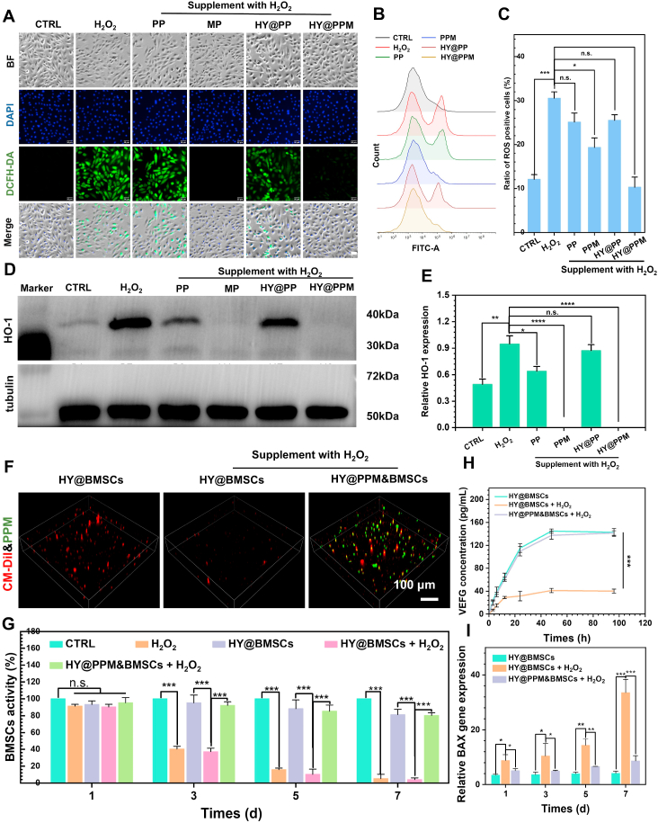
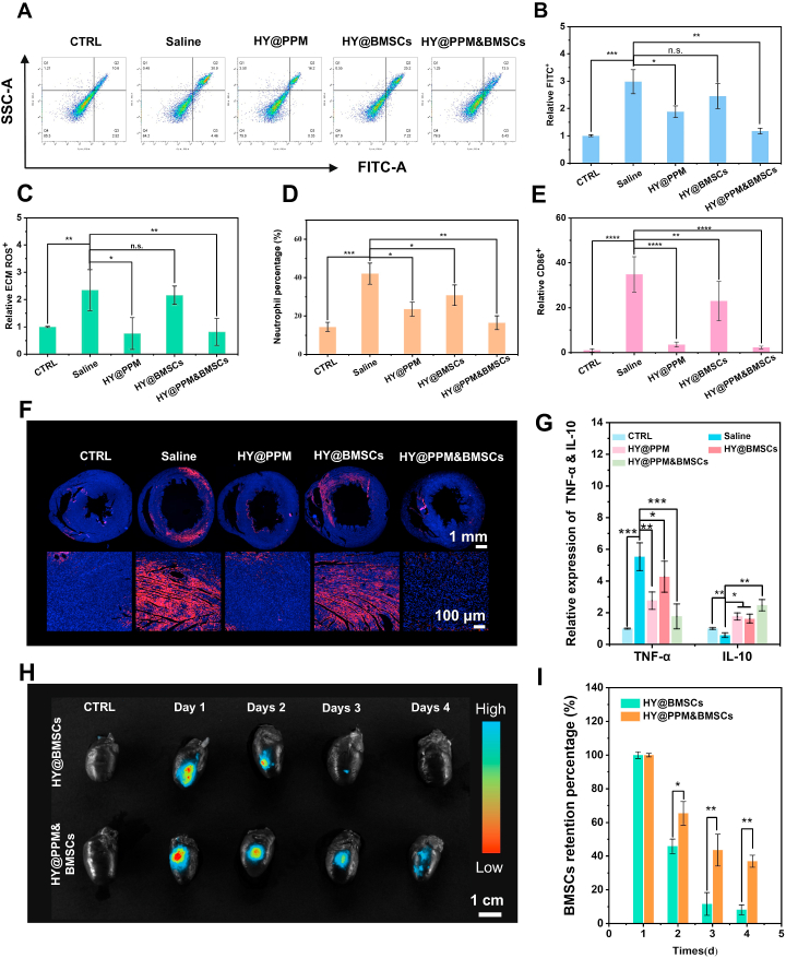
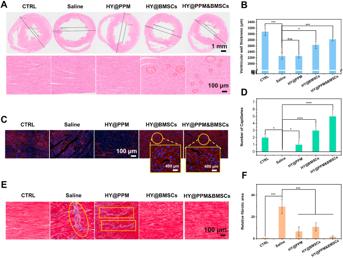
Mesenchymal stem cells (MSCs) have demonstrated significant therapeutic potential in heart failure (HF) treatment. However, their clinical application is impeded by low retention rate and low cellular activity of MSCs caused by high inflammatory and reactive oxygen species (ROS) microenvironment. In this study, monascus pigment (MP) nanoparticle (PPM) was proposed for improving adverse microenvironment and assisting in transplantation of bone marrow-derived MSCs (BMSCs). Meanwhile, in order to load PPM and reduce the mechanical damage of BMSCs, injectable hydrogels based on Schiff base cross-linking were prepared. The PPM displays ROS-scavenging and macrophage phenotype-regulating capabilities, significantly enhancing BMSCs survival and activity in HF microenvironment. This hydrogel demonstrates superior biocompatibility, injectability, and tissue adhesion. With the synergistic effects of injectable, adhesive hydrogel and the microenvironment-modulating properties of MP, cardiac function was effectively improved in the pericardial sac of rats. Our results offer insights into advancing BMSCs-based HF therapies and their clinical applications.
Keywords: BMSCs; Heart failure; Hydrogel; Microenvironment; Monascus pigment.
© 2024 The Authors.
Conflict of interest statement
The authors declare the following personal relationships which may be considered as potential competing interests: Zhilu Yang is an editorial board member for Bioactive Materials and was not involved in the editorial review or the decision to publish this article. Xiangbo Yang and Qisen Ji are currently employed by Ya'an Xunkang Pharmaceutical Co., LTD.
Figures

Similar articles
-
Reactive Oxygen Species Scavenging Hydrogel Regulates Stem Cell Behavior and Promotes Bone Healing in Osteoporosis.Tissue Eng Regen Med. 2023 Oct;20(6):981-992. doi: 10.1007/s13770-023-00561-w. Epub 2023 Sep 12. Tissue Eng Regen Med. 2023. PMID: 37697063 Free PMC article.
-
Injectable Polypeptide Hydrogels with Tunable Microenvironment for 3D Spreading and Chondrogenic Differentiation of Bone-Marrow-Derived Mesenchymal Stem Cells.Biomacromolecules. 2016 Dec 12;17(12):3862-3871. doi: 10.1021/acs.biomac.6b00884. Epub 2016 Nov 4. Biomacromolecules. 2016. PMID: 27775890
-
[Influence of the stiffness of three-dimensionally bioprinted extracellular matrix analogue on the differentiation of bone mesenchymal stem cells into skin appendage cells].Zhonghua Shao Shang Za Zhi. 2020 Nov 20;36(11):1013-1023. doi: 10.3760/cma.j.cn501120-20200811-00375. Zhonghua Shao Shang Za Zhi. 2020. PMID: 33238684 Chinese.
-
Single-Cell Profiles and Clinically Useful Properties of Human Mesenchymal Stem Cells of Adipose and Bone Marrow Origin.Am J Sports Med. 2019 Jun;47(7):1722-1733. doi: 10.1177/0363546519848678. Epub 2019 May 17. Am J Sports Med. 2019. PMID: 31100005 Review.
-
Advances in Injectable Hydrogel Strategies for Heart Failure Treatment.Adv Healthc Mater. 2023 Jul;12(19):e2300029. doi: 10.1002/adhm.202300029. Epub 2023 Mar 27. Adv Healthc Mater. 2023. PMID: 36913661 Review.
Cited by
-
Exploring the landscape of exosomes in heart failure: a bibliometric analysis.Int J Surg. 2025 May 1;111(5):3356-3372. doi: 10.1097/JS9.0000000000002248. Int J Surg. 2025. PMID: 39869380 Free PMC article. Review.
-
Effects of silver doping at the A-site on the structure, surface morphology, and magnetic behavior of La1-x Ag x SrMn2O5+δ (x = 0.1 and 0.2).RSC Adv. 2025 Jul 2;15(28):22616-22628. doi: 10.1039/d5ra02729b. eCollection 2025 Jun 30. RSC Adv. 2025. PMID: 40606197 Free PMC article.
-
Metal halide perovskites for energy applications: recent advances, challenges, and future perspectives.RSC Adv. 2025 Jun 26;15(27):21811-21837. doi: 10.1039/d5ra02730f. eCollection 2025 Jun 23. RSC Adv. 2025. PMID: 40575558 Free PMC article. Review.
-
Stem cells in the treatment of myocardial injury-induced cardiomyopathy: mechanisms and efficient utilization strategies.Front Pharmacol. 2025 Jun 18;16:1600604. doi: 10.3389/fphar.2025.1600604. eCollection 2025. Front Pharmacol. 2025. PMID: 40606613 Free PMC article. Review.
-
Impact of nanoemulsion of Ajwain-cardamom essential oils on Mortadella sausage quality during chilling (4°C) storage.Heliyon. 2025 Jan 2;11(1):e41643. doi: 10.1016/j.heliyon.2025.e41643. eCollection 2025 Jan 15. Heliyon. 2025. PMID: 39866456 Free PMC article.
References
-
- Heidenreich P.A., Bozkurt B., Aguilar D., Allen L.A., Byun J.J., Colvin M.M., Deswal A., Drazner M.H., Dunlay S.M., Evers L.R., Fang J.C., Fedson S.E., Fonarow G.C., Hayek S.S., Hernandez A.F., Khazanie P., Kittleson M.M., Lee C.S., Link M.S., Milano C.A., Nnacheta L.C., Sandhu A.T., Stevenson L.W., Vardeny O., Vest A.R., Yancy C.W. AHA/ACC/HFSA guideline for the management of heart failure: a report of the American college of cardiology/American heart association joint committee on clinical practice Guidelines. Circulation. 2022;145(18):e895–e1032. 2022. - PubMed
-
- Borlaug B.A., Sharma K., Shah S.J., Ho J.E. Heart failure with preserved ejection fraction: JACC scientific statement. J. Am. Coll. Cardiol. 2023;81(18):1810–1834. - PubMed
-
- Redfield M.M., Borlaug B.A. Heart failure with preserved ejection fraction: a review. JAMA. 2023;329(10):827–838. - PubMed
LinkOut - more resources
Full Text Sources
Research Materials
Miscellaneous
