Case Reports
. 2024 Aug 31;19(11):5389-5392.
doi: 10.1016/j.radcr.2024.08.001. eCollection 2024 Nov.

Intramedullary cavernous malformation: A case report

Affiliations
Case Reports

Intramedullary cavernous malformation: A case report

Nicola Maria Lucarelli et al. Radiol Case Rep. .

Abstract

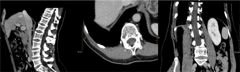
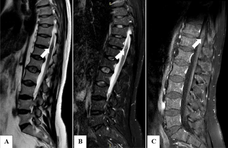
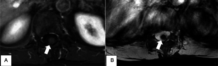
Cavernous malformations are rare vascular anomalies of the central nervous system, occurring in the spinal cord in just 5% of cases. Despite being documented in the literature, intramedullary cavernous malformations are exceedingly rare and often challenging to distinguish from other intramedullary lesions. We report a case of a 42-year-old patient with back pain, right-sided dysesthesias, and impaired proprioception in the distal limbs for approximately 3 months. Magnetic resonance imaging, crucial for differential diagnosis, identified intramedullary cavernous malformations at T11-12. Several conditions can hide the real cause of back pain; however, magnetic resonance imaging can reveal common conditions (such as discal hernia) and rare findings like cavernous malformations. Magnetic resonance imaging remains the study of choice for diagnosing and characterizing intramedullary cavernous malformations.

Keywords: Cavernoma; Cavernous; Intramedullary; MRI; Spinal cord; Vascular malformation.


Figures

Fig 1:
Fig. 1
Sagittal, axial, and coronal CT images at the T11-12 level showing the ICM was not visible and no calcifications were present.
Fig 2:
Fig. 2
(A) Sagittal T2-weighted MR image showing a well-defined lobulated lesion with a center of mixed signal and a hypointense rim (arrow). (B) Sagittal T2-weighted MR image with fat suppression magnifying the typical ICM characteristics (arrow). (C) Sagittal T1-weighted-SPIR MR image with intravenous contrast material showing slight gadolinium enhancement of the lesion (arrow).
Fig 3:
Fig. 3
(A) Axial T1-weighted-SPIR MR image with intravenous contrast material showing slight gadolinium enhancement of the lesion (arrow). (B) Axial T2-weighted-FFE MRI image showing a perilesional signal drop due to magnetic susceptibility artefacts, more pronounced in GRE sequences (arrow).

References

    1. Utomo SA, Bajamal AH, Yueniwati Y, Parenrengi MA, Fauziah D. Multiple spinal intramedullary cavernous angiomas with bleeding episode mimicking an intramedullary tumor. J Radiol Case Rep. 2022;16(3):15–22. doi: 10.3941/jrcr.v16i3.4251. - DOI - PMC - PubMed
    1. Liu W, Wang C, Wang B, Zhang Y, Jia W. Intramedullary spinal cavernous malformations with high ossification: a case report and review of the literature. Chin Neurosurg J. 2023;9(1):11. doi: 10.1186/s41016-023-00323-6. - DOI - PMC - PubMed
    1. Mariniello G, De Liso M, Russo C, Del Vecchio W, De Divitiis O, Bruno F, et al. Radiation-induced brain cavernomas in elderly: review of the literature and a rare case report. Acta Biomed. 2019;90(5-S):77–83. doi: 10.23750/abm.v90i5-S.8328. - DOI - PMC - PubMed
    1. Rauschenbach L, Santos AN, Engel A, Olbrich A, Benet A, Li Y, et al. Functional neurological outcome of spinal cavernous malformation surgery. Eur Spine J. 2023;32(5):1714–1720. doi: 10.1007/s00586-023-07640-5. - DOI - PubMed
    1. Reith W, Kettner M. Spinale Gefäßmissbildungen [Spinal cord vascular diseases] Radiologe. 2021;61(3):258–262. doi: 10.1007/s00117-021-00828-0. German. - DOI - PubMed

Publication types

LinkOut - more resources