. 2024 Aug 30;10(17):e37087.
doi: 10.1016/j.heliyon.2024.e37087. eCollection 2024 Sep 15.

Silencing miR-155-5p expression improves intestinal damage through inhibiting inflammation and ferroptosis in necrotizing enterocolitis

Affiliations

Silencing miR-155-5p expression improves intestinal damage through inhibiting inflammation and ferroptosis in necrotizing enterocolitis

Le Zhang et al. Heliyon. .

Abstract

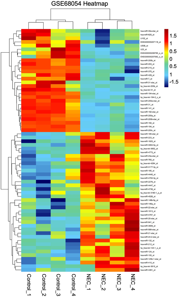
Background: Necrotizing enterocolitis (NEC) is a condition characterized by acquired damage to the mucosal lining, predominantly affecting premature infants. Bioinformatics assessments uncovered a notable rise in miR-155-5p expression in the intestinal tissues of infants suffering from NEC. Nevertheless, the development of NEC's underlying mechanisms and the role of miR-155-5p are still not well understood. This research aimed to explore the role of miR-155-5p in NEC and to elucidate its underlying mechanisms.

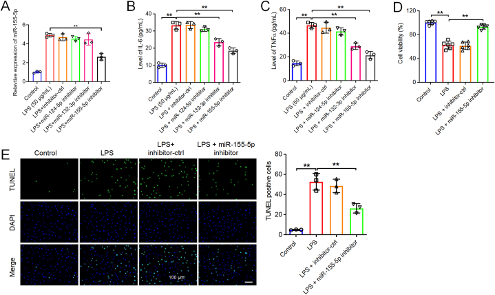
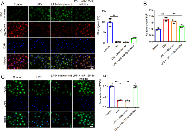
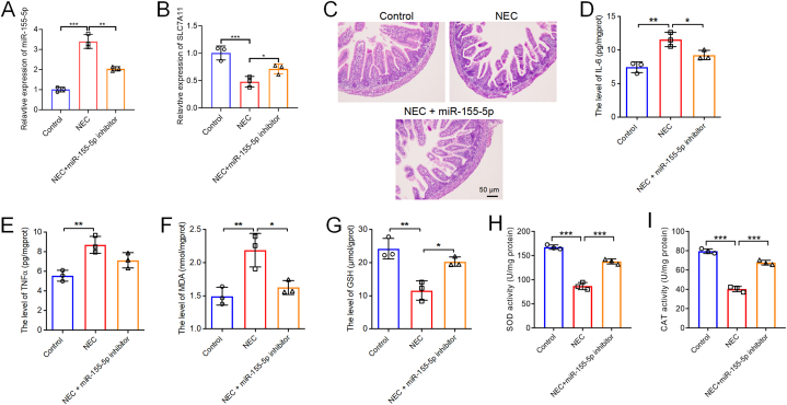
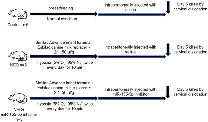
Methods: To replicate NEC in vitro, lipopolysaccharide (LPS) was employed, whereas an in vivo rat model of NEC was established using formula feeding and exposure to hypoxia. Subsequently, levels of inflammatory cytokines, cell survival, and apoptosis rates were assessed. Various biochemical indicators such as glutathione (GSH), superoxide dismutase (SOD), catalase (CAT), and malondialdehyde (MDA) were measured utilizing a purchased diagnostic kit. For the assessment of reactive oxygen species (ROS) and mitochondrial membrane potential (MMP) within FHC cells, analysis by flow cytometry was conducted. Additionally, the technique of Western blotting was utilized to analyze the levels of ferroptosis-associated proteins. Moreover, hematoxylin and eosin (H&E) staining was carried out to observe the histopathological alterations in the intestinal tissue samples from rats with necrotizing enterocolitis (NEC).

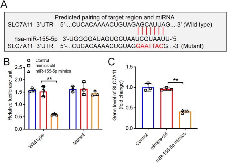
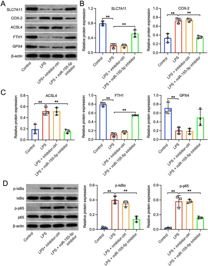
Results: Reducing miR-155-5p improved the survival of FHC cells exposed to LPS, decreased cell apoptosis, inflammation, and ferroptosis, and mitigated intestinal damage in NEC rats. Furthermore, SLC7A11 was found to be a direct target of miR-155-5p. The inhibition of miR-155-5p decreased LPS-induced inflammation and ferroptosis in both FHC cells and NEC rats by promoting SLC7A11 expression. This effect was evidenced by increased levels of ferroptosis-related proteins FTH1 and GPX4, decreased COX-2 and ACSL4 levels, lower lipid peroxidation marker MDA, reduced antioxidant markers GSH, SOD, and CAT, fewer IL-6 and TNF-α, and suppression of the IκBα/NF-κB p65 signaling pathway.

Conclusions: In conclusion, reducing miR-155-5p could improve intestinal damage in NEC by inhibiting inflammation and ferroptosis. These findings may provide theoretical insights for the development of new therapies for NEC.

Keywords: Ferroptosis; Inflammation; MiR-155–5p; Necrotizing enterocolitis.


Conflict of interest statement

The authors declare that there are no conflicts of interest.

Figures

Fig. 1
Fig. 1
Identification of DEmiRNAs in NEC. (A, B) The DEmiRNAs in intestinal tissues between patients with NEC and healthy controls were presented in the heatmap and volcano plot.
Fig. 2
Fig. 2
Downregulation of miR-155-5p promoted the viability and suppressed the apoptosis and inflammation of LPS-treated FHC cells. (A) FHC cells were stimulated with 50 μg/ml LPS for 6 h, and then transfected with inhibitor-ctrl, miR-124–5p inhibitor, miR-132–3p inhibitor or miR-155–5p inhibitor for 48 h. The levels of miR-155–5p in FHC cells were assessed by RT-qPCR. n = 3. (B and C) The level of IL-6 and TNF-α was evaluated by ELISA. n = 3. (D and E) FHC cells were stimulated with 50 μg/ml LPS for 6 h, and then transfected with inhibitor-ctrl or miR-155–5p inhibitor for 48 h. The viability and apoptosis of FHC cells were determined by CCK-8 (n = 6) and TUNEL (n = 3) assays respectively. Scale bar = 100 μm. **P < 0.01.
Fig. 3
Fig. 3
Downregulation of miR-155-5p suppressed the oxidative stress of LPS-treated FHC cells. FHC cells were stimulated with 50 μg/ml LPS for 6 h, and then transfected with inhibitor-ctrl or miR-155–5p inhibitor for 48 h. (A) ROS level in FHC cells were evaluated by flow cytometry. Levels of MDA (B), GSH (C), SOD (D) and CAT (E) in FHC cells were assessed by corresponding kit. n = 3, **P < 0.01, ***P < 0.001.
Fig. 4
Fig. 4
Downregulation of miR-155-5p suppressed the mitochondrial damage and ferroptosis in LPS-treated FHC cells. FHC cells were stimulated with 50 μg/ml LPS for 6 h, and then transfected with inhibitor-ctrl or miR-155–5p inhibitor for 48 h. (A) MMP in FHC cells was detected by JC-1 assay. (B) The iron levels in FHC cells were assessed by commercial kits. (C) The level of Fe2+ in FHC cells was detected by fluorescence intensity of PGSK. Scale bar = 50 μm. n = 3, **P < 0.01.
Fig. 5
Fig. 5
SLC7A11 was a direct target of miR-155-5p. (A) The putative binding sites of miR-155–5p on SLC7A11. (B) Luciferase assay of FHC cells co-transfected with SLC7A11-WT or SLC7A11-MT reporter plasmid and miR-155–5p mimic or mimics-ctrl. (C) The SLC7A11 level in FHC cells transfected with miR-155–5p mimics was assessed by RT-qPCR. n = 3, **P < 0.01.
Fig. 6
Fig. 6
Downregulation of miR-155-5p suppressed the ferroptosis in LPS-treated FHC cells via regulating SLC7A11 and IκBα/NF-κB p65 pathway. FHC cells were stimulated with 50 μg/ml LPS for 6 h, and then transfected with inhibitor-ctrl or miR-155–5p inhibitor for 48 h (A, B, C) Western blot assay was applied to determine the expressions of SLC7A11, COX-2, ACSL4, FTH1, GPX4 in FHC cells. (D) Western blot assay was conducted to detect the expressions of p-IκBα, IκBα, p-p65 and p65 in FHC cells. n = 3, **P < 0.01.
Fig. 7
Fig. 7
Downregulation of miR-155-5p attenuated intestinal injury, inflammatory response, and oxidative stress in NEC rats. (A) The relative expression of miR-155–5p in intestinal tissues of NEC rat and NEC + miR-155–5p inhibitor rat was evaluated by RT-qPCR. (B) The relative expression of SLC7A11 in intestinal tissues of NEC rat and NEC + miR-155–5p inhibitor rat was evaluated by RT-qPCR. (C) H&E staining showed the pathological changes in the intestine tissues. Scale bar = 50 μm. (D, E) The level of IL-6, TNF-α in intestinal tissues were assessed by ELISA. (F to I) Levels of MDA, GSH, SOD and CAT in intestinal tissues were assessed by corresponding kit. n = 3, *P < 0.05, **P < 0.01, ***P < 0.001.
Fig. 8
Fig. 8
Downregulation of miR-155-5p attenuated intestinal injury in NEC rats via modulating SLC7A11 affecting the IκBα/NF-κB p65 pathway. (A, B, C) Western blot assay was applied to determine the expressions of SLC7A11, COX-2, ACSL4, GPX4 in intestine tissues. (D) Western blot assay was conducted to detect the expressions of p-IκBα, IκBα, p-p65 and p65 in intestine tissues. n = 3, *P < 0.05, **P < 0.01.
figs1
figs1
figs2
figs2
Image 1

References

    1. Alganabi M., Lee C., Bindi E., et al. Recent advances in understanding necrotizing enterocolitis. F1000Research. 2019;8 - PMC - PubMed
    1. Gephart S.M., McGrath J.M., Effken J.A., et al. Necrotizing enterocolitis risk: state of the science. Adv. Neonatal Care. 2012;12(2):77–87. quiz 8-9. - PMC - PubMed
    1. Fan H., Chen Z., Lin R., et al. Bacteroides fragilis strain ZY-312 defense against cronobacter sakazakii-induced necrotizing enterocolitis in vitro and in a neonatal rat model. mSystems. 2019;4(4) - PMC - PubMed
    1. Chen Y., Koike Y., Chi L., et al. Formula feeding and immature gut microcirculation promote intestinal hypoxia, leading to necrotizing enterocolitis. Disease models & mechanisms. 2019;12(12) - PMC - PubMed
    1. Nair J., Lakshminrusimha S. Role of NO and other vascular mediators in the etiopathogenesis of necrotizing enterocolitis. Front. Biosci. 2019;11(1):9–28. - PubMed

LinkOut - more resources