. 2024 Aug 10;10(17):e36118.
doi: 10.1016/j.heliyon.2024.e36118. eCollection 2024 Sep 15.

Wound healing potential of silver nanoparticles from Hybanthus enneaspermus on rats

Affiliations

Wound healing potential of silver nanoparticles from Hybanthus enneaspermus on rats

Liang Cheng et al. Heliyon. .

Abstract

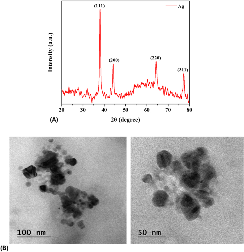
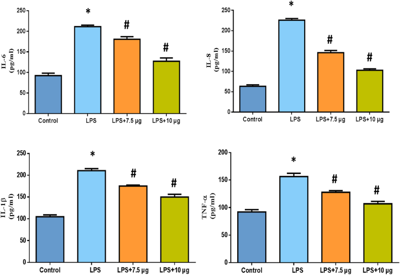
In this study, we green synthesized silver nanoparticles (Ag Nps) from Hybanthus enneaspermus leaves (HE-Ag NPs) and evaluated their antimicrobial and wound-healing properties. The synthesized HE-Ag NPs were characterized using various techniques, revealing face-centered polygonal structures, a well-dispersed appearance, and an average particle size of 42-51 nm. The antimicrobial effects of HE-Ag NPs and their embedded cotton fabrics were tested against several pathogens, showing effective inhibition of growth. The cytotoxicity and anti-inflammatory properties of HE-Ag NPs were assessed using MTT assays on L929 and RAW 264.7 cells and by measuring inflammatory cytokine levels in LPS-treated RAW 264.7 cells. HE-Ag NPs did not affect the viability of L929 and RAW 264.7 cells and significantly reduced inflammatory cytokine levels. In vivo studies using an excision wound model demonstrated that HE-Ag NPs-loaded ointment significantly increased hydroxyproline, total protein, and antioxidant levels and enhanced the wound contraction rate. These findings suggest that HE-Ag NPs have potent antimicrobial properties and promote wound healing, indicating their potential for use in topical ointments for wound care.

Keywords: Excision wound; Hybanthus enneaspermus; Hydroxyproline; RAW 264.7 cells; Silver nanoparticles.


Conflict of interest statement

The authors declare that there are no conflicts of interest.

Figures

Image 1
Graphical abstract
Fig. 1
Fig. 1
(A) UV–visible spectroscopy and (B) FT-IR analysis of the HE-Ag NPs.
Fig. 2
Fig. 2
(A) XRD and (B) TEM analysis of the HE-Ag NPs.
Fig. 3
Fig. 3
(A, B & C) SEM and EDAX (D) analysis of the HE-Ag NPs-embedded cotton fabrics. The appearance of the HE-Ag NPs embedded in the cotton fabrics was investigated using SEM along with EDAX. The SEM images indicated the formation of dispersed HE-Ag NPs and their occurrence on the cotton fabrics (A, B & C). The EDAX analysis revealed that HE-Ag-coated cotton fabrics have the highest Ag ions (D).
Fig. 4
Fig. 4
Anti-bacterial activity of the HE-Ag NPs. The synthesized HE-Ag NPs at various concentrations (20, 40, and 60 μg) considerably inhibited the pathogen's growth. The highest inhibition zone was noted on the K. pneumoniae and P. aeruginosa against 60 μg of synthesized HE-Ag NPs.
Fig. 5
Fig. 5
Anti-bacterial activity of the HE-Ag NPs-coated cotton fabrics. The cotton fabrics, which are coated with HE-Ag NPs revealed considerable anti-microbial effects against the tested pathogens. Control (C), Mixed cotton (MC), Cotton control (CC), and Mixed cotton control (MCC). The maximum inhibition was observed against K. pneumoniae and P. aeruginosa.
Fig. 6
Fig. 6
Effect of HE-Ag NPs on the viability of L929 and RAW 264.7 cells. Results were taken from triplicate assays and given as a mean ± SD of triplicates. The values are analyzed using the one-way ANOVA and Tukey's post hoc assay.
Fig. 7
Fig. 7
Effect of HE-Ag NPs on the levels of pro-inflammatory cytokines in the LPS-induced RAW 264.7 cells. Results were obtained from triplicate assays and given as a mean ± SD of triplicates. The values are analyzed using the one-way ANOVA and Tukey's post hoc assay. Note: ‘*’ represents that data are significant at p < 0.05 from the control and ‘#’ represents that the data are significant at p < 0.01 from the LPS-induced group.
Fig. 8
Fig. 8
Effect of HE-Ag NPs on the total protein and hydroxyproline levels (μg/100 mg of tissue) in wound tissues of the placebo. Values are expressed as mean ± SD for experiments. The values are analyzed using the one-way ANOVA and Tukey's post hoc assay. Note: ‘*’ represents that data are significant at p < 0.05 from group I and ‘#’ represents that the data are significant at p < 0.01 from LPS-induced group II.
Fig. 9
Fig. 9
Effect of HE-Ag NPs on the oxidative and anti-oxidative parameter levels. Results were obtained from expressed as mean ± SD for experiments. The values are analyzed using the one-way ANOVA and Tukey's post hoc assay. Note: ‘*’ represents that data are significant at p < 0.05 from group I and ‘#’ represents that the data are significant at p < 0.01 from LPS-induced group II.
Fig. 10
Fig. 10
Effect of HE-Ag NPs on the in vivo wound healing activity. The topical administration of ointment with HE-Ag NPs increased the wound contraction rate in rats with excision wounds. Group I: Control, Group II: HE-Ag NPs-loaded ointment, and Group III: Standard drug.
Fig. 11
Fig. 11
Effect of HE-Ag NPs on the in vivo wound contraction rate. Results were obtained as a mean ± SD. The values are analyzed using the one-way ANOVA and Tukey's post hoc assay. Note: ‘*’ represents that data are significant at p < 0.05 from group I and ‘#’ represents that the data are significant at p < 0.01 from LPS-induced group II.
Fig. 12
Fig. 12
Effect of HE-Ag NPs on wound histopathology. The untreated rats showed considerable changes such as fibrosis, infiltration of inflammatory cells, and inflammation (Group I). The wound tissues of the HE-Ag NPs-loaded ointment-treated rats had well-organized collagen fiber, reduced inflammatory cell infiltration, and no symptoms of inflammation in the regenerated tissues (Group II). Similar results were also noted on the wound tissues of the povidone iodine-treated rats (Group III).

References

    1. Christophers E., Schröder J.M. Evolution of innate defense in human skin. Exp. Dermatol. 2022;31:304–311. doi: 10.1111/exd.14482. - DOI - PubMed
    1. Varaprasad K., Jayaramudu T., Kanikireddy V., Toro C., Sadiku E.R. Alginate-based composite materials for wound dressing application: a mini-review. Carbohydr. Polym. 2020;236 doi: 10.1016/j.carbpol.2020.116025. - DOI - PubMed
    1. J Son Y., John W.T., Zhou Y., Mao W., Yim E.K., Yoo H.S. Biomaterials and controlled release strategy for epithelial wound healing. Biomater. Sci. 2019;7:4444–4471. doi: 10.1039/C9BM00456D. - DOI - PubMed
    1. Childs D.R., Murthy A.S. Overview of wound healing and management. Surgical Clinics. 2017;97:189–207. doi: 10.1016/j.suc.2016.08.013. - DOI - PubMed
    1. Dev S.K., Choudhury P.K., Srivastava R., Sharma M. Antimicrobial, anti-inflammatory and wound healing activity of polyherbal formulation. Biomed. Pharmacother. 2019;111:555–567. doi: 10.1016/j.biopha.2018.12.075. - DOI - PubMed

LinkOut - more resources