. 2024 Sep 11:19:9373-9393.
doi: 10.2147/IJN.S479959. eCollection 2024.

Multiomic Profiling and Neuroprotective Bioactivity of Salvia Hairy Root-Derived Extracellular Vesicles in a Cellular Model of Parkinson's Disease

Affiliations

Multiomic Profiling and Neuroprotective Bioactivity of Salvia Hairy Root-Derived Extracellular Vesicles in a Cellular Model of Parkinson's Disease

Vincenzo Vestuto et al. Int J Nanomedicine. .

Abstract

Purpose: Extracellular vesicles (EVs) are promising tools for nanomedicine and nanobiotechnology. The purification of mammalian-derived EVs involves intensive processes, and their therapeutic application raises multiple safety and regulatory issues. Plants have the potential to serve as nonconventional sources of therapeutically relevant EVs. In this context, we recently identified hairy roots (HRs) of medicinal plants as a novel biotechnological platform to produce EVs for human health.

Methods: Herein, we report the purification, omics profiling, and bioactivity of EVs isolated from HRs of the medicinal plants S. sclarea and S. dominica. EVs were isolated from conditioned media of HR cultures using differential ultracentrifugation (dUC) and size exclusion chromatography (SEC). The isolated EVs were characterized by nanoparticle tracking analysis (NTA) and electron microscopy. The proteomic and metabolomic profiles of the EVs were determined using mass spectrometry. Uptake studies and bioactivity assays, including confocal microscopy, MTT, flow cytometry, ROS quantification, and untargeted metabolomics analyses, were conducted in SH-SY5Y cells treated with the neurotoxin 6-hydroxydopamine (6-OHDA) to evaluate the therapeutic potential of EVs in an in vitro model of Parkinson's disease.

Results: S. sclarea HRs released nanosized round-shaped EVs with a distinctive molecular signature. HR EVs from S. sclarea and S. dominica revealed conserved cargo of secondary metabolites, predominantly triterpenoids, which are known for their antioxidant properties. We showed that HR EVs are safe, enter the cells, and strongly inhibit apoptosis in a cellular model of Parkinson's disease. Cellular metabolomics revealed that EVs preserved metabolic homeostasis and mitigated cellular oxidative stress when co-administered with 6-OHDA. Mechanistically, HR EVs inhibited 6-OHDA autoxidation and substantially reduced the accumulation of its oxidative products, which are responsible for 6-OHDA-induced toxicity.

Conclusion: Collectively, our findings provide compelling evidence that EVs isolated from the hairy roots of Salvia species are promising, non-mammalian alternative for the design of novel therapies targeting neurological disorders.

Keywords: Parkinson’s disease; Salvia extracellular vesicles; hairy roots; nanomedicine; neuroprotection; non-mammalian EV source.


Conflict of interest statement

The authors declare that they have no conflicts of interest in this work.

Figures

None
Graphical abstract
Figure 1
Figure 1
Isolation and characterization of EVs from Salvia sclarea HR conditioned medium. (A) Representative image of S. sclarea HR culture employed as EV biofactory. (B) Conditioned media were collected from two-month-old HRs and used for the purification of EVs by differential ultracentrifugation (dUC) or size exclusion chromatography (SEC). (C) Protein profile analysis of HR-derived EVs visualized through SDS-PAGE and silver staining. (D and E) Nanoparticle Tracking Analysis (NTA) measurements depict the size distribution of S. sclarea HR EVs purified by dUC (dilution= 1: 100) and SEC (dilution= 1:5), respectively. (F) Scanning electron image showing a group of S. sclarea HR EVs. (G) and (H) Transmission electron microscopy (TEM) close-up images displaying the round-shaped morphology of S. sclarea HR EVs. (I) Amplification of rolB, rolC and virD2 genes, located in the pRi15834 agrobacterium-type Ri plasmid was carried out through PCR in HR EVs. pRi15834 plasmid extracted from A. rhizogenes was employed as template for positive PCR control. Scale bars: 200 nm in (F), 100 nm in (G), 200 nm in (H).
Figure 2
Figure 2
Results of gene ontology analysis of the HR EV and HR proteomes. Proteins identified in the HR EVs (A) and HR (B) proteomes were classified based on their biological functions. The number of proteins identified, fold enrichment, and the FDR are indicated.
Figure 3
Figure 3
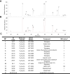
Metabolomic analysis of EVs of S. sclarea and S. dominica hairy roots. (A and B) Representative chromatograms showing the metabolomic profiles of S. sclarea and S. dominica HR EVs, respectively. (C) List of specialized metabolites detected in HR EVs of S. sclarea and S. dominica by UHPLC-HR-ESI-Orbitrap/MS. MSI status# in c: Metabolomics Standards Initiative.
Figure 4
Figure 4
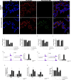
Evaluating neuroprotective effects of S. sclarea and S. dominica EVs against 6-OHDA-induced SH-SY5Y. (AD) Representative confocal microscopy imaging of SH-SY5Y control cells. (EH) Representative confocal microscopy analysis of SH-SY5Y cells treated with BODIPY -stained EVs (1.2 × 10⁸ particles/mL) of S. sclarea HRs for 24 h. Cells have been stained with phalloidin-TRITC (actin, red). Nuclei were stained with DAPI. Magnification 63×/1.4 numerical aperture. Scale bar = 20 mm in (D) and (H). (IM) Neuroprotective effects of S. sclarea and S. dominica upon administration of 1 µM mitoxantrone (I), 40 µM Aβ (1–42) (J), 400 µM H2O2 (K) and 100 µM 6-OHDA (L). The viability variations were determined by calculating the percentage of viable cells in treated cultures in comparison to untreated ones. (M and N) Evaluation of apoptosis in SH-SY5Y cells by propidium iodide assay through flow cytometry upon co-administration of S. sclarea and S. dominica HR EVs, respectively. Data are expressed as percentage of hypodiploid nuclei. (O and P) PINK1 and PARK2 mRNA levels by qRT-PCR in SH-SY5Y cells upon co-administration of S. sclarea and S. dominica EVs, respectively. GAPDH was used as housekeeping control. The 2–∆∆CT method was employed to calculate the expression fold changes relative to untreated cells. (Q) Spectrophotometric fluorescence intensity measurement showing the protective role of HR EVs in SH-SY5Y treated with 100 µM 6-OHDA. All results are showed as mean ± standard deviation from three independent experiments conducted with dUC and SEC-purified EVs. *, **, ***Denote respectively p < 0.05, p < 0.01 and p < 0.001 vs Ctrl; # and ###Denote respectively p < 0.05 and p < 0.001 vs 6-OHDA.
Figure 5
Figure 5
Untargeted metabolomics profiling to study the neuroprotective effects of HR EVs. (A and B) Principal component analysis of metabolomic data demonstrating good separation of 6-OHDA-treated groups with respect to treatments with S. sclarea and S. dominica HR EVs, respectively. (C and D) Enrichment analysis to functionally classify the differentially expressed metabolites in 6-OHDA compared to treatments with S. sclarea and S. dominica HR EVs, respectively. The enrichment ratio, beside the plots, is indicated by the proportional diameter size of the black circles.
Figure 6
Figure 6
Biochemical effects of S. sclarea and S. dominica HR EVs against 6-OHDA autoxidation. The formation of p-quinone was monitored at 490 nm in DMEM without serum with S. sclarea (A) or S. dominica (B) HR EVs using NAC (1 mM) as a positive control. The melanin formation assay was performed in a cell-free system, and the product was spectrophotometrically measured every 1 h. Melanin formation was monitored at 405 nm in DMEM without serum with S. sclarea (C) or S. dominica (d) HR EVs, using Fe2+ (300 µM) as a positive control. The results are presented as mean ± standard deviation (SD) from two independent experiments. *, **, *** denote respectively p < 0.05, p < 0.01 and p < 0.001 vs Ctrl; # and ## denote respectively p < 0.05, p < 0.01 and p < 0.001 vs 6-OHDA.

References

    1. Théry C, Witwer KW, Aikawa E, et al. Minimal information for studies of extracellular vesicles 2018 (MISEV2018): A position statement of the international society for extracellular vesicles and update of the MISEV2014 guidelines. J Extracell Vesicles. 2018;7(1):1535750. doi:10.1080/20013078.2018.1535750 - DOI - PMC - PubMed
    1. Welsh JA, Goberdhan DCI, O’Driscoll L, et al. Minimal information for studies of extracellular vesicles (MISEV2023): From basic to advanced approaches. J Extracell Vesicles. 2024;13(2):e12404. doi:10.1002/jev2.12404 - DOI - PMC - PubMed
    1. Chen Y, Xu Y, Zhong H, et al. Extracellular vesicles in inter-kingdom communication in gastrointestinal cancer. Am J Cancer Res. 2021;11(4):1087–1103. - PMC - PubMed
    1. Raposo G, Stahl PD. Extracellular vesicles: A new communication paradigm? Nat Rev Mol Cell Biol. 2019;20(9):509–510. doi:10.1038/s41580-019-0158-7 - DOI - PubMed
    1. Cai Q, Qiao L, Wang M, et al. Plants send small RNAs in extracellular vesicles to fungal pathogen to silence virulence genes. Science. 2018;360(6393):1126–1129. doi:10.1126/science.aar4142 - DOI - PMC - PubMed

MeSH terms