. 2024 Aug 11:9:100250.
doi: 10.1016/j.jtauto.2024.100250. eCollection 2024 Dec.

Vascular injury derived apoptotic exosome-like vesicles trigger autoimmunity

Affiliations

Vascular injury derived apoptotic exosome-like vesicles trigger autoimmunity

Sandrine Juillard et al. J Transl Autoimmun. .

Abstract

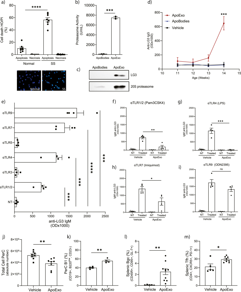
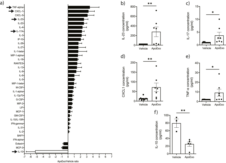
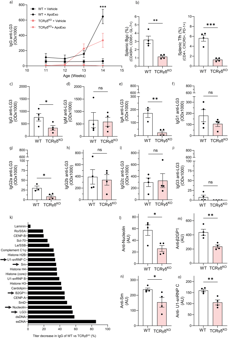
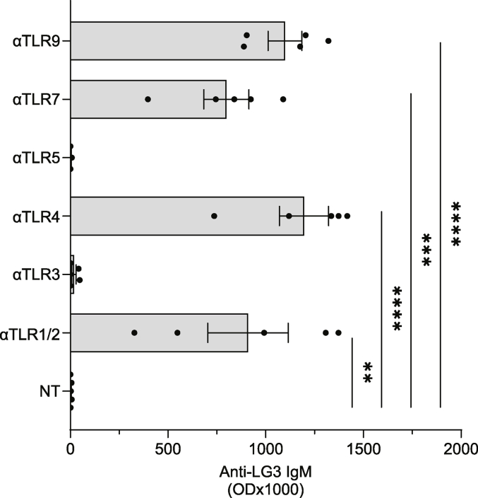
According to a central tenet of classical immune theory, a healthy immune system must avoid self-reactive lymphocyte clones but we now know that B cells repertoire exhibit some level of autoreactivity. These autoreactive B cells are thought to rely on self-ligands for their clonal selection and survival. Here, we confirm that healthy mice exhibit self-reactive B cell clones that can be stimulated in vitro by agonists of toll-like receptor (TLR) 1/2, TLR4, TLR7 and TLR9 to secrete anti-LG3/perlecan. LG3/perlecan is an antigen packaged in exosome-like structures released by apoptotic endothelial cells (ApoExos) upon vascular injury. We demonstrate that the injection of ApoExos in healthy animals activates the IL-23/IL-17 pro-inflammatory and autoimmune axis, and produces several autoantibodies, including anti-LG3 autoantibodies and hallmark autoantibodies found in systemic lupus erythematosus. We also identify γδT cells as key mediators of the maturation of ApoExos-induced autoantibodies in healthy mice. Altogether we show that ApoExos released by apoptotic endothelial cells display immune-mediating functions that can stimulate the B cells in the normal repertoire to produce autoantibodies. Our work also identifies TLR activation and γδT cells as important modulators of the humoral autoimmune response induced by ApoExos.

Keywords: Anti-LG3; ApoExos; Autoantibodies; Systemic lupus erythematosus (SLE); Toll-like receptors (TLR).


Conflict of interest statement

The authors declare that they have no known competing financial interests or personal relationships that could have appeared to influence the work reported in this paper.

Figures

Fig. 1
Fig. 1
B cells producing anti-LG3 are found in the normal B cell repertoire and are affected by ApoExos. (a) Levels of apoptosis (HO) or necrosis (PI) of murine aortic endothelial cells after 9 h in normal or serum starvation (SS) media. (b) Proteasome caspase-like activity assay kit revealed an increased proteasome activity in ApoExos compared to apoptotic bodies (ApoBodies). (c) Western blot showed increased in LG3 and 20S proteasome in ApoExos compare to ApoBodies. (d) ApoExos, but not ApoBodies or vehicle injections, produced anti-LG3 IgG autoantibodies (p = 0.0001). (e) LG3-specific B cells isolated from the peritoneal cavity (PerC) of vehicle-injected wild type (WT) mice are present in the normal repertoire and are activated in a TLR-dependent manner (TLR1/2, p = 0.0001; TLR4, p = 0.0005; TLR7, p < 0.0001; and TLR9, p = 0.0001) to secrete anti-LG3 IgM. ApoExos injection decreases anti-LG3 IgM production after stimulation with the aforementioned TLR agonists (TLR1/2, p = 0.0034 (f); TLR4, p = 0.0004 (g); TLR7, p = 0.0278 (h) and TLR9, trend only (i)) and in (j) total number of PerC cells compared to vehicle-injected WT mice (p = 0.0038). The percentage of (k) B1 cells among all B cells from the PerC, (l) splenic geminal center B cells (Bgc) and (m) follicular T cells (Tfh) increased after ApoExos injection (respectively, p = 0.0012, p = 0.0069 and p = 0.0151). Data are expressed as means ± SEMs, and statistical comparisons were carried out with a two-tailed Student's t-test. ****p < 0.0001; ***p < 0.001; **p < 0.01; *p < 0.05; OD, optical density; α, agonist.
Fig. 2
Fig. 2
ApoExos activate the IL-23/IL-17 autoimmune axis. (a) Ratio of cytokine levels in the sera of WT mice that received an ApoExos or vehicle injection, as measured by Luminex-based multiplex assay. (b) IL-23 (p = 0.0049), (c) IL-17 (p = 0.0132), (d) CXCL1 (p = 0.0042), (e) TNF-a (p = 0.0249), and (f) IL-10 (p = 0.0027) cytokines levels measured by ELISA in sera from WT mice after ApoExos or vehicle injection. Data were pooled from 3 independent experiments (n = 6 for each condition) and were expressed as means ± SEM. The statistical comparison between ApoExos and vehicle were carried out with a Student's t-test. ****p < 0.0001; ***p < 0.001; **p < 0.01; *p < 0.05.
Fig. 3
Fig. 3
ApoExos trigger the production of autoantibodies. (a) ELISA was performed on the sera from ApoExos- or vehicle-injected WT mice (n = 5), and a significant increase of circulating total IgG was observed (p=0.0481). (b) Microarray experiments of multiple autoantigens showed an SLE-associated autoantibody profile in mice injected with ApoExos, but not in those injected with vehicle. Autoantibodies detected were IgG. Each row of the heatmap represents one animal for a total of 12 ApoExos-injected WT mice and 10 vehicle-injected WT mice. The results were compared by ANOVA and minimal q value of represented data was 0,05. Blue and yellow are low and high level respectively. ELISA of sera from ApoExos- or vehicle-injected mice confirmed the increased levels of SLE-associated autoantibodies (i.e., (c) anti-LG3 (p=0.0019), (d) antinuclear antibodies (ANA) (p=0.0009), (e) anti-dsDNA (p=0.0212), (f) anti-Sm/nRNP (p=0.0124), (g) anti-Sm (p=0.0008), (h) anti-SSA (Ro) (p=0.0195), (i) anti-SSB (La) (p=0.0002), (j) anti-cardiolipin (CL) (p=0.0001) and (k) anti-β2GPI (p<0.0001)), while autoantibodies related to autoimmunity in transplantation were not affected (i.e., (l) anti-AT1R, (m) anti-vimentin and (n) anti-fibronectin). Statistical significance was assessed by Student's t-test. ****p < 0.0001; ***p < 0.001; **p < 0.01; *p < 0.05; ns not significant. Results are expressed as indicated: AU, Arbitrary Units; OD, Optical Density. (For interpretation of the references to colour in this figure legend, the reader is referred to the Web version of this article.)
Fig. 4
Fig. 4
γδT cells mediate anti-LG3 class switching after ApoExos injection. (a) TCRγδKO mice injected with ApoExos produce more circulating anti-LG3 IgG than those injected with the vehicle, although the levels remained below those of ApoExos-injected WT mice. (b) TCRγδKO mice injected with ApoExos have a decreased percentage of germinal center B cell (Bgc) and follicular helper T cell (Tfh) in total alive splenocytes harvested 21 days after the first exposure to ApoExos compared to WT mice also injected with ApoExos (respectivly p=0.0046 and p=0.0005). ELISA on serum from both strains injected with ApoExos showed unchanged levels of circulating anti-LG3 (d) IgM in the absence of γδT cells while (c) anti-LG3 IgG were significantly decreased (p = 0.05). (e) The levels of circulating anti-LG3 IgA were also significantly lower in TCRγδKO mice than WT ones after ApoExos injection (p = 0.005). An assessment of anti-LG3 IgG subclasses showed unchanged levels of (f) IgG1, (h) IgG2b, (i) IgG2c and (j) IgG3, while (g) IgG2a significantly decreased (p = 0.005). (k) Circulating autoantibodies from ApoExos injected in WT mice profiled using an antigen microarray and compared to the profile from ApoExos injected TCRγδKO mice. Arrows identify antibodies showing significant decrease that are also showed in independent graphs (l) anti-nucleolin (p=0.0229), (m) anti-β2GPI (p=0.0057), (n) anti-Sm (p=0.0466) and (o) anti-U1-sn-RNP (p=0.0057). Statistical significance was assessed by Student's t-test. ****p < 0.0001; ***p < 0.001; **p < 0.01; *p < 0.05; ns not significant. OD, Optical Density; AU, Arbitrary Unit.
Fig. S1
Fig. S1
Splenic B cells from WT mice produce anti-LG3 IgM in vitro with TLR agonists. Stimulation with agonists of TLR1/2 (p = 0.0023), TLR4 (p < 0.0001), TLR7 (p = 0.0001), and TLR9 (p < 0.0001), trigger the production of anti-LG3 IgM while TLR3 and TLR5 agonists did not. Statistical significance was assessed by Student's t-test. ****p < 0.0001; ***p < 0.001; **p < 0.01; *p < 0.05. OD, optical density; α, agonist.
Fig. S2
Fig. S2
Cytokine levels ratio in the sera of WT mice IV injected with apoptotic bodies (ApoBodies) or vehicle 21 days post-exposure, as measured by luminex-based assay multiplex assay.
Fig. S3
Fig. S3
Gating strategy for representative samples from ApoExos or vehicle injected WT mice. Gating on single cell and alive cells preceded all demonstrated gating. (a) Follicular helper T cell (Tfh) and (b) Germinal center B cell (Bgc) panels are assessed on the flow cytometer Fortessa while (c) B1 cell panel is assessed on an LSRII cytometer as described in the material and method section. Tfh are CD4+, CXCR5+, PD-1+, Bgc are CD45R+, CD95+, GL-7+ and B1 cells are CD19+, CD45Rlow, CD23. Analyses are done on Flowjo software (Ashland, OR, USA).
Fig. S4
Fig. S4
Autoantigen microarray IgG profiling showed an SLE-associated autoantibody profile in mice injected with ApoExos, but not in those injected with vehicle or apoptotic bodies (ApoBodies). Each row of the heatmap represents one animal for a total of 12 ApoExos-injected WT mice, 10 vehicle-injected WT mice and 11 ApoBodies-injected WT mice. The results were compared by ANOVA and minimal q value of represented data was 0,05. Blue and yellow are low and high level respectively.
Fig. S5
Fig. S5
ApoExos injected mice and vehicle injected ones had similar level of blood urea nitrogen (BUN) concentration (a) as well as negative urinary proteinuria (b). H&E and Siriusred staining of vehicle- and ApoExos-injected WT mice showed no difference in infiltration, tubular injury score or fibrosis 21 days after the first injection. Representative example of the kidney cortex of vehicule- or ApoExos injected mice (c). Statistical significance was assessed by Student's t-test. ns, not significant.

References

    1. Avrameas S., et al. Naturally occurring B-cell autoreactivity: a critical overview. J. Autoimmun. 2007;29(4):213–218. - PubMed
    1. Palma J., et al. Natural antibodies - facts known and unknown. Cent. Eur. J. Immunol. 2018;43(4):466–475. - PMC - PubMed
    1. Baumgarth N. B-1 cell heterogeneity and the regulation of natural and antigen-induced IgM production. Front. Immunol. 2016;7:324. - PMC - PubMed
    1. Baumgarth N. The double life of a B-1 cell: self-reactivity selects for protective effector functions. Nat. Rev. Immunol. 2011;11(1):34–46. - PubMed
    1. Baumgarth N. A hard(y) look at B-1 cell development and function. J. Immunol. 2017;199(10):3387–3394. - PMC - PubMed

LinkOut - more resources