. 2024 Nov;11(41):e2405265.
doi: 10.1002/advs.202405265. Epub 2024 Sep 17.

Establishment of a Magnetically Controlled Scalable Nerve Injury Model

Affiliations

Establishment of a Magnetically Controlled Scalable Nerve Injury Model

Tuo Yang et al. Adv Sci (Weinh). 2024 Nov.

Abstract

Animal models of peripheral nerve injury (PNI) serve as the fundamental basis for the investigations of nerve injury, regeneration, and neuropathic pain. The injury properties of such models, including the intensity and duration, significantly influence the subsequent pathological changes, pain development, and therapeutic efficacy. However, precise control over the intensity and duration of nerve injury remains challenging within existing animal models, thereby impeding accurate and comparative assessments of relevant cases. Here, a new model that provides quantitative and off-body controllable injury properties via a magnetically controlled clamp, is presented. The clamp can be implanted onto the rat sciatic nerve and exert varying degrees of compression under the control of an external magnetic field. It is demonstrated that this model can accurately simulate various degrees of pathology of human patients by adjusting the magnetic control and reveal specific pathological changes resulting from intensity heterogeneity that are challenging to detect previously. The controllability and quantifiability of this model may significantly reduce the uncertainty of central response and inter-experimenter variability, facilitating precise investigations into nerve injury, regeneration, and pain mechanisms.

Keywords: animal model; magnetic control; neuropathic pain; peripheral nerve injury (PNI).


Conflict of interest statement

X.L., W.W., and S.C. are inventors of CN Patent Application ZL 201910709478.4, ZL 202010727396.5, ZL 202110257070.5, and ZL 202210815021.3, which covers the mClamp, magnetic control unit and are assigned to Jilin university. The remaining authors declare no competing interests.

Figures

Figure 1
Figure 1
3D‐printed magnetically controlled clamp. A) Clamp mounting illustration and design details of the internal structure of the mClamp. B) Finite element geometry model with permanent magnets, 3D printed RG material, and air. C) Magnetic flux density distribution with 54 mT external magnetic field intensity. D) Stress distribution on a 3D‐printed structure with an external magnetic field of 54 mT. E) Locking mechanism stress distribution at locking state. F) Illustration of the magnetically controlled locking mechanism. The magnetic induction force (blue arrow) exerts a rotational torque on the magnetic beads. With movement along the support direction restricted, the magnetic beads can only generate a lateral tightening force (red arrow) for clamping purposes. The lower side of the support is connected to the bottom, creating a counteracting transverse force (red arrow) that balances out the clamping force. G) In vivo micro‐CT scanning of the compressed sciatic nerve and mClamp under the condition of the final compression intensities of control, mild, moderate, and severe intensities. Scale bar is 5 mm.
Figure 2
Figure 2
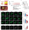
mClamp exerted spatiotemporal controllable compression to the rat sciatic nerve. A) Schematic of experimental design for implantation of the mClamp. B) Exterior of the implanted mClamp on rat sciatic nerve. Scale bar is 2 mm. C) Grouping and timeline. D) Schematic for the evaluation of the area of different sections of the sciatic nerve. Compression points are indicated by red arrowheads. E–G) Immunostaining of NF 200 for different sections of sciatic nerve trucks (E) and their quantification (F,G) at 12 weeks after compression. Compression points are indicated by red arrowheads. Scale bar is 50 µm. n = 3, 3, 3, 5, and 4 rats for the sham, control, mild, moderate, and severe group. H,I) Immunostaining of ATF3 in affected DRGs (H) and their quantification (I) at 12 weeks after compression. Scale bar is 500 µm. n = 5 rats per group. All data are expressed as the mean ± s.e.m. Statistical comparisons were conducted with one‐way ANOVA followed by Bonferroni's post hoc test.
Figure 3
Figure 3
mSNI induced controllable nociceptor hypersensitivity, motor and sensory dysfunction. A,B) mSNI rats exhibited graded mechanical (A) and thermal (B) pain hypersensitivity. n = 5 rats per group. C) Schematic of experimental design for whole‐mount patch clamp recordings of rat DRG and a small‐diameter DRG neuron with attached recording pipette under infrared phase contrast microscope. D) Quantification of rheobase. E,F) Representative traces (E) and quantification (F) of current‐evoked action potentials. n = 6 neurons from at least three separate rats (D,F). G,H) mSNI rats exhibited graded motor dysfunction in SFI (G) and dSFI (H) evaluations. n = 5 rats per group. All data are expressed as the mean ± s.e.m. Statistical comparisons were conducted with two‐way ANOVA followed by Bonferroni's post hoc test (A,B,F,G,H) or one‐way ANOVA followed by Bonferroni's post hoc test (D).
Figure 4
Figure 4
mSNI induced controllable histopathological changes. A,B) Electron micrographs (A) and their quantification (B) of the cross section at 5 mm distal to the injury site 12 weeks after compression. Areas of degeneration as well as axons in demyelination are indicated by red arrowheads. Axons in remyelination are indicated by blue arrowheads. Scale bar is 2 µm (upper) and 200 nm (lower). For axon diameter and G‐ratio, n = 15, 16, 33, 68, and 61 axons from 5, 6, 6, 7, and 6 rats. For axon density, n = 5, 6, 6, 7, and 6 rats. For myelin thickness, n = 28, 26, 60, 100, and 77 axons from 5, 6, 6, 7, and 6 rats. C) Immunostaining of the axon (NF 200, green) and myelin sheath (S100β, red) of the injured sciatic nerve 12 weeks after compression. Scale bar is 20 µm. D,E) Quantification of demyelinated axons at compression site (D) and ratio of demyelinated axons (E). n = 4 rats per group. F) Immunostaining of the myosin, including Laminin (red), myosin type 1 (MyHC‐1, blue), and myosin type 2A (MyHC‐IIA, green). Scale bar is 100 µm. G,H) Quantification of type 2 (G) and type 2BX (H) myosin. n = 5, 6, 7, 5, and 6 rats. I,J) Immunostaining for NMJ (I) and their quantification (J). Nerve fiber and presynaptic membrane (NF‐L and SYN, green), postsynaptic membrane (α‐BTX, red). Scale bar is 50 µm (I). n = 3 rats per group. All data are expressed as the mean ± s.e.m. Statistical comparisons were conducted with one‐way ANOVA followed by Bonferroni's post hoc test.

References

    1. a) Basbaum A. I., Bautista D. M., Scherrer G., Julius D., Cell 2009, 139, 267; - PMC - PubMed
    2. b) Julius D., Basbaum A. I., Nature 2001, 413, 203; - PubMed
    3. c) Donnelly C. R., Jiang C., Andriessen A. S., Wang K., Wang Z., Ding H., Zhao J., Luo X., Lee M. S., Lei Y. L., Maixner W., Ko M. C., Ji R. R., Nature 2021, 591, 275; - PMC - PubMed
    4. d) Gold M. S., Gebhart G. F., Nat. Med. 2010, 16, 1248; - PMC - PubMed
    5. e) Serger E., Luengo‐Gutierrez L., Chadwick J. S., Kong G., Zhou L., Crawford G., Danzi M. C., Myridakis A., Brandis A., Bello A. T., Muller F., Sanchez‐Vassopoulos A., De Virgiliis F., Liddell P., Dumas M. E., Strid J., Mani S., Dodd D., Di Giovanni S., Nature 2022, 607, 585. - PubMed
    1. a) Haroutounian S., Nikolajsen L., Bendtsen T. F., Finnerup N. B., Kristensen A. D., Hasselstrom J. B., Jensen T. S., Pain 2014, 155, 1272; - PubMed
    2. b) Zhong W., Ma X., Xing Y., Kim D. K., Wang A., Jiang W., Xu T., Neurosci. Lett. 2020, 716, 134643; - PubMed
    3. c) Challa S. R., Int. J. Neurosci. 2015, 125, 170; - PubMed
    4. d) Finnerup N. B., Kuner R., Jensen T. S., Physiol. Rev. 2021, 101, 259; - PubMed
    5. e) Scheib J., Hoke A., Nat. Rev. Neurol. 2013, 9, 668. - PubMed
    1. a) Dahlin L. B., Zimmerman M., Calcagni M., Hundepool C. A., van Alfen N., Chung K. C., Nat. Rev. Dis. Primers 2024, 10, 37; - PubMed
    2. b) Padua L., Coraci D., Erra C., Pazzaglia C., Paolasso I., Loreti C., Caliandro P., Hobson‐Webb L. D., Lancet Neurol. 2016, 15, 1273; - PubMed
    3. c) Padua L., Aprile I., Caliandro P., Mondelli M., Pasqualetti P., Tonali P. A., Neurology 2002, 59, 1643. - PubMed
    1. a) Gracely R. H., Lynch S. A., Bennett G. J., Pain 1992, 51, 175; - PubMed
    2. b) Meacham K., Shepherd A., Mohapatra D. P., Haroutounian S., Curr. Pain Headache Rep. 2017, 21, 28. - PubMed
    1. Nabaei V., Chandrawati R., Heidari H., Biosens. Bioelectron. 2018, 103, 69. - PubMed

LinkOut - more resources