. 2024 Oct 22;9(10):e0038724.
doi: 10.1128/msystems.00387-24. Epub 2024 Sep 17.

Genetic requirements for uropathogenic E. coli proliferation in the bladder cell infection cycle

Affiliations

Genetic requirements for uropathogenic E. coli proliferation in the bladder cell infection cycle

Daniel G Mediati et al. mSystems. .

Abstract

Uropathogenic Escherichia coli (UPEC) requires an adaptable physiology to survive the wide range of environments experienced in the host, including gut and urinary tract surfaces. To identify UPEC genes required during intracellular infection, we developed a transposon-directed insertion-site sequencing approach for cellular infection models and searched for genes in a library of ~20,000 UTI89 transposon-insertion mutants that are specifically required at the distinct stages of infection of cultured bladder epithelial cells. Some of the bacterial functional requirements apparent in host bladder cell growth overlapped with those for M9-glycerol, notably nutrient utilization, polysaccharide and macromolecule precursor biosynthesis, and cell envelope stress tolerance. Two genes implicated in the intracellular bladder cell infection stage were confirmed through independent gene deletion studies: neuC (sialic acid capsule biosynthesis) and hisF (histidine biosynthesis). Distinct sets of UPEC genes were also implicated in bacterial dispersal, where UPEC erupts from bladder cells in highly filamentous or motile forms upon exposure to human urine, and during recovery from infection in a rich medium. We confirm that the dedD gene linked to septal peptidoglycan remodeling is required during UPEC dispersal from human bladder cells and may help stabilize cell division or the cell wall during envelope stress created by host cells. Our findings support a view that the host intracellular environment and infection cycle are multi-nutrient limited and create stress that demands an array of biosynthetic, cell envelope integrity, and biofilm-related functions of UPEC.

Importance: Urinary tract infections (UTIs) are one of the most frequent infections worldwide. Uropathogenic Escherichia coli (UPEC), which accounts for ~80% of UTIs, must rapidly adapt to highly variable host environments, such as the gut, bladder sub-surface, and urine. In this study, we searched for UPEC genes required for bacterial growth and survival throughout the cellular infection cycle. Genes required for de novo synthesis of biomolecules and cell envelope integrity appeared to be important, and other genes were also implicated in bacterial dispersal and recovery from infection of cultured bladder cells. With further studies of individual gene function, their potential as therapeutic targets may be realized. This study expands knowledge of the UTI cycle and establishes an approach to genome-wide functional analyses of stage-resolved microbial infections.

Keywords: TraDIS; UPEC; UTI; cystitis; intracellular infection; stage-resolved model.


Conflict of interest statement

The authors declare no conflict of interest.

Figures
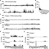
Fig 1
Fig 1
TraDIS mapping of an E. coli UTI89 transposon-insertion library. (A) The distribution of TraDIS mapped-reads in 150 bp windows along the ~5.06 Mbp UTI89 chromosome and ~114 kb plasmid, pUTI89. (B) Frequency distribution of the gene insertion index—the number of Tn-insertions within each gene divided by that gene’s length (in bp)—in the UTI89 Tn-insertion library. (C) The number of mapped-reads in 150 bp windows along the UTI89 chromosome following daily passaging (1/400) in LB media. Data were combined from both Tn-ends to generate these plots. (D) Examples of the read-count distribution (150 bp windows) from one culture replicate, encompassing 4,053,040–4,066,540 bp, showing a decrease in abundance of insertion mutants in waaV and waaL, and (E) 4,394,185–4,413,535 bp, showing an increase in abundance of insertion mutants in cytR over time.
Fig 2
Fig 2
Cell culture model of UTI89 infection of BECs for TraDIS analysis. (A) The stages of infection and the pre- and post-infection samples that were subsequently analyzed by TraDIS are represented by sample tubes. PI = post-infection. (B) Optical density measurements (OD600nm) of samples harvested in 20- or 100-min windows during the urine dispersal phase in the infection model. Error bars indicate the SD. (C) Phase-contrast micrograph of a dispersion sample shows a mixture of rod-shaped and highly filamentous UPEC. (D) Flow cytometry frequency distributions of cell size (represented by the side-scatter peak area), showing populations of high-scatter filamentous cells during dispersal.
Fig 3
Fig 3
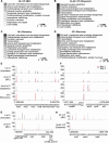
TraDIS read counts within selected UTI89 genomic loci at the indicated stages of BEC infection and recovery. (A–D) COGs detailed for statistically significant genes found within the TraDIS datasets in all stages of the infection and recovery process (adjusted p < 0.05). (E and F) Mapped read counts in the indicated genomic regions in all stages of the infection and recovery process that were sampled by TraDIS. The two experimental replicates are shown with blue and red bars. (G) Mapped read counts from the Inoculate (top panel) and IBC (bottom panel) samples encompassing the indicated tol and pal genes. (H) Mapped read counts from the IBC (top panel) and Dispersal (bottom panel) samples encompassing genes from the cedA region. Each plot has a bin size of 50 bp.
Fig 4
Fig 4
Identification of genes important for growth in M9-glycerol compared to LB. (A) Plot of each gene’s p-value against its corresponding logFC value, resulting from the comparison of transposon-insertion counts in M9-glycerol and LB media. The dashed lines indicate the stringent threshold criteria (p ≤ 0.05 and logFC < −2). (B) COGs detailed for the 42 statistically significant genes found to be required for growth in M9-glycerol (adjusted p < 0.05). (C–E) Representative read counts from one biological replicate at selected loci (read counts determined in 150 bp windows). The position of the annotated gene is indicated (below). (B) Coordinates 2,225,678–2,239,628 bp. (C) Coordinates 4,431,758–4,445,258 bp. (D) Coordinates 3,288,114–3,301,914 bp.
Fig 5
Fig 5
Growth of selected E. coli gene deletion mutants. (A) The relative log growth rates (µ) determined from growth curves (normalized as a percentage of WT UTI89) grown in LB or M9-glycerol. (B) The maximum OD600nm (% of WT) over the 24 h growth period. Panels (C) and (D) represent the same approach applied to E. coli K-12 strain BW25113 and the indicated mutants. Error bars indicate the SD from n = 2 independent replicates. *p-value ≤ 0.05 (two-sided t-test compared to WT).
Fig 6
Fig 6
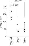
Deletion of hisF or neuC affects bladder cell infection. Gentamicin-protected bacteria (intracellular) of the indicated strains were recovered at the IBC stage and cfu, per area of the infection surface, were counted. Error bars indicate the SD (n = 3). Significance was calculated using a two-sided t-test.
Fig 7
Fig 7
Deletion of dedD affects bacterial growth during infection of human BECs. (A) Fluorescence composite image of mixed UTI89 WT (isogenic parent; mCherry—magenta) and ΔdedD (GFP—green) strains (pre-infection, 0 h). (B) The relative biomass (total cell area) was measured after dispersal (20 h urine treatment) from infection (49 h PI; n = 3). (C) LB co-culture assay, indicating a growth advantage of ΔdedD compared to WT; the example image at 49 h PI shows ΔdedD (GFP) dominant. Cell area data were normalized at each timepoint using a WT (mCherry) vs WT (GFP) control co-culture, normalized to a ratio of 1 (n = 3). Cultures were passaged at 9 h and 29 h (1:1,000 dilution into fresh LB).

References

    1. Justice SS, Hung C, Theriot JA, Fletcher DA, Anderson GG, Footer MJ, Hultgren SJ. 2004. Differentiation and developmental pathways of uropathogenic Escherichia coli in urinary tract pathogenesis. Proc Natl Acad Sci U S A 101:1333–1338. doi:10.1073/pnas.0308125100 - DOI - PMC - PubMed
    1. Lewis AJ, Richards AC, Mulvey MA. 2016. Invasion of host cells and tissues by uropathogenic bacteria. Microbiol Spectr 4. doi:10.1128/microbiolspec.UTI-0026-2016 - DOI - PMC - PubMed
    1. Mulvey MA, Schilling JD, Hultgren SJ. 2001. Establishment of a persistent Escherichia coli reservoir during the acute phase of a bladder infection. Infect Immun 69:4572–4579. doi:10.1128/IAI.69.7.4572-4579.2001 - DOI - PMC - PubMed
    1. Iosifidis G, Duggin IG. 2020. Distinct morphological fates of uropathogenic Escherichia coli intracellular bacterial communities: dependency on urine composition and pH. Infect Immun 88:e00884-19. doi:10.1128/IAI.00884-19 - DOI - PMC - PubMed
    1. Söderström B, Pittorino MJ, Daley DO, Duggin IG. 2022. Assembly dynamics of FtsZ and DamX during infection-related filamentation and division in uropathogenic E. coli. Nat Commun 13:3648. doi:10.1038/s41467-022-31378-1 - DOI - PMC - PubMed

MeSH terms

Substances

LinkOut - more resources